Tuftsin signals through its receptor neuropilin-1 via the transforming growth factor beta pathway
- PMID: 24033337
- PMCID: PMC3805743
- DOI: 10.1111/jnc.12404
Tuftsin signals through its receptor neuropilin-1 via the transforming growth factor beta pathway
Abstract
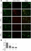
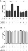
Tuftsin (Thr-Lys-Pro-Arg) is a natural immunomodulating peptide found to stimulate phagocytosis in macrophages/microglia. Tuftsin binds to the receptor neuropilin-1 (Nrp1) on the surface of cells. Nrp1 is a single-pass transmembrane protein, but its intracellular C-terminal domain is too small to signal independently. Instead, it associates with a variety of coreceptors. Despite its long history, the pathway through which tuftsin signals has not been described. To investigate this question, we employed various inhibitors to Nrp1's coreceptors to determine which route is responsible for tuftsin signaling. We use the inhibitor EG00229, which prevents tuftsin binding to Nrp1 on the surface of microglia and reverses the anti-inflammatory M2 shift induced by tuftsin. Furthermore, we demonstrate that blockade of transforming growth factor beta (TGFβ) signaling via TβR1 disrupts the M2 shift similar to EG00229. We report that tuftsin promotes Smad3 phosphorylation and reduces Akt phosphorylation. Taken together, our data show that tuftsin signals through Nrp1 and the canonical TGFβ signaling pathway. Despite the 40-year history of the tetrapeptide tuftsin (TKPR), a macrophage and microglial activator, its mechanism of action has not been defined. Here, we report that the tuftsin-mediated anti-inflammatory M2 shift in microglia is caused specifically by tuftsin binding to the receptor neuropilin-1 (Nrp1) and signaling through TGFβ receptor-1, a coreceptor of Nrp1. We further show that tuftsin signals via the canonical TGFβ pathway and promotes TGFβ release from target cells.
Keywords: anti-inflammatory; cytokines; microglia; neuropilin 1; tuftsin.
© 2013 International Society for Neurochemistry.
Figures


Similar articles
-
Tuftsin-driven experimental autoimmune encephalomyelitis recovery requires neuropilin-1.Glia. 2016 Jun;64(6):923-36. doi: 10.1002/glia.22972. Epub 2016 Feb 16. Glia. 2016. PMID: 26880314 Free PMC article.
-
Tuftsin binds neuropilin-1 through a sequence similar to that encoded by exon 8 of vascular endothelial growth factor.J Biol Chem. 2006 Mar 3;281(9):5702-10. doi: 10.1074/jbc.M511941200. Epub 2005 Dec 21. J Biol Chem. 2006. PMID: 16371354
-
Ablation of Neuropilin 1 from glioma-associated microglia and macrophages slows tumor progression.Oncotarget. 2016 Mar 1;7(9):9801-14. doi: 10.18632/oncotarget.6877. Oncotarget. 2016. PMID: 26755653 Free PMC article.
-
Neuropilin 1: function and therapeutic potential in cancer.Cancer Immunol Immunother. 2014 Feb;63(2):81-99. doi: 10.1007/s00262-013-1500-0. Epub 2013 Nov 22. Cancer Immunol Immunother. 2014. PMID: 24263240 Free PMC article. Review.
-
The interaction of Neuropilin-1 and Neuropilin-2 with tyrosine-kinase receptors for VEGF.Adv Exp Med Biol. 2002;515:81-90. doi: 10.1007/978-1-4615-0119-0_7. Adv Exp Med Biol. 2002. PMID: 12613545 Review.
Cited by
-
Glial and myeloid heterogeneity in the brain tumour microenvironment.Nat Rev Cancer. 2021 Dec;21(12):786-802. doi: 10.1038/s41568-021-00397-3. Epub 2021 Sep 28. Nat Rev Cancer. 2021. PMID: 34584243 Free PMC article. Review.
-
Suppressing Inflammation for the Treatment of Diabetic Retinopathy and Age-Related Macular Degeneration: Dazdotuftide as a Potential New Multitarget Therapeutic Candidate.Biomedicines. 2023 May 27;11(6):1562. doi: 10.3390/biomedicines11061562. Biomedicines. 2023. PMID: 37371657 Free PMC article. Review.
-
Small Molecule Neuropilin-1 Antagonists Combine Antiangiogenic and Antitumor Activity with Immune Modulation through Reduction of Transforming Growth Factor Beta (TGFβ) Production in Regulatory T-Cells.J Med Chem. 2018 May 10;61(9):4135-4154. doi: 10.1021/acs.jmedchem.8b00210. Epub 2018 Apr 24. J Med Chem. 2018. PMID: 29648813 Free PMC article.
-
Novel Compounds Targeting Neuropilin Receptor 1 with Potential To Interfere with SARS-CoV-2 Virus Entry.ACS Chem Neurosci. 2021 Apr 21;12(8):1299-1312. doi: 10.1021/acschemneuro.0c00619. Epub 2021 Mar 31. ACS Chem Neurosci. 2021. PMID: 33787218 Free PMC article.
-
In silico identification and validation of inhibitors of the interaction between neuropilin receptor 1 and SARS-CoV-2 Spike protein.bioRxiv [Preprint]. 2020 Sep 23:2020.09.22.308783. doi: 10.1101/2020.09.22.308783. bioRxiv. 2020. PMID: 32995772 Free PMC article. Preprint.
References
Publication types
MeSH terms
Substances
Grants and funding
LinkOut - more resources
Full Text Sources
Other Literature Sources
Miscellaneous

