

Pigmented lesions of the oral cavity: an update
- PMID: 24034073
- PMCID: PMC3775277
- DOI: 10.1016/j.cden.2013.07.006
Pigmented lesions of the oral cavity: an update
Abstract
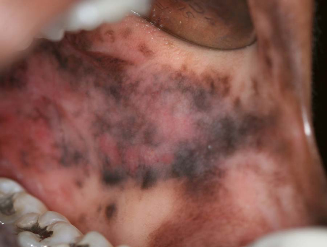
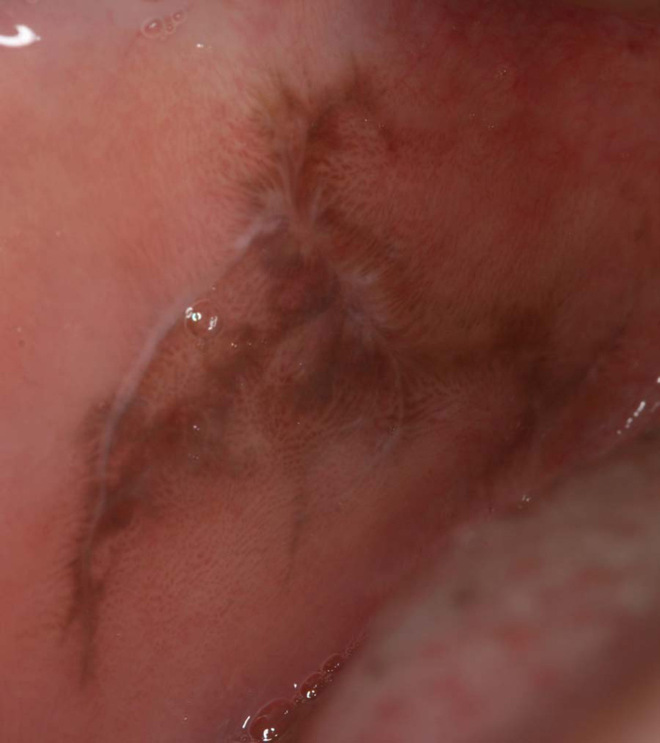
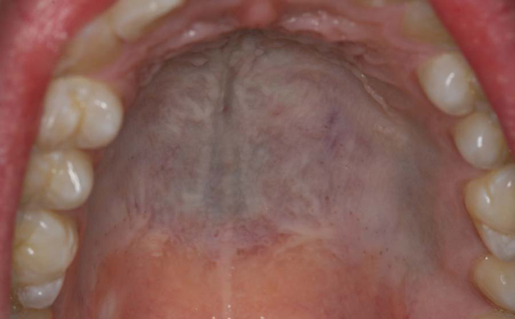
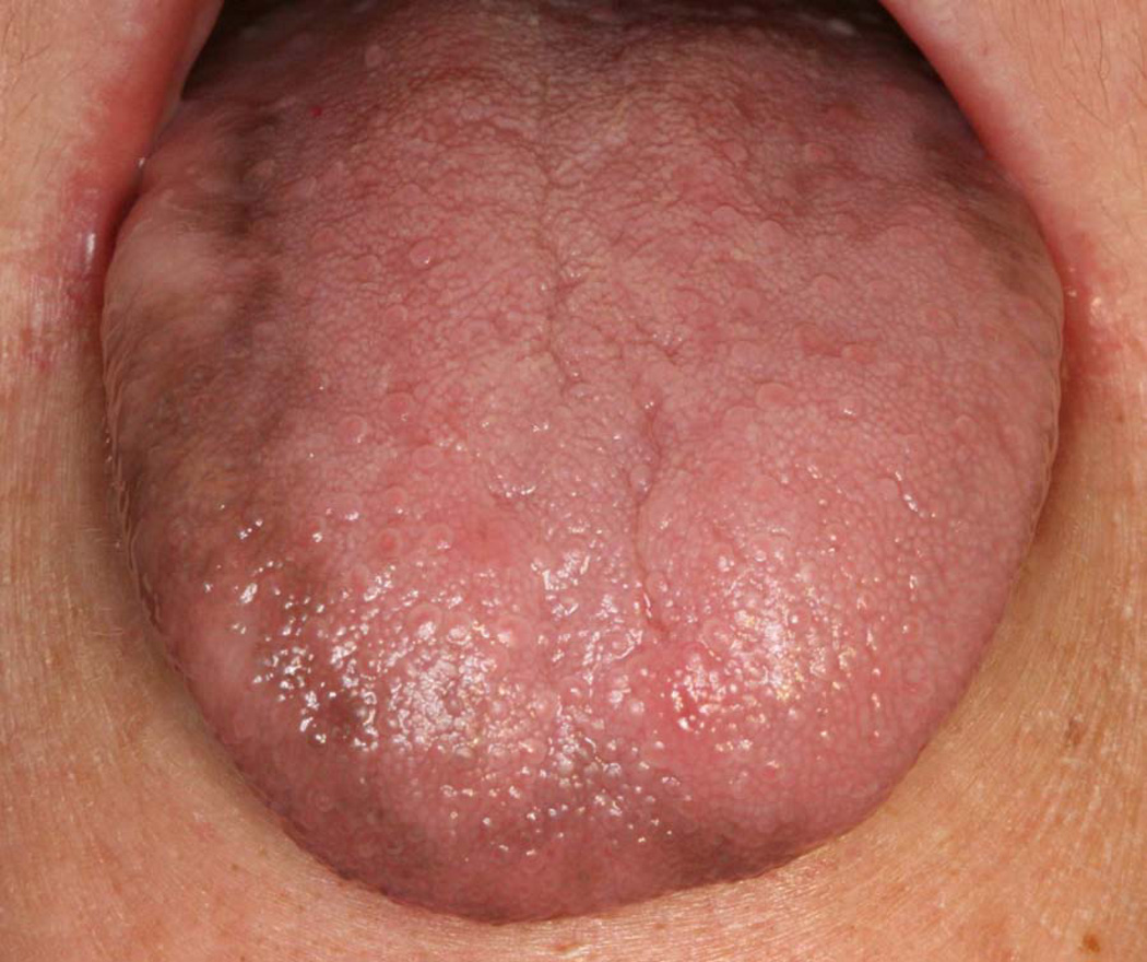
Oral pigmentation may be focal, multifocal, or diffuse. The lesions may be blue, purple, brown, gray, or black. They may be macular or tumefactive. Some are localized harmless accumulations of melanin, hemosiderin, or exogenous metal; others are harbingers of systemic or genetic disease; and some can be associated with life-threatening medical conditions that require immediate intervention. The differential diagnosis for any pigmented lesion is extensive, and can include examples of endogenous and exogenous pigmentation. Although biopsy is a helpful and necessary aid in the diagnosis of focally pigmented lesions, with diffuse presentations lesions require a thorough history and laboratory studies to establish a definitive diagnosis.
Keywords: Cushing disease; Hypoadrenocorticism; Malignant melanoma; Melanin; Pigmentation.
Copyright © 2013 Elsevier Inc. All rights reserved.
Figures

References
-
- Lenane P, Powell FC. Oral pigmentation. J Eur Acad Dermatol Venereol. 2000;14:448–465. - PubMed
-
- Westerhof W. The discovery of the human melanocyte. Pigment Cell Res. 2006;19:183–193. - PubMed
-
- Kaugars GE, Heise AP, Riley WT, Abbey LM, Svirsky JA. Oral melanotic macules. A review of 353 cases. Oral Surg Oral Med Oral Pathol. 1993;76:59–61. - PubMed
-
- Fornatora ML, Reich RF, Haber S, Solomon F, Freedman PD. Oral melanoacanthoma: a report of 10 cases, review of the literature, and immunohistochemical analysis for HMB-45 reactivity. Am J Dermatopathol. 2003;25:12–15. - PubMed
-
- Taybos G. Oral changes associated with tobacco use. Am J Med Sci. 2003;326:179–182. - PubMed
Publication types
MeSH terms
Grants and funding
LinkOut - more resources
Full Text Sources
Other Literature Sources
Medical
