. 2013 Sep 11;14(3):306-17.
doi: 10.1016/j.chom.2013.08.013.

Identifying Yersinia YopH-targeted signal transduction pathways that impair neutrophil responses during in vivo murine infection

Affiliations

Identifying Yersinia YopH-targeted signal transduction pathways that impair neutrophil responses during in vivo murine infection

Hortensia G Rolán et al. Cell Host Microbe. .

Abstract

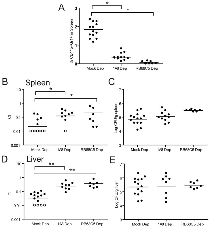
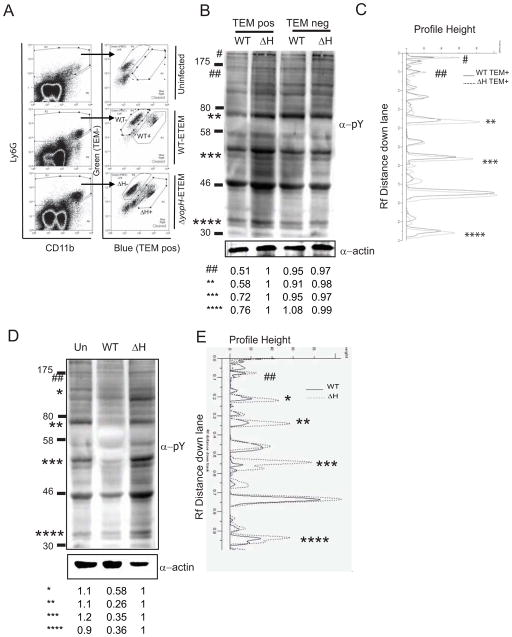
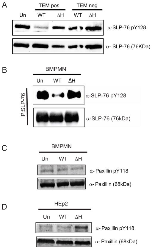
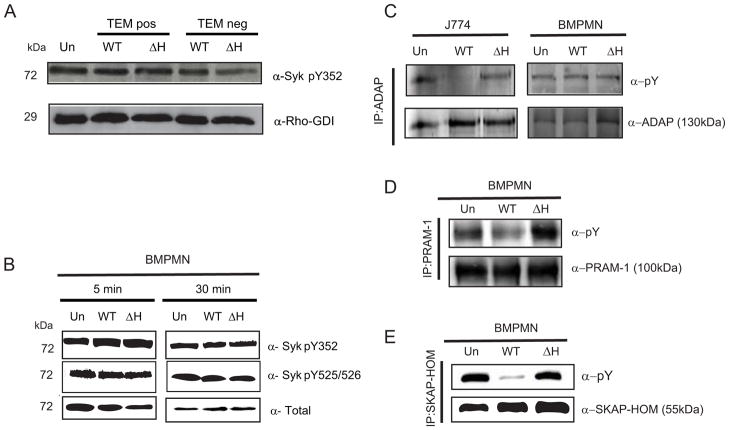
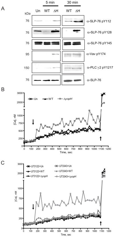
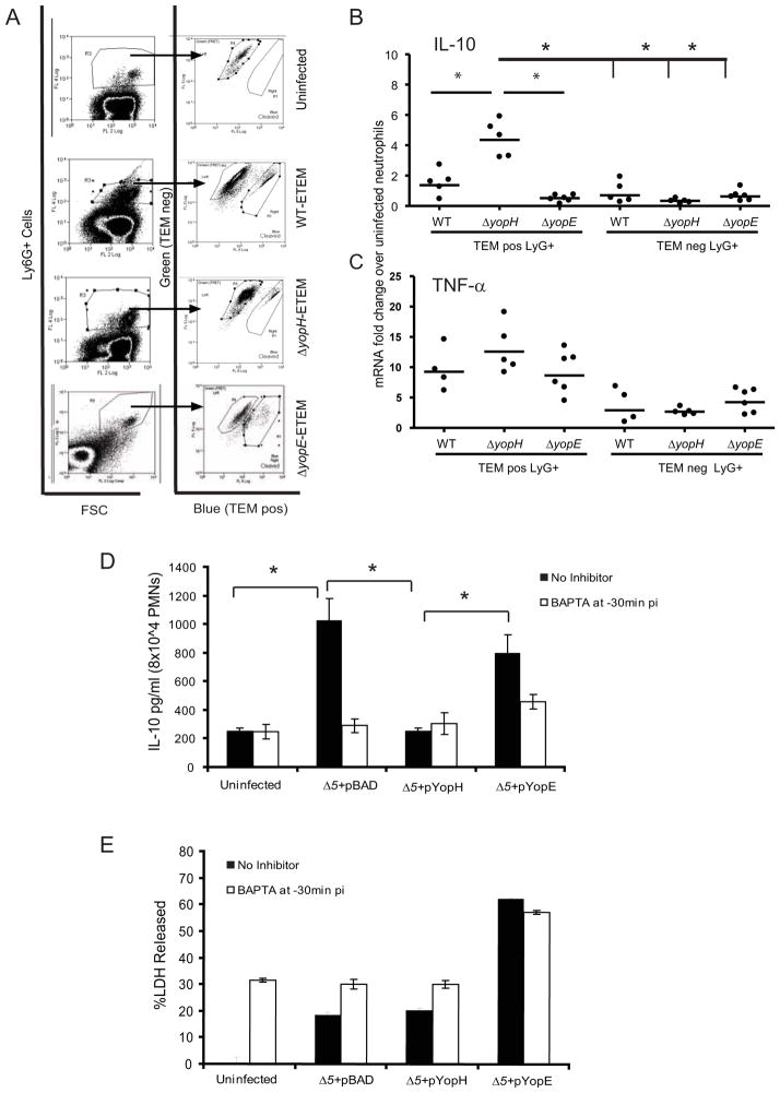
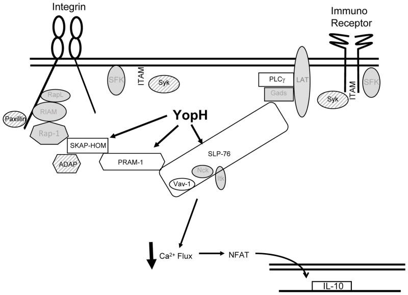
Identifying molecular targets of Yersinia virulence effectors, or Yops, during animal infection is challenging because few cells are targeted by Yops in an infected organ, and isolating these sparse effector-containing cells is difficult. YopH, a tyrosine phosphatase, is essential for full virulence of Yersinia. Investigating the YopH-targeted signal transduction pathway(s) in neutrophils during infection of a murine host, we find that several host proteins, including the essential signaling adaptor SLP-76, are dephosphorylated in the presence of YopH in neutrophils isolated from infected tissues. YopH inactivated PRAM-1/SKAP-HOM and the SLP-76/Vav/PLCγ2 signal transduction axes, leading to an inhibition of calcium response in isolated neutrophils. Consistent with a failure to mount a calcium response, IL-10 production was reduced in neutrophils containing YopH from infected tissues. Finally, a yopH mutant survived better in the absence of neutrophils, indicating that neutrophil inactivation by YopH by targeting PRAM-1/SKAP-HOM and SLP-76/Vav/PLCγ2 signaling hubs may be critical for Yersinia survival.


Figures

Figure 1
Figure 1. PMN depletion restores growth of a ΔyopH strain in competition with WT
BALB/c were infected IV with an equal mixture of WT-Kan and ΔyopH after mock, 1A8 or RB68C5 injection and sacrificed 3 days p.i. (A) Percentage of Gr1+CD11b+ cells in spleens of untreated mice and mice treated with 1A8 or RB68C5. Competitive index (C.I) in the (B) spleens and (D) livers of BALB/c-depleted mice. The total number of bacteria recovered in the (C) spleen and (E) liver. Each dot represents a mouse; horizontal bars represent the average (A) or geometric mean (B–E); open circles indicate that no ΔyopH was recovered in 100 colonies tested. The experiment was repeated twice and all the data is shown. C.I. = (ΔyopH/WTKan)output/(ΔyopH/WTKan)input. Significance was calculated using one-way ANOVA followed by Tukey’s post test (*P < 0.05 or **P < 0.01). See also Table S1.
Figure 2
Figure 2. YopH-dependent dephosphorylation in PMNs isolated from spleens of infected animals
(A–C) Spleens from mice infected with WT-ETEM or ΔyopH-ETEM were harvested 5 days p.i. and TEMpos and TEMneg PMNs were collected. (B–C) Western blot analysis of 1×10⁵ TEMpos PMNs and TEMneg PMNs probed with a α-phosphotyrosine antibody (4G10), striped and reprobed with α-actin antibody. (D–E) BMPMNs infected with WT or ΔyopH strains were lysed and probed with 4G10, stripped and re-probed for actin. (B–E) GeneTools software was used to quantify the phosphorylation levels of bands of interest, denoted by *, by normalizing them to actin controls and then to TEMpos ΔH lane. Each blot is representative of at least 2 independent experiments.* and # denotes bands with reduced intensities; ‘Un’ denotes uninfected PMNs; see also Figure S1.
Figure 3
Figure 3. SLP-76, but not paxillin, is dephosphorylated by YopH during animal infection
(A) TEMpos PMNs and iMo (Gr1+CD11b+) from mice intravenously infected with WT-ETEM or ΔyopH-ETEM for 5 days were collected by FACS. Lysates from 1×10⁵ cells were probed for total SLP-76 and phosphorylation at Y128. (B) BMPMNs (5×10⁵/lane) were infected with WT or ΔyopH for 5 min at a MOI of 50:1. Immunoprecipitated SLP-76 was analyzed by Western blot for phosphorylated SLP-76 Y128, and total SLP-76. (C–D) BMPMNs (C) or HEp2 cells (D) were infected at MOI 50:1 for 30 min with WT or ΔyopH, lysed and probed for phosphorylation at paxillin Y118 or total paxillin. Blots shown are representative of at least 2 independent experiments; see also Figure S2.
Figure 4
Figure 4. YopH dephosphorylates PRAM-1, SKAP-HOM, but not Syk or ADAP, in PMNs
(A)TEMpos Gr1+CD11b+ and TEMneg Gr1+CD11b+ PMNs and iMO from spleens of mice infected with WT-ETEM or ΔyopH-ETEM for 5 days were collected, lysed and probed for phosphorylation at Y352 in Syk or Rho-GDI as loading control. (B) BMPMNs were infected for 5 or 30 min at a MOI of 50:1 with either WT or ΔyopH and then were lysed, resolved by SDS-PAGE and analyzed by Western blot for phosphorylation of Syk pY352, Syk PY525/526 and total. (C) J774 (5×10⁵cells/lane) and BMPMNs (2×10⁶cells/lane) were infected with WT or ΔyopH for 30 min at a MOI of 50:1. Lysates were immunoprecipitated with antibodies to ADAP, resolved by SDS-PAGE and analyzed by Western blots for phosphotyrosine and total ADAP. (D–E) BMPMNs (5×10⁵/lane) were infected with WT or ΔyopH for 30 min at a MOI of 50:1. Lysates were immunoprecipitated with antibodies to PRAM-1 (D) or SKAP-HOM (E), resolved by SDS-PAGE and analyzed by Western blot for phosphotyrosine and total PRAM-1 and SKAP-HOM. All blots shown are representative of at least 2 independent experiments; see also Figures S3.
Figure 5
Figure 5. YopH inactivates the SLP-76/Vav/PLCγ2 signaling axis and prevents calcium flux in PMNs
(A) BMPMNs were infected for 5 or 30 minutes at a MOI of 50:1 with either WT or ΔyopH and then were lysed, resolved by SDS-PAGE and analyzed by Western blot for phosphorylation of SLP-76 pY112, SLP-76 pY128, SLP-76 pY145, Vav-1 pY174, phospho PLCγ2, and total SLP-76. All data shown are representative of at least 2 independent experiments. (B–C) BMPMNs were loaded with Fura-2 and (C) with 1μM U73122 (a PLC inhibitor) or with 1μM of its inactive analog, U73343. After recording basal levels for 2 (B) or 3 (C) min in a SpectraMax M5 plate reader at 37°C, cells were infected (indicated by the arrow) with WT or ΔyopH strains at MOI of 50:1 for 12 min and then ionomycin (indicated by second arrow) was added. Fluorescence was recorded over time to calculate intracellular calcium. Each experiment was repeated three times and a representative experiment is shown.
Figure 6
Figure 6. YopH inhibits IL-10 secretion in PMNs
(A–C) Mice were infected IV with either WT-ETEM, ΔyopH-ETEM or ΔyopE-ETEM for 5 days. TEMposLy6G+(1A8) and TEMnegLy6G+(1A8) PMNs from spleens of each mouse were sorted by FACS (A). RNA was isolated and transcript levels of (B) IL-10 and (C) TNF-α were determined using real-time PCR. Each dot represents one animal. (D–E) BMPMNs were untreated or pretreated with 25μM BAPTA for 30 minutes and then infected with Δ5+pBAD, Δ5+pYopH or Δ 5+pYopE at MOI 10:1 for 4 hours before gentamicin was added. The supernatants were collected 12 hours after the addition of gentamicin and IL-10 (D) and LDH (E) levels were determined. Three independent experiments were performed in triplicate. The average +/− STD of one representative experiment is shown. Statistical significance was calculated using one-way ANOVA with Tukey’s post test * indicates P<0.05; see also Figures S4.
Figure 7
Figure 7. Model of YopH targets in PMNs
YopH targets SLP-76 and/or PRAM-1/SKAP-HOM signal transduction pathways in PMNs. White shapes represent direct or indirect targets of YopH. Calcium flux is blocked and cytokine production altered. Syk, ADAP and paxillin (hatched shapes) were tested, but were not dephosphorylated by YopH in PMNs. Gray shapes represent molecules in pathway not tested.

References

    1. Alenghat FJ, Baca QJ, Rubin NT, Pao LI, Matozaki T, Lowell CA, Golan DE, Neel BG, Swanson KD. Macrophages require Skap2 and Sirpalpha for integrin-stimulated cytoskeletal rearrangement. Journal of cell science. 2012;125:5535–5545. - PMC - PubMed
    1. Alonso A, Bottini N, Bruckner S, Rahmouni S, Williams S, Schoenberger SP, Mustelin T. Lck dephosphorylation at Tyr-394 and inhibition of T cell antigen receptor signaling by Yersinia phosphatase YopH. J Biol Chem. 2004;279:4922–4928. - PubMed
    1. Andersson K, Magnusson KE, Majeed M, Stendahl O, Fallman M. Yersinia pseudotuberculosis-induced calcium signaling in neutrophils is blocked by the virulence effector YopH. Infect Immun. 1999;67:2567–2574. - PMC - PubMed
    1. Auerbuch V, Isberg RR. Growth of Yersinia pseudotuberculosis in Mice Occurs Independently of Toll-Like Receptor 2 expression and induction of Interleukin-10. Infect Immun. 2007;75:3561–3570. - PMC - PubMed
    1. Black DS, Bliska JB. Identification of p130Cas as a substrate of Yersinia YopH (Yop51), a bacterial protein tyrosine phosphatase that translocates into mammalian cells and targets focal adhesions. EMBO J. 1997;16:2730–2744. - PMC - PubMed

Publication types

MeSH terms