. 2013 Sep 14:14:619.
doi: 10.1186/1471-2164-14-619.

Efficient disruption of Zebrafish genes using a Gal4-containing gene trap

Affiliations

Efficient disruption of Zebrafish genes using a Gal4-containing gene trap

Jorune Balciuniene et al. BMC Genomics. .

Abstract

Background: External development and optical transparency of embryos make zebrafish exceptionally suitable for in vivo insertional mutagenesis using fluorescent proteins to visualize expression patterns of mutated genes. Recently developed Gene Breaking Transposon (GBT) vectors greatly improve the fidelity and mutagenicity of transposon-based gene trap vectors.

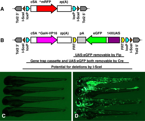
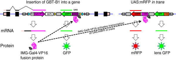
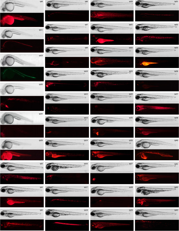
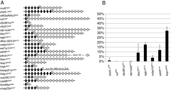
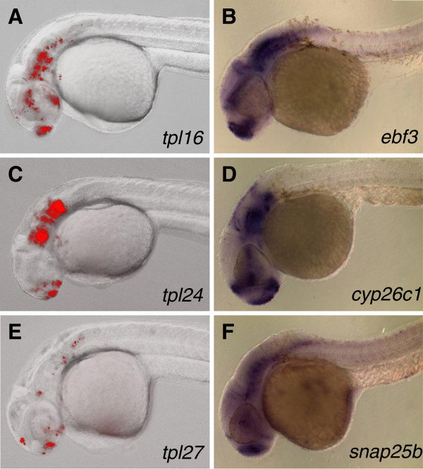
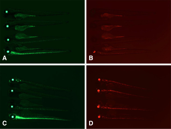
Results: We constructed and tested a bipartite GBT vector with Gal4-VP16 as the primary gene trap reporter. Our vector also contains a UAS:eGFP cassette for direct detection of gene trap events by fluorescence. To confirm gene trap events, we generated a UAS:mRFP tester line. We screened 270 potential founders and established 41 gene trap lines. Three of our gene trap alleles display homozygous lethal phenotypes ranging from embryonic to late larval: nsf( tpl6), atp1a3a(tpl10) and flr(tpl19). Our gene trap cassette is flanked by direct loxP sites, which enabled us to successfully revert nsf( tpl6), atp1a3a(tpl10) and flr(tpl19) gene trap alleles by injection of Cre mRNA. The UAS:eGFP cassette is flanked by direct FRT sites. It can be readily removed by injection of Flp mRNA for use of our gene trap alleles with other tissue-specific GFP-marked lines. The Gal4-VP16 component of our vector provides two important advantages over other GBT vectors. The first is increased sensitivity, which enabled us to detect previously unnoticed expression of nsf in the pancreas. The second advantage is that all our gene trap lines, including integrations into non-essential genes, can be used as highly specific Gal4 drivers for expression of other transgenes under the control of Gal4 UAS.

Conclusions: The Gal4-containing bipartite Gene Breaking Transposon vector presented here retains high specificity for integrations into genes, high mutagenicity and revertibility by Cre. These features, together with utility as highly specific Gal4 drivers, make gene trap mutants presented here especially useful to the research community.


Figures

Figure 1
Figure 1
Design of GBT-B1 trap. (A) Parental vector GBT-R15 [27,28]. cSA denotes carp β-actin splice acceptor, ^mRFP denotes AUG-less mRFP, and zp(A) denotes zebrafish β-actin 3’ untranslated region and transcriptional termination / polyadenylation cassette. Tol2 5’ and Tol2 3’ are miniTol2 transposon arms as described in [37]. (B) Features of GBT-B1 trap. ^Gal4-VP16 denotes AUG-less Gal4-VP16, and the 14XUAS:eGFP cassette is from [34]. (C) Embryos injected with GBT-B1 without Tol2 transposase mRNA. (D) Embryos injected with GBT-B1 and Tol2 transposase mRNA.
Figure 2
Figure 2
Confirmation of GBT-B1 gene traps by transactivation of UAS:mRFP. A gene trap event is depicted on the left, the UAS:mRFP tester transgene on the right. Solid black boxes and lines denote exons, pale pink boxes and lines denote untranslated regions of the insertionally mutated gene (IMG). Color scheme and shape used is identical to Figure 1. T denotes miniTol2 inverted repeats, UAS denotes 14 X Gal4 UAS, Cry denotes lens-specific γCry promoter from pT2/γCry:GFP cassette pDB387 in [19].
Figure 3
Figure 3
Expression patterns recovered from the gene trap screen. Each gene trap line is represented by two images: brightfield and fluorescence. Most of the lines are represented by expression pattern of the fluorescent reporter observed in 3dpf embryos, except for those lines were reporter’s expression pattern is best visible in embryos at earlier stages of development. The latter include lines tpl8, tpl11, tpl19, tpl30, tpl34 and tpl39 for which embryos were imaged at 1 dpf, as well as lines tpl4, tpl6, tpl16, tpl21, tpl22, tpl25, tpl27, tpl28, tpl40 and tpl42 that are represented by 2 dpf embryos.
Figure 4
Figure 4
Characterization of GBT-B1 gene trap events at the molecular level. (A) Schematic illustration of molecularly characterized gene trap events. Gene trap integration is indicated by an open triangle. Exons upstream of gene trap integration are shown as black (coding exons) or grey (non-coding exons) boxes. Exons downstream of gene trap integration are shown as open boxes. Integrations into jam3b and fam46bb are not shown because they do not form in-frame fusions with the upstream exon. (B) Levels of wild-type transcript present in homozygous gene trap mutants.
Figure 5
Figure 5
Comparison between patterns of UAS-driven mRFP fluorescence and endogenous gene expression by whole mount in situ hybridization. Embryos containing ebf3tpl16, cyp26c1tpl24 and snap25btpl27 gene traps and Tg(UAS:mRFP)tpl2 reporter where photographed on Zeiss AxioImager microscope at 30–32 hpf (A, C, and E respectively). Expression of endogenous genes detected by whole mount in situ hybridization using antisense probes against ebf3(B), cyp26c1(D) and snap25b(F) on 30 hpf embryos.
Figure 6
Figure 6
Manipulation of the nsfatpl6 gene trap using Cre and Flp recombinases. Embryos containing the nsfatpl6 gene trap and UAS:mRFPtpl2 were injected with 25 pg of Cre (A, B) or 600 pg of eFlp (C, D) mRNA. Groups of four representative embryos, with the bottom embryo representing low recombinase activity. Note simultaneous loss of GFP and RFP in panels A and B (injection of Cre RNA), and loss of GFP without loss of RFP in panels C and D (injection of eFlp RNA).

References

    1. Walker C, Streisinger G. Induction of mutations by gamma-rays in pregonial germ cells of zebrafish embryos. Genetics. 1983;103(1):125–136. - PMC - PubMed
    1. Haffter P, Granato M, Brand M, Mullins MC, Hammerschmidt M, Kane DA, Odenthal J, van Eeden FJ, Jiang YJ, Heisenberg CP. et al.The identification of genes with unique and essential functions in the development of the zebrafish, Danio rerio. Development. 1996;123:1–36. - PubMed
    1. Mullins MC, Hammerschmidt M, Haffter P, Nusslein-Volhard C. Large-scale mutagenesis in the zebrafish: in search of genes controlling development in a vertebrate. Curr Biol. 1994;4(3):189–202. doi: 10.1016/S0960-9822(00)00048-8. - DOI - PubMed
    1. Driever W, Solnica-Krezel L, Schier AF, Neuhauss SC, Malicki J, Stemple DL, Stainier DY, Zwartkruis F, Abdelilah S, Rangini Z. et al.A genetic screen for mutations affecting embryogenesis in zebrafish. Development. 1996;123:37–46. - PubMed
    1. Patton EE, Zon LI. The art and design of genetic screens: zebrafish. Nat Rev Genet. 2001;2(12):956–966. doi: 10.1038/35103567. - DOI - PubMed

Publication types

Substances