. 2013 Oct 18;46(15):2674-81.
doi: 10.1016/j.jbiomech.2013.07.043. Epub 2013 Aug 30.

Applied osmotic loading for promoting development of engineered cartilage

Affiliations

Applied osmotic loading for promoting development of engineered cartilage

Sonal R Sampat et al. J Biomech. .

Abstract

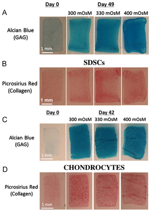
This study investigated the potential use of static osmotic loading as a cartilage tissue engineering strategy for growing clinically relevant grafts from either synovium-derived stem cells (SDSCs) or chondrocytes. Bovine SDSCs and chondrocytes were individually encapsulated in 2% w/v agarose and divided into chondrogenic media of osmolarities 300 (hypotonic), 330 (isotonic), and 400 (hypertonic, physiologic) mOsM for up to 7 weeks. The application of hypertonic media to constructs comprised of SDSCs or chondrocytes led to increased mechanical properties as compared to hypotonic (300mOsM) or isotonic (330mOsM) media (p<0.05). Constant exposure of SDSC-seeded constructs to 400mOsM media from day 0 to day 49 yielded a Young's modulus of 513±89kPa and GAG content of 7.39±0.52%ww on day 49, well within the range of values of native, immature bovine cartilage. Primary chondrocyte-seeded constructs achieved almost as high a Young's modulus, reaching 487±187kPa and 6.77±0.54%ww (GAG) for the 400mOsM condition (day 42). These findings suggest hypertonic loading as a straightforward strategy for 3D cultivation with significant benefits for cartilage tissue engineering strategies. In an effort to understand potential mechanisms responsible for the observed response, cell volume measurements in response to varying osmotic conditions were evaluated in relation to the Boyle-van't Hoff (BVH) law. Results confirmed that chondrocytes behave as perfect osmometers; however SDSCs deviated from the BVH relation.

Keywords: Cartilage; Chondrocytes; Static osmotic loading; Synovium-derived stem cells; Tissue engineering.


Conflict of interest statement

Conflict of interest statement

The authors certify that there is no conflict of interest related to the work presented in this manuscript.

Figures

Fig. 1
Fig. 1
Schematic of the experimental groups. After 3D encapsulation in agarose, SDSC- and chondrocyte-seeded constructs were cultured with serum-free chondrogenic media of osmolarities 300, 330, and 400 mOsM. TGF-β3 was removed at Day 14 for chondrocytes and Day 21 for SDSCs, which were previously demonstrated to create functionally relevant tissue.
Fig. 2
Fig. 2
Multi-differentiation potential of juvenile bovine SDSCs in 2D monolayer culture. (A) and (B): Adipogenesis of SDSCs. After 7 days of expansion in serum-containing media, SDSCs were incubated in adipogenesis medium for an additional 14 days before staining with Oil Red-O. Lipid vacuoles are visible in (B). (C): SDSCs were expanded in serum-containing media before staining with Oil Red-O as a negative control. (D) and (E): Osteogenesis of SDSCs. After 7 days of expansion in serum-containing media, SDSCs were incubated in osteogenic media for an additional 14 days before staining with Alizarin Red. Calcified nodules are visible in (E). (F): SDSCs were expanded in serum-containing media before staining with Alizarin Red as a negative control. Bars: 50 μm. (A) and (D): Stereoscopic images; (B), (C), (E), and (F): 20× magnification.
Fig. 3
Fig. 3
Representative gross morphology of SDSC-seeded constructs at day 49. The 300 mOsM group (left) had final average dimensions of diameter = 3.85±0.07 mm, thickness = 2.49±0.02 mm, the 330 mOsM group (middle) had final average dimensions of diameter = 3.84±0.06 mm, thickness = 2.52±0.12 mm, and the 400 mOsM group (right) had final average dimensions of diameter = 4.40±0.13 mm, thickness = 3.02±0.08 mm.
Fig. 4
Fig. 4
(A) Equilibrium Young’s modulus (EY), (B) Dynamic modulus (G*), (C) glycosaminoglycan (GAG) (%ww), and (D) DNA content (μg) for SDSC-seeded constructs cultured over 7-week study period. (E) Equilibrium Young’s modulus (EY), (F) Dynamic modulus (G*), (G) glycosaminoglycan (GAG) (%ww), and (H) DNA content (μg) for primary chondrocyte-seeded constructs cultured over a 6-week period. Solid line indicates release time point (*p<0.05). The results of each study were subsequently repeated and consistent. (For clearer interpretation of the hatching patterns in this figure legend, the reader is referred to the web version of this article for the high quality image.)
Fig. 5
Fig. 5
Representative GAG and collagen staining (A) Alcian Blue staining (GAG) and (B) Picrosirius Red staining (collagen) for constructs seeded with SDSCs at days 0 and 49. (C) Alcian Blue staining (GAG) and (D) Picrosirius Red staining (collagen) for constructs seeded with primary chondrocytes at days 0 and 42.
Fig. 6
Fig. 6
Representative immunohistochemical staining for (A) COMP (green), (B) type II collagen (green), (C) type VI collagen (green), and (D) TRPV4 (green) for SDSC constructs and (E) COMP (green), (F) type II collagen (green), (G) type VI collagen (green), and (H) TRPV4 (green) for chondrocyte constructs cultured in 400 mOsM media at final time point. (I) Negative control image. Cell nuclei (A–I) stained with TOTO-3 (red). (For interpretation of the references to color in this figure legend, the reader is referred to the web version of this article.)
Fig. 7
Fig. 7
Boyle–van’t Hoff (BVH) plots of average normalized data for (A) chondrocytes (4% deviation from BVH line) and (B) SDSCs (8% deviation from BVH line). True Boyle–van’t Hoff line (red) (vBVH = (1 − vb)x+vb) plotted to determine inactive cell volume (vb). (For interpretation of the references to color in this figure legend, the reader is referred to the web version of this article.)

References

    1. Archer CW, Dowthwaite GP, Francis-West P. Development of synovial joints. Birth Defects Research Part C. 2003;69:144–155. - PubMed
    1. Barbero A, Ploegert S, Heberer M, Martin I. Plasticity of clonal populations of dedifferentiated adult human articular chondrocytes. Arthritis and Rheumatism. 2003;48:1315–1325. - PubMed
    1. Bian L, Crivello KM, Ng KW, Xu D, Williams DY, et al. Influence of temporary chondroitinase ABC-induced glycosaminoglycan suppression on maturation of tissue-engineered cartilage. Tissue Engineering Part A. 2009;15:2065–2072. - PMC - PubMed
    1. Bilgen B, Orsini E, Aaron RK, Ciombor DM. FBS suppresses TGF-beta1-induced chondrogenesis in synoviocyte pellet cultures while dexamethasone and dynamic stimuli are beneficial. Journal of Tissue Engineering and Regenerative Medicine. 2007;1:436–442. - PubMed
    1. Bush PG, Hall AC. The osmotic sensitivity of isolated and in situ bovine articular chondrocytes. Journal of Orthopaedic Research. 2001;19:768–778. - PubMed

Publication types

LinkOut - more resources