. 2013 Oct 24;20(10):1245-54.
doi: 10.1016/j.chembiol.2013.07.017. Epub 2013 Sep 12.

Modulation of curli assembly and pellicle biofilm formation by chemical and protein chaperones

Affiliations

Modulation of curli assembly and pellicle biofilm formation by chemical and protein chaperones

Emma K Andersson et al. Chem Biol. .

Abstract

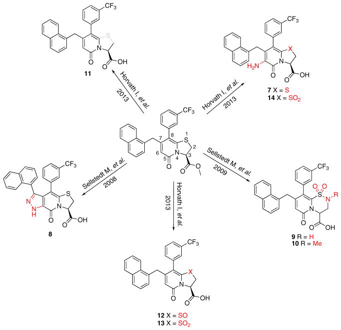
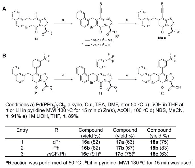
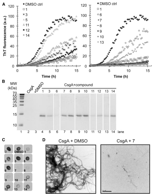
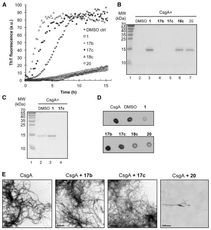
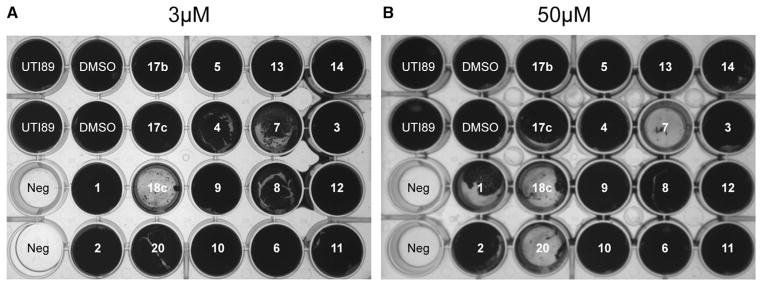
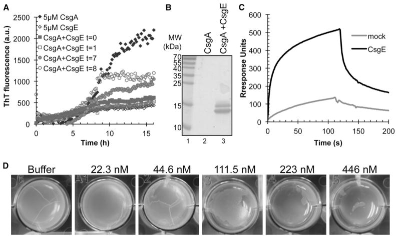
Enteric bacteria assemble functional amyloid fibers, curli, on their surfaces that share structural and biochemical properties with disease-associated amyloids. Here, we test rationally designed 2-pyridone compounds for their ability to alter amyloid formation of the major curli subunit CsgA. We identified several compounds that discourage CsgA amyloid formation and several compounds that accelerate CsgA amyloid formation. The ability of inhibitor compounds to stop growing CsgA fibers was compared to the same property of the CsgA chaperone, CsgE. CsgE blocked CsgA amyloid assembly and arrested polymerization when added to actively polymerizing fibers. Additionally, CsgE and the 2-pyridone inhibitors prevented biofilm formation by Escherichia coli at the air-liquid interface of a static culture. We demonstrate that curli amyloid assembly and curli-dependent biofilm formation can be modulated not only by protein chaperones, but also by "chemical chaperones."


Figures

Figure 1
Figure 1. Compounds Synthesized to Analyze Central Fragment Alterations and Substituents
With 1 as starting point, the peptidomimetic backbone was extended by introducing an amine in the pyridone ring resulting in compound 7. A rigidified tricyclic structure, compound 8, was included as well as analogs where the five-membered thiazolino group had been exchanged for six-membered sultams in compounds 9 and 10. Compound 11 is a desulfurized ring-opened analog and compounds 12 and 13 are analogs where the sulfur had been oxidized to the corresponding sulfoxide or sulfone. Compound 14 has both the extended peptidomimetic backbone and oxidized sulfur. See also Table S1.
Figure 2
Figure 2. Synthesis of the Acetylene Spacer Analogs and the Brominated Analog of 1
(A) Synthesis of acetylene spacer analogs 18a, 18b, and 18c. The intermediates 17a, 17b, and 17c, with bromine (Br) residues at position 6 were also hydrolyzed and tested for biological activity. (B) Bromination of 1 to obtain compound 20. See also Table S2.
Figure 3
Figure 3. In Vitro Effect of Inhibitory Compounds on CsgA Polymerization
(A) ThT fluorescence of freshly purified 5 μM CsgA with or without 50 μM compound. Reduction in ThT fluorescence corresponds to inhibition of CsgA polymerization. (B) CsgA solubility after overnight incubation with or without compound by SDS-PAGE. Compounds that inhibit CsgA polymerization kept CsgA in a soluble SDS-sensitive state. (C) CsgA incubated overnight with compounds was spotted onto a nitrocellulose membrane and probed with a gammabody grafted with the CsgA R1 sequence that detects CsgA amyloid structure. (D) Transmission electron microscopy of freshly purified CsgA incubated for 6 hr revealed abundant fibers, whereas no curli fibers were detected by when freshly purified CsgA was incubated for 6 hr with the inhibitor compound 7. Scale bars represent 0.5 μM. See also Figure S1.
Figure 4
Figure 4. In Vitro Effect of Accelerating Compounds on CsgA Polymerization
(A) ThT fluorescence of freshly purified 5 μM CsgA with or without 50 μM compound. The lag phase of the fluorescence curve as a consequence of ThT binding to polymerizing CsgA was shortened when accelerating compounds 17b and 17c were present. (B) SDS-PAGE of CsgA incubated overnight with or without compound. No soluble CsgA was detected after incubation with accelerators 17b and 17c (lanes 4 and 5, respectively). (C) SDS-PAGE of CsgA incubated for 8h with or without compound. Incubation with the accelerating compound 17c results in a SDS-insoluble amyloid after 8 hr (lane 4) whereas pure CsgA is still partly soluble (lane 2). In the presence of the inhibitory compound 1, CsgA is soluble (lane 3). (D) CsgA incubated overnight with compounds was spotted onto a nitrocellulose membrane and probed with the Tessier gammabody grafted with the CsgA R1 sequence that detects CsgA amyloid structure. (E) Transmission electron microscopy of CsgA. Negative staining of freshly purified CsgA incubated for 6 hr (left). Freshly purified CsgA incubated for 6h with accelerators 17b or 17c appeared similar to CsgA alone. Fibers were not detectable when CsgA was incubation with the chemically similar, yet functionally inhibitory, compound 20. Scale bars represent 0.5 μM. See also Figure S1.
Figure 5
Figure 5. Inhibition of Pellicle Biofilm by Chemical Compounds
(A) Effect of the compounds at a concentration of 3 μM on the formation of curli-dependent pellicle biofilm, incubated at 26°C for 48 hr. Compound 18c was the most potent inhibitor. (B) Effect of the compounds at a concentration of 50 μM on the formation of curli-dependent pellicle biofilm, incubated at 26°C for 48 hr. Compounds 7, 18c, and 20 were correspondingly efficient in vitro. See also Figure S2.
Figure 6
Figure 6. Effect of CsgE on CsgA Polymerization and Pellicle Formation
(A) Immuno-gold labeling of overexpressed CsgE. MC4100 csgE mutant cells with empty vector (left) or overexpressing CsgE (middle and right) were incubated with CsgE antibody (left and middle) or buffer (right) followed by incubation with gold particle conjugated secondary antibody prior to uranyl acetate staining and TEM. Scale bar represents 200 nm. (B) CsgE interacts with CsgA fibers. Surface plasmon resonance sensograms of 1 μM purified CsgE (solid line) or a mock purification (dotted line) were injected over immobilized sonicated CsgA fibers on a CM5 sensor chip. Flow of phosphate buffer was resumed after 120 s. (C) CsgE added to freshly purified CsgA or CsgA incubated for 1 hr inhibits CsgA polymerization. CsgE added to CsgA incubated for 7 hr or 8 hr efficiently inhibits further polymerization. (D) SDS-PAGE after CsgA overnight incubation with or without equimolar concentration of the chaperone CsgE. No CsgA is left in solution when incubated alone (lane 2) whereas CsgA incubated with CsgE (lane 3) was still in a soluble form. (E) Extracellular addition of CsgE prevents formation of curli-dependent pellicle biofilm in a concentration-dependent manner.

References

    1. Andersson EK, Chapman M. Small molecule disruption of B. subtilis biofilms by targeting the amyloid matrix. Chem Biol. 2013;20:5–7. - PMC - PubMed
    1. Ban T, Hamada D, Hasegawa K, Naiki H, Goto Y. Direct observation of amyloid fibril growth monitored by thioflavin T fluorescence. J Biol Chem. 2003;278:16462–16465. - PubMed
    1. Barnhart MM, Chapman MR. Curli biogenesis and function. Annu Rev Microbiol. 2006;60:131–147. - PMC - PubMed
    1. Berg V, Sellstedt M, Hedenström M, Pinkner JS, Hultgren SJ, Almqvist F. Design, synthesis and evaluation of peptidomimetics based on substituted bicyclic 2-pyridones-targeting virulence of uropathogenic E. coli. Bioorg Med Chem. 2006;14:7563–7581. - PubMed
    1. Bian Z, Normark S. Nucleator function of CsgB for the assembly of adhesive surface organelles in Escherichia coli. EMBO J. 1997;16:5827–5836. - PMC - PubMed

Publication types

MeSH terms