. 2013 Sep 30;26(6):666-673.
doi: 10.1016/j.devcel.2013.07.016. Epub 2013 Sep 12.

Adrenocortical zonation results from lineage conversion of differentiated zona glomerulosa cells

Affiliations

Adrenocortical zonation results from lineage conversion of differentiated zona glomerulosa cells

Bethany D Freedman et al. Dev Cell. .

Abstract

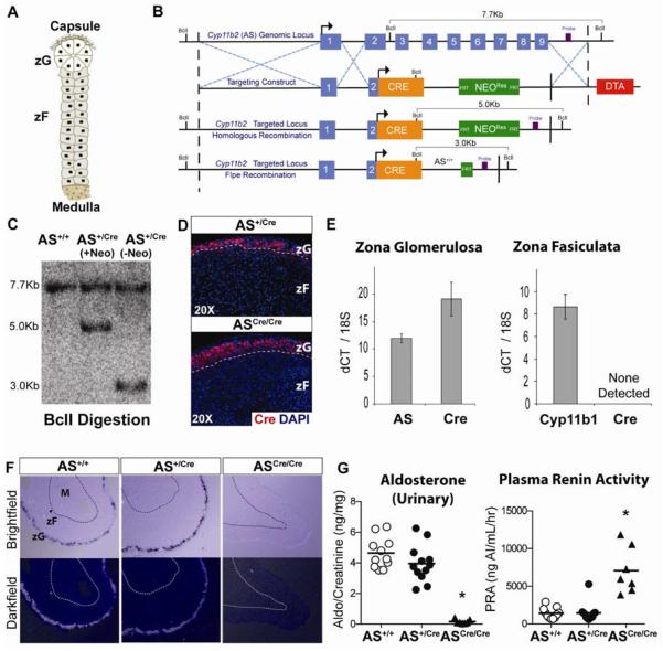
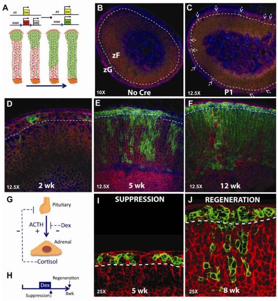
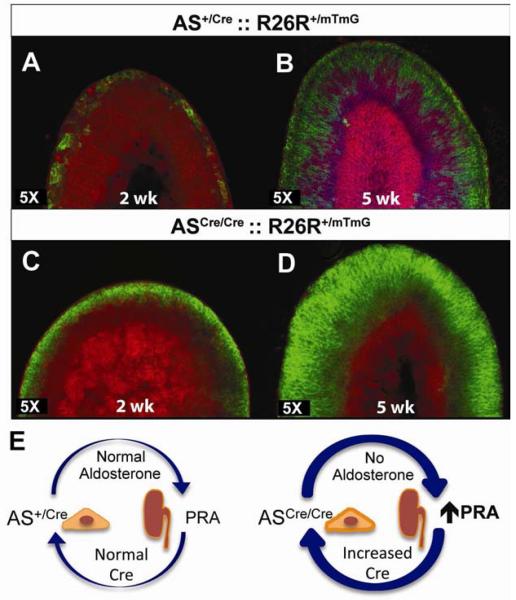
Lineage conversion of differentiated cells in response to hormonal feedback has yet to be described. To investigate this, we studied the adrenal cortex, which is composed of functionally distinct concentric layers that develop postnatally, the outer zona glomerulosa (zG) and the inner zona fasciculata (zF). These layers have separate functions, are continuously renewed in response to physiological demands, and are regulated by discrete hormonal feedback loops. Their cellular origin, lineage relationship, and renewal mechanism, however, remain poorly understood. Cell-fate mapping and gene-deletion studies using zG-specific Cre expression demonstrate that differentiated zG cells undergo lineage conversion into zF cells. In addition, zG maintenance is dependent on the master transcriptional regulator Steroidogenic Factor 1 (SF-1), and zG-specific Sf-1 deletion prevents lineage conversion. These findings demonstrate that adrenocortical zonation and regeneration result from lineage conversion and may provide a paradigm for homeostatic cellular renewal in other tissues.


Figures

Figure 1
Figure 1. Zona Glomerulosa is normally regulated in AS+/Cre mice
(A) Schematic of adrenal cortex showing the relationship of zG to zF cells. (B) Cre recombinase was targeted to the Cyp11b2 (AS) genomic locus by homologous recombination in mouse ES cells. The resultant transgenic mice were crossed with FLP deleter mice to excise the FRT-flanked NeomycinRes cassette. (C) Southern blot analysis confirmed homologous recombination. (D) Cre recombinase is restricted to the zG demonstrated by immunostaining in both AS+/Cre and ASCre/Cre mice. (E) Laser capture microdissection and isolation of zG and zF RNA, followed by qRT-PCR analysis, confirmed restriction of Cre expression to zG cells, Mean±SEM. (F) In Situ Hybridization demonstrates comparable AS expression in wild-type AS+/+ and heterozygous AS+/Cre adrenals and the lack of AS expression in homozygous ASCre/Cre adrenals. (G) Aldosterone and PRA levels are unchanged in AS+/Cre mice compared with controls. ASCre/Cre mice show undetectable levels of aldosterone and a compensatory rise in PRA levels (Mean, * p<0.0001). See also Figure S1.
Figure 2
Figure 2. zF cells arise from zG cells through lineage conversion
(A) Schematic of genetic loci for AS+/Cre and the R26RmT/mG reporter along with the expected result for centripetal migration over time. (B–F) Confocal analysis of mTomato and mGFP expression from AS+/CreR26RmT/mG mice from birth (P1) through 12 weeks. Arrows demarcate GFP+ cells. (G) Schematic showing positive (+) and negative (−) feedback regulation. (H) Mice received dexamethasone from 3–5 weeks of age and adrenals were analyzed at the peak of suppression (I) and following adrenal regeneration (J). See also Figure S2.
Figure 3
Figure 3. Accelerated lineage marking in AS deficient mice
(A–D) Confocal analysis of adrenals from AS+/CreR26R+/mTmG (A, B) and ASCre/CreR26R+/mTmG (C, D) mice at 2 (A,C) and 5 (B,D) weeks of age. Female adrenal that retains the X-Zone (bright pink) is shown in B. (E) Schematic showing aldosterone deficiency leads to secondary elevation of PRA levels through hormonal feedback. See also Figure S3.
Figure 4
Figure 4. SF-1 is required for zG maintenance and subsequent lineage conversion into zF
Histological and confocal analyses of 12 week old AS+/CreSf-1+/+ (A–C) and AS+/CreSf-1fl/fl (F–H) adrenal glands. Analysis of SF-1 expression in GFP+ zG cells revealed lack of co-expression in the majority of cells (J) compared with control zG cells (E). Despite deletion of Sf-1 in GFP+ zG cells, zF cells retained SF-1 expression (J) and showed normal levels of P450scc, in contrast to zG cells, which showed decreased P450scc (K). (L) Analysis of plasma aldosterone levels in 7 week old mice from both groups were unchanged, however, PRA levels were increased ~3 fold in mice with Sf-1-deficient zG cells (Mean±SEM,* p<0.05). (M) Schematic illustration showing zG-dependent and independent models of adrenal lineage development. See also Figure S4.

Comment in

References

    1. Beuschlein F, Looyenga BD, Bleasdale SE, Mutch C, Bavers DL, Parlow AF, Nilson JH, Hammer GD. Activin induces x-zone apoptosis that inhibits luteinizing hormone-dependent adrenocortical tumor formation in inhibin-deficient mice. Molecular and cellular biology. 2003;23:3951–3964. - PMC - PubMed
    1. Beuschlein F, Mutch C, Bavers DL, Ulrich-Lai YM, Engeland WC, Keegan C, Hammer GD. Steroidogenic factor-1 is essential for compensatory adrenal growth following unilateral adrenalectomy. Endocrinology. 2002;143:3122–3135. - PubMed
    1. Bland ML, Jamieson CA, Akana SF, Bornstein SR, Eisenhofer G, Dallman MF, Ingraham HA. Haploinsufficiency of steroidogenic factor-1 in mice disrupts adrenal development leading to an impaired stress response. Proc Natl Acad Sci U S A. 2000;97:14488–14493. - PMC - PubMed
    1. Buaas FW, Gardiner JR, Clayton S, Val P, Swain A. In vivo evidence for the crucial role of SF1 in steroid-producing cells of the testis, ovary and adrenal gland. Development. 2012;139:4561–4570. - PMC - PubMed
    1. Crawford PA, Sadovsky Y, Milbrandt J. Nuclear receptor steroidogenic factor 1 directs embryonic stem cells toward the steroidogenic lineage. Molecular and cellular biology. 1997;17:3997–4006. - PMC - PubMed

Publication types

MeSH terms