Review
. 2013 Nov 29;167(1-2):168-80.
doi: 10.1016/j.vetmic.2013.08.013. Epub 2013 Aug 28.

West Nile viral infection of equids

Affiliations
Review

West Nile viral infection of equids

J Angenvoort et al. Vet Microbiol. .

Abstract

West Nile virus (WNV) is a flavivirus transmitted between certain species of birds and mosquito vectors. Tangential infections of equids and subsequent equine epizootics have occurred historically. Although the attack rate has been estimated to be below 10%, mortality rates can approach 50% in horses that present clinical disease. Symptoms are most commonly presenting in the form of encephalitis with ataxia as well as limb weakness, recumbency and muscle fasciculation. The most effective strategy for prevention of equine disease is proper vaccination with one of the numerous commercially available vaccines available in North America or the European Union. Recently, WNV has been increasingly associated with equine epizootics resulting from novel non-lineage-1a viruses in expanding geographic areas. However, specific experimental data on the virulence of these novel virus strains is lacking and questions remain as to the etiology of the expanded epizootics: whether it be a function of inherent virulence or ecological and/or climactic factors that could precipitate the altered epidemiological patterns observed.

Keywords: Equids; Flavivirus; Horses; West Nile virus.


Figures

Fig. 1
Fig. 1
Structure of West Nile. (a) The WNV virion is composed of an icosahedral core formed by positively charged capsid (C) proteins of 12 kDa molecular mass (red). This core is enveloped by a host-cell derived membrane in which the major envelope protein E (53 kDa, blue dimers) and the membrane protein M (8 kDa, blue capsules) are integrated. C-proteins itself enclose an RNA genome encoding for a single open reading frame (ORF) of 10.302 nucleotides and non-coding regions of 96 and 631 nucleotides at the 5′ and 3′ ends, respectively. (b) The WNV genome encodes a polyprotein that is post-translationally processed by viral and cellular proteases into five non-structural proteins (NS1, NS2A/NS2B, NS3, NS4A/NS4B and NS5) and into three structural proteins (C, prM/M and E). E protein and prM/M protein (18–20 kDa) virus. (For interpretation of the references to color in Figs. 1 and 2 legends, the reader is referred to the web version of this article.)
Fig. 2
Fig. 2
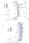
This figure is reused from the Journal of Virology, 2011, volume 85 (6), pp. 2964–2974, DOI 10.1128/jvi.01963-10 (May et al., 2011), reproduced/amended with permission from American Society for Microbiology and kindly provided by Alan Maximum clade credibility (MCC) tree of WNV genomes constructed using BEAST version 1.5.3 software. Colors of branches indicate geographic locations per the color key. Branch lengths correspond to lengths of time, as measured by the scale bar, and the 95% HPD range of divergence dates is shown as a bar. The letters at the nodes correspond to data in Table 1. (A) All isolates (with the branch of isolates from the Americas collapsed). (B) Cluster 4 isolates only (May et al., 2011). Barrett.
Fig. 3
Fig. 3
Bird/mosquito transmission cycle of WNV is transmitted in an enzootic passeriform bird–mosquito (Culex spp. mosquitoes) cycle (green shading). Bridge vectors (such as Aedes spp.) can transmit the virus to humans and equines (gray shading) that are designated as “dead-end” hosts in that they can become infected but are not capable of serving as a source of infection for additional mosquito vectors. Some peridomestic avian hosts (red shading and dashed ovals; such as corvids) are capable of developing high titers following infection with certain WNV strains and facilitate epidemic amplification. WNV.
Fig. 4
Fig. 4
Equine WNF cases reported worldwide officially reported to WAHID/OIE or PROMED or published in literature since 1959. Lineage color codes were assigned if at least one horse isolate of this lineage was found or if this lineage was the only published lineage occurring in this country (for example in humans, birds, mosquitoes).
Fig. 5
Fig. 5
Virus Infection and immune response in horses. WNV infected viremia is observed in the first six days post-infection and can last 1–6 days (Bunning et al., 2002; Minke et al., 2004; Seino et al., 2007). Antibodies can be detected from day 7 or 8 post-infection. Detection of IgM is possible for less than 3 months (as reviewed in Castillo-Olivares and Wood, 2004), whereas neutralizing antibodies persist for at least 15 months (Ostlund et al., 2001). Clinical symptoms are thought to occur mostly after the end of viremia (Castillo-Olivares and Wood, 2004), nevertheless the onset of symptoms in naturally acquired infections can only be estimated. Following mild infections, equines usually recover after two to seven days, but may need more than 20 days up to several months for complete reconstitution to their original condition (Trock et al., 2001; Venter et al., 2009). In some cases residual signs of the infection may even persist (Salazar et al., 2004).
Fig. 6
Fig. 6
Serum viremia profiles for 47 horses experimentally infected with WNV NY99 by feeding of infected mosquitoes. No horse had a demonstrable viremia beyond 6.5 days post-feeding.

References

    1. Abutarbush SM, O’Connor BP, Clark C, Sampieri F, Naylor JM. Clinical West Nile virus infection in 2 horses in western Canada. Can Vet J [La Revue Veterinaire Canadienne] 2004;45:315–317. - PMC - PubMed
    1. Aliota MT, Jones SA, Dupuis AP, 2nd, Ciota AT, Hubalek Z, Kramer LD. Characterization of Rabensburg virus, a flavivirus closely related to West Nile virus of the Japanese encephalitis antigenic group. PloS One. 2012;7:e39387. - PMC - PubMed
    1. APHIS. Annual trends in US distribution of equine West Nile virus cases. APHIS; 2012. West Nile virus distribution maps, 1999–2012. http://www.aphis.usda.gov/vs/nahss/equine/wnv/wnv_distribution_maps.htm.
    1. Autorino GL, Battisti A, Deubel V, Ferrari G, Forletta R, Giovannini A, Lelli R, Murri S, Scicluna MT. West Nile virus epidemic in horses, Tuscany region, Italy. Emerg Infect Dis. 2002;8:1372–1378. - PMC - PubMed
    1. Bagnarelli P, Marinelli K, Trotta D, Monachetti A, Tavio M, Del Gobbo R, Capobianchi M, Menzo S, Nicoletti L, Magurano F, Varaldo P. Human case of autochthonous West Nile virus lineage 2 infection in Italy, September 2011. Euro Surveill. 2011;16 pii 20002. - PubMed

Publication types