. 2013 Nov;183(5):1425-1436.
doi: 10.1016/j.ajpath.2013.07.022. Epub 2013 Sep 10.

Transthyretin is dysregulated in preeclampsia, and its native form prevents the onset of disease in a preclinical mouse model

Affiliations

Transthyretin is dysregulated in preeclampsia, and its native form prevents the onset of disease in a preclinical mouse model

Satyan S Kalkunte et al. Am J Pathol. 2013 Nov.

Abstract

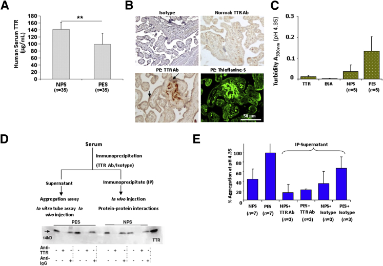
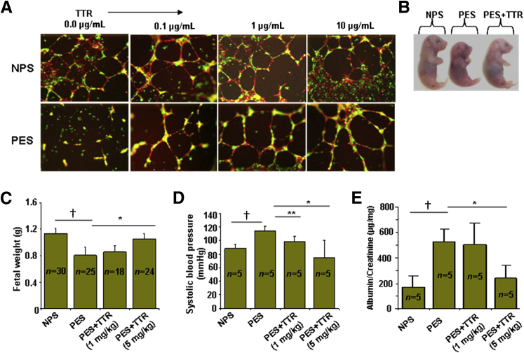
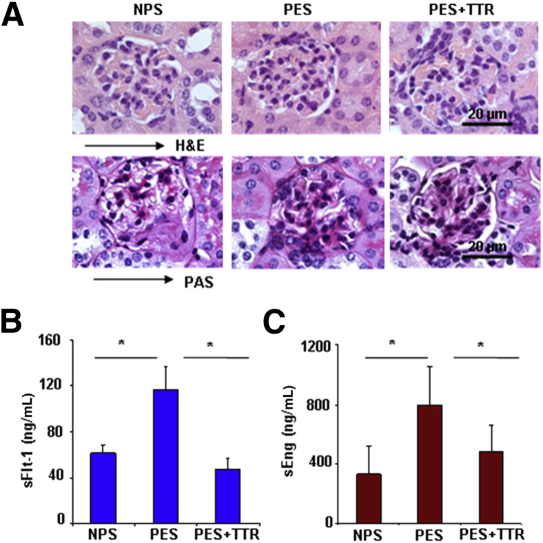
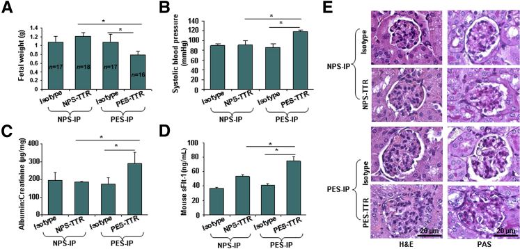
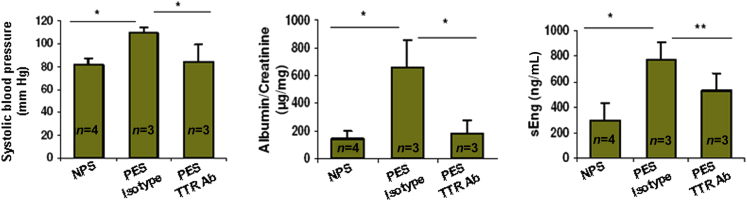
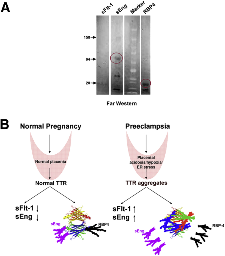
Preeclampsia is a major pregnancy complication with potential short- and long-term consequences for both mother and fetus. Understanding its pathogenesis and causative biomarkers is likely to yield insights for prediction and treatment. Herein, we provide evidence that transthyretin, a transporter of thyroxine and retinol, is aggregated in preeclampsia and is present at reduced levels in sera of preeclamptic women, as detected by proteomic screen. We demonstrate that transthyretin aggregates form deposits in preeclampsia placental tissue and cause apoptosis. By using in vitro approaches and a humanized mouse model, we provide evidence for a causal link between dysregulated transthyretin and preeclampsia. Native transthyretin inhibits all preeclampsia-like features in the humanized mouse model, including new-onset proteinuria, increased blood pressure, glomerular endotheliosis, and production of anti-angiogenic factors. Our findings suggest that a focus on transthyretin structure and function is a novel strategy to understand and combat preeclampsia.


Figures

Figure 1
Figure 1
Surface enhanced laser desorption ionization-time-of-flight (SELDI-TOF) and biochemical analyses of transthyretin in preeclampsia serum. A: Preeclampsia serum (PES) (n = 53) and normal pregnancy serum (NPS) (n = 16) were analyzed by SELDI-TOF in the molecular mass range of 2 to 200 kDa. In the entire molecular weight (MW) range, the time of flight scatterplot shows most significant changes (reduction) in the median intensities of a 14-kDa protein group in PES when compared with NPS samples. B: Isoelectric point (pI) and molecular weight of a protein spot (approximately 14 kDa) separated by two-dimensional gel electrophoresis from a representative NPS sample are shown. The mass fragmentation pattern of the corresponding trypsin-digested spot and molecular mass (m/z) of the peptide fragments are also shown. The sequence coverage obtained by comparison with the ExPASy database (P02766; Swiss Institute of Bioinformatics, http://www.expasy.org, last accessed August 20, 2011) identified the protein spot (approximately 14 kDa) to be transthyretin. C: Normalized Protein Chip array profiles are shown of a representative NPS sample immunodepleted with a transthyretin-neutralizing antibody or an isotype-matched antibody. Successful immunodepletion of transthyretin (pink highlighted area) confirmed that the protein with a molecular mass of approximately 14 kDa was transthyretin. D: Comparison of transthyretin protein peaks (molecular mass of approximately 14,000 Da) in NPS, PES, and commercial human transthyretin (cTTR) is shown.
Figure 2
Figure 2
Transthyretin in preeclampsia is dysregulated and forms aggregates. A: Analysis of serum transthyretin in normal pregnancy serum (NPS) and preeclampsia serum (PES) by ELISA. Transthyretin is present at significantly reduced levels in PES (P < 0.001). B: Immunohistochemical (IHC) analysis using transthyretin-specific antibody (Ab) shows strong transthyretin staining in the extravillous domain of human placental section from preeclampsia, not normal pregnancy, in addition to staining in the trophoblast layer (arrows) lining the placental villi. Adjacent section of the same preeclampsia placenta shows intense fluorescence for amyloid-specific thioflavin S staining that overlaps with extracellular transthyretin-positive deposits. Additional placental tissue sections are shown in Supplemental Figure S2. C: Comparative analysis of aggregation of purified transthyretin, NPS, PES, and albumin indicated by turbidity measurements (330 nm) is shown as average values of multiple serum samples analyzed. All of the serum samples used were normalized to 0.4 mg/mL transthyretin, as determined by ELISA. PES showed a higher propensity to aggregate, as reflected by higher turbidity. On an average, eight different samples of NPS and PES were analyzed. D: Experimental flow chart for transthyretin IP and depletion is presented. SDS-PAGE immunoblotting shows the presence or absence of transthyretin monomer in the immunoprecipitate or supernatant of PES or NPS, respectively. E: Turbidity (330 nm) of supernatants obtained from transthyretin-depleted NPS or PES samples was measured and expressed as percentage aggregation. Antibody-mediated transthyretin depletion abolished aggregation associated with PES. ∗∗P < 0.01.
Figure 3
Figure 3
Exogenous transthyretin inhibits preeclampsia-associated features in vitro and in vivo. A: Serum-induced and transthyretin-modified endovascular interaction between endothelial cells (human umbilical cord endothelial cells, cell tracker red) and first-trimester trophoblasts (HTR8, cell tracker green) was analyzed by three-dimensional tube formation on Matrigel. Native transthyretin rescued tube formation disrupted by preeclampsia serum (PES). B: Reversal of PES-induced intrauterine growth restriction (IUGR) by exogenous transthyretin in IL-10−/− mice. A representative image of gd 17 fetus is shown. A total of five animals were used in each condition. C: The average fetal weight from IL-10−/− mice receiving different treatments was evaluated. D: Systolic blood pressure in pregnant mice on gd 17 was evaluated in response to different treatments. E: Proteinuria in pregnant mice was assessed on gd 17 in response to various treatments. The results are expressed as a ratio of albumin/creatinine excretion (μg/mg). All values represent means ± SD of at least 5 to 30 animals per group, depending on the experiment. P < 0.01, ∗∗P < 0.05 between PES and transthyretin treatment groups; P < 0.05 between NPS and PES groups.
Figure 4
Figure 4
Exogenous transthyretin rescues glomerular integrity and inhibits production of sFlt-1 and sEng in preeclamptic IL-10−/− mice. A: Histopathological analysis of H&E- or periodic acid schiff (PAS)-stained renal tissues from NPS, PES, or PES+ transthyretin-treated (5 mg/kg) pregnant IL-10−/− mice are shown. Original magnification, ×100. The H&E stain shows the normalization of PES-induced capillary occlusion, enlarged glomeruli, and swollen endothelial cells by transthyretin treatment that is comparable to NPS-treated control mice. PAS-based staining indicates that transthyretin treatment reverses PES-induced inflammation of capillary endothelial cells (endotheliosis). These pathological changes are absent in the NPS-treated mice. A representative image from staining of at least three animals per group is shown. B: Serum levels of mouse sFlt-1 from pregnant IL-10−/− mice obtained on gd 17 from different treatment groups are shown. Treatment with 5 mg/kg transthyretin reverses the PES-induced excess production of sFlt-1 in IL-10−/− mice. C: Serum levels of mouse sEng from pregnant IL-10−/− mice obtained on gd 17 from different treatment groups are shown. Treatment with transthyretin reverses the preeclampsia serum (PES)-induced excess production of sEng to levels comparable to normal pregnancy serum (NPS) treatment control group. All values are expressed as means ± SD obtained from at least five animals per treatment group. P < 0.05 represents the significance between different treatment groups.
Figure 5
Figure 5
Immunoprecipitated (IP) transthyretin from preeclampsia serum (PES) induces disease features in IL-10−/− mice. Pregnant mice were injected (i.p.) on gd 10 with transthyretin immunoprecipitate obtained from 100 μL normal pregnancy serum (NPS) or PES each, as described in Materials and Methods. A: The average weight of fetal units derived from pregnant mice on gd 17 receiving different treatments is shown. B: The systolic blood pressure of pregnant mice on gd 17 was evaluated in response to different treatments. C: Proteinuria (albumin/creatinine ratio, in μg/mg) is shown in response to various treatments. PES-transthyretin caused significant proteinuria compared with NPS-transthyretin. Isotype-matched IgG-mediated immunoprecipitation from PES resulted in values similar to NPS, confirming the validity of transthyretin-specific antibody. D: Transthyretin immunoprecipitate from PES induced excess production of mouse sFlt-1 in pregnant IL-10−/− mice. E: Histological analysis using H&E or periodic acid schiff (PAS) staining of renal tissue from pregnant IL-10−/− mice in response to transthyretin immunoprecipitates from NPS and PES is shown. A representative image from staining of at least three animals per group is presented. All values represent means ± SD of at least 3 to 18 animals per group. P < 0.05 between treatment groups indicated.
Figure 6
Figure 6
Transthyretin-immunodepleted preeclampsia serum (PES) fails to induce preeclampsia-like features. Transthyretin-depleted PES (100 μL) was injected on gd10 in IL-10−/− mice, and its effects on fetal size, blood pressure, proteinuria, and serum levels of sEng were compared with injection of an equal volume of normal pregnancy serum (NPS) or isotype-matched IgG-depleted PES. Immunodepletion of transthyretin in PES fails to cause significant changes in blood pressure, proteinuria, or sEng levels, suggesting that dysregulated transthyretin in PES is responsible for induction of preeclampsia pathological characteristics in IL-10−/− mice. All values represent means ± SD of at least three to four animals per group. P < 0.05 between treatment groups tested.
Figure 7
Figure 7
Transthyretin binds to soluble endoglin and its possible mode of action. A: Far Western blot showing the binding of transthyretin with sEng and RBP-4 (red circle) and lack of binding to sFlt-1. Purified sFlt-1, sEng, and RBP-4 were used as prey proteins, and purified human transthyretin was used as probing bait protein. A representative immunoblot from multiple independent experiments is shown. B: Proposed model showing the role of upstream factors, such as hypoxia, acidosis, and stress, in triggering aggregation of transthyretin that induces production of anti-angiogenic factors and impairs the binding and scavenging of preeclampsia-causing soluble factors.

Similar articles

Cited by

References

    1. Roberts J.M., Hubel C.A. Is oxidative stress the link in the two-stage model of pre-eclampsia? Lancet. 1999;354:788–789. - PubMed
    1. Redman C.W., Sargent I.L. Latest advances in understanding preeclampsia. Science. 2005;308:1592–1594. - PubMed
    1. Dekker G., Sibai B. Primary, secondary and tertiary prevention of pre-eclampsia. Lancet. 2001;357:209–215. - PubMed
    1. Steegers E.A., Dadelszen P., Duvekot J.J., Pijnenborg R. Pre-eclampsia. Lancet. 2010;376:631–644. - PubMed
    1. Parikh S.M., Karumanchi S.A. Putting pressure on pre-eclampsia. Nat Med. 2008;14:810–812. - PubMed

Publication types

MeSH terms