. 2013 Nov;183(5):1596-1607.
doi: 10.1016/j.ajpath.2013.07.031. Epub 2013 Sep 13.

VEGFR-3 neutralization inhibits ovarian lymphangiogenesis, follicle maturation, and murine pregnancy

Affiliations

VEGFR-3 neutralization inhibits ovarian lymphangiogenesis, follicle maturation, and murine pregnancy

Joseph M Rutkowski et al. Am J Pathol. 2013 Nov.

Abstract

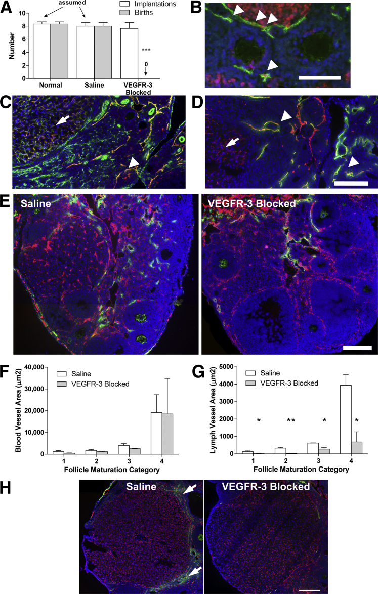
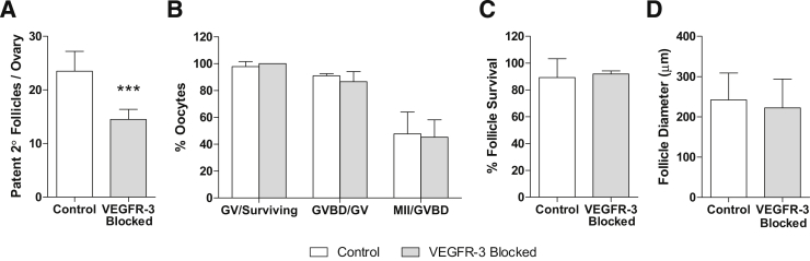
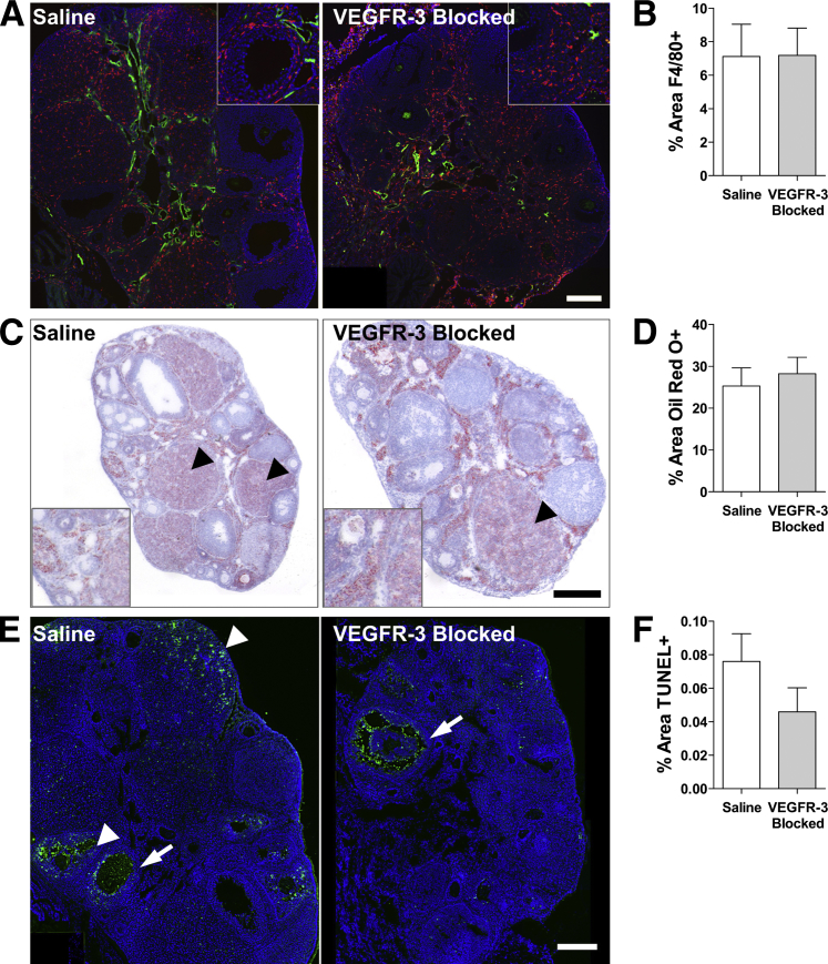
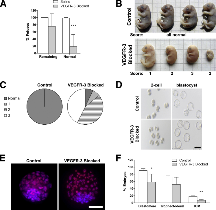
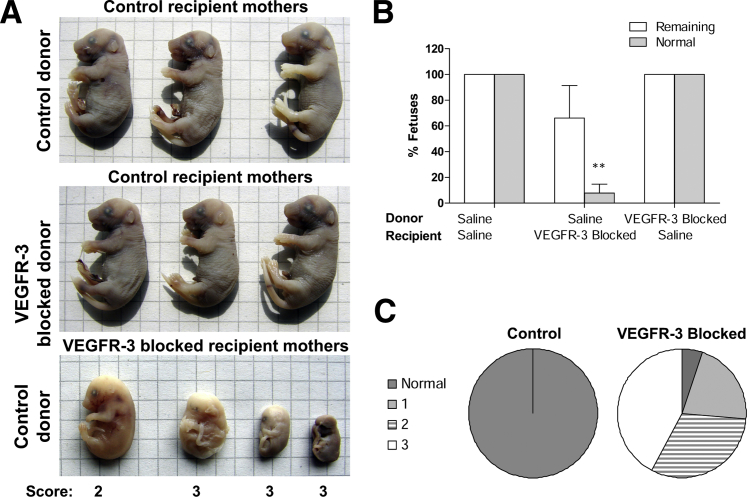
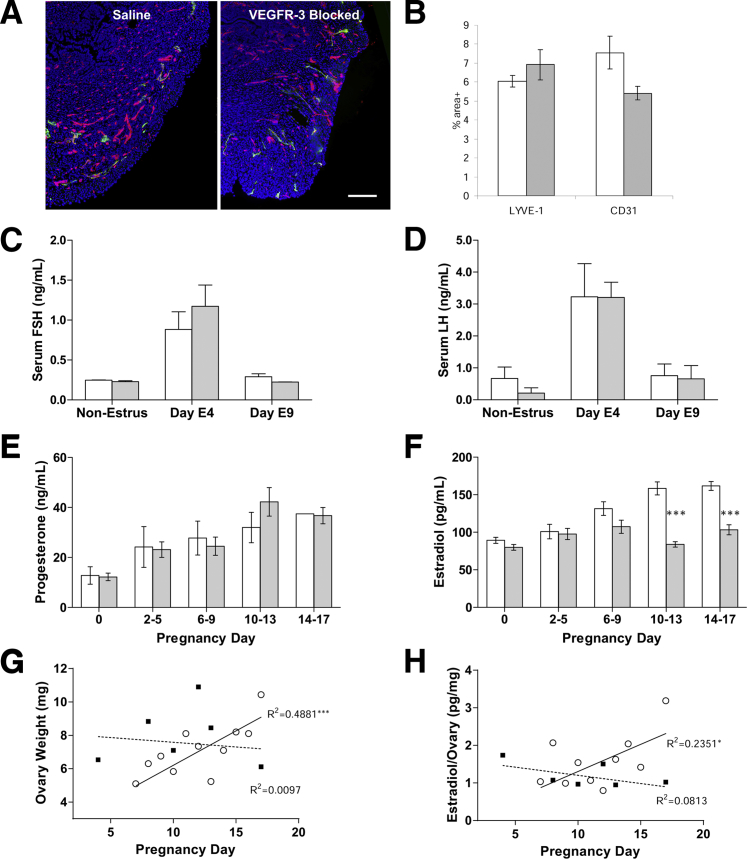
Lymphatic vessels surround follicles within the ovary, but their roles in folliculogenesis and pregnancy, as well as the necessity of lymphangiogenesis in follicle maturation and health, are undefined. We used systemic delivery of mF4-31C1, a specific antagonist vascular endothelial growth factor receptor 3 (VEGFR-3) antibody to block lymphangiogenesis in mice. VEGFR-3 neutralization for 2 weeks before mating blocked ovarian lymphangiogenesis at all stages of follicle maturation, most notably around corpora lutea, without significantly affecting follicular blood angiogenesis. The numbers of oocytes ovulated, fertilized, and implanted in the uterus were normal in these mice; however, pregnancies were unsuccessful because of retarded fetal growth and miscarriage. Fewer patent secondary follicles were isolated from treated ovaries, and isolated blastocysts exhibited reduced cell densities. Embryos from VEGFR-3-neutralized dams developed normally when transferred to untreated surrogates. Conversely, normal embryos transferred into mF4-31C1-treated dams led to the same fetal deficiencies observed with in situ gestation. Although no significant changes were measured in uterine blood or lymphatic vascular densities, VEGFR-3 neutralization reduced serum and ovarian estradiol concentrations during gestation. VEGFR-3-mediated lymphangiogenesis thus appears to modulate the folliculogenic microenvironment and may be necessary for maintenance of hormone levels during pregnancy; both of these are novel roles for the lymphatic vasculature.


Figures

Figure 1
Figure 1
Ovarian and follicular lymphangiogenesis (but not blood angiogenesis) is inhibited by VEGFR-3 blockade in the ovary, preventing successful pregnancy. A: Number of embryo implantations (white bars) in the uterus of normal, saline-injected, and VEGFR-3–blocked dams and live births (gray bars) assessed after delivery. Dams of viable pups were nursing, and uterine implantations were assumed to equal pups born. B: Ki-67+ (red) lymphatic endothelial cell (LYVE-1; green) nuclei in lymphatic vessels surrounding follicles (arrowheads). C: Blood vessels (CD31; green) are mostly negative for VEGFR-3 (red), with the exception of those within the corpora lutea (arrow). Because lymphatic vessels also express CD31, the strong VEGFR-3 colocalization likely marks lymphatic vessels (arrowhead). D: Ovarian lymphatic vessels (LYVE-1; green) are positive for VEGFR-3 (red) (arrowheads). LYVE-1+/VEGFR-3+ vessels within the corpora lutea are blood vessels (arrows). E: The extent of lymphatic vasculature (LYVE-1; green) surrounding follicles within the ovary was reduced in VEGFR-3–blocked ovaries; the blood vasculature and blood angiogenesis (CD31; red) were unaffected by the treatment. F: Quantification of blood and lymphatic vessel coverage in preantral follicles (maturation category 1), small follicles with formed atrium (maturation category 2), large follicles with significant atrium (maturation category 3), and corpora lutea (maturation category 4) demonstrated no changes in blood vessel density with VEGFR-3 blockade, despite some vessels expressing VEGFR-3. G: There was a significant reduction in lymphatic vessels at each maturation state with VEGFR-3 blockade. Note the normal increase in vascularization with folliculogenesis for both lymphatic and blood vessels; lymphangiogenesis was specifically blocked by VEGFR-3 neutralization. H: Lymphatic vessels (LYVE-1; green) are clearly visualized around the hormone-producing corpora lutea (category 4) (arrows) in normal ovaries (right), but lacking in VEGFR-3–blocked ovaries (left). P < 0.05, ∗∗P < 0.01, and ∗∗∗P < 0.001. Scale bar = 200 μm.
Figure 2
Figure 2
Secondary follicles, once retrieved from the ovaries of treated mice, mature normally in vitro. A: Fewer patent follicles were successfully separated from collected ovaries in treated mice. B: Follicles and their oocytes separated from ovaries of VEGFR-3–treated mice survive normally; germinal vesicle oocytes from treated and control mice mature to germinal vesicle breakdown and metaphase II stages using the in vitro drop culture technique at equal rates. C: Survival of secondary follicles directly treated with the VEGFR-3–blocking antibody in vitro was not inhibited. D: Maturation potential, as determined by follicle size, was also unaffected by direct VEGFR-3 treatment on the follicles. ∗∗∗P < 0.001. GV, germinal vesicle; GVBD, germinal vesicle breakdown; MII, metaphase II; 2°, secondary.
Figure 3
Figure 3
Macrophage recruitment, lipid accumulation, and cell apoptosis in the ovary appear to be unaffected by VEGFR-3 blockade. A: Macrophages (F4/80; red) present in the ovary are limited to follicular peripheries in both control and treated ovaries (LYVE-1; green). B: Total F4/80+ area within the ovary remains unchanged. C: The ovaries contain large amount of lipids (Oil Red O; red), particularly in the corpora lutea (arrowheads). D: Lipid accumulation (the Oil Red O+ area) in the ovary is not significantly changed between conditions. E: Apoptotic cells (TUNEL; green) are limited to interior granulosa cells (arrows) of regressing follicles and regular apoptosis of regressing corpora lutea and corpus hemorrhagicum (arrowheads) in both treatments. F: No difference was measured in the percent area of apoptotic cell nuclei within the ovary with mF4-31C1 treatment. Scale bars: 200 μm (A and E); 300 μm (B); insets, 2× zoom of main image to visualize localization.
Figure 4
Figure 4
Pregnancies occur after lymphangiogenesis is blocked, but embryonic development is severely impaired in the womb. A: In mice treated with mF4-31C1 before fertilization, few fetal bodies remained within the uterus at pregnancy day 17. The percentage is based on number of fetuses per implantation spot. B: Fetuses extracted at pregnancy day 17 from treated animals display a marked deficiency from control animals. They were scored as normal or as normal sized but discolored (1), deficient size and underdeveloped (2), and identifiable cell masses (3). C: The majority of fetal bodies are severely underdeveloped masses. D: Two-cell embryos were extracted from fertilized VEGFR-3–treated mice and cultured in vitro. Both two-cell embryos and blastomeres were identical in appearance to those taken from control animals. E: Effects of VEGFR-3 blockade prior to fertilization are manifest in blastocyst cell density. No differences were observed in the number of trophectodermal cells (propidium iodide and bisbenzimide; red/blue) that would later become the placenta, but fewer cells were identified in the inner cell mass (ICM) (bisbenzimide; blue) that would later develop into the fetus. F: Quantification indicated a significant reduction in cell numbers in each mass, compared with control. P < 0.05, ∗∗P < 0.01, and ∗∗∗P < 0.001. Scale bars: 100 μm (D); 50 μm (E). Grid = 4 mm.
Figure 5
Figure 5
VEGFR-3 blockade in the maternal gestational environment controls pregnancy success. A: Both normal and treated embryos implanted into normal, pseudo-pregnant recipient dams developed into normal, viable fetuses by pregnancy day 17. In pseudo-pregnant dams pretreated with mF4-31C1, no embryos developed into normal fetuses, despite normal implantations. B: Quantification of normal and remaining fetuses in implantation experiments. C: The distribution of fetal quality at pregnancy day 17 after normal embryo transfer to mF4-31C1–treated recipients closely replicates that of in situ fetal development in VEGFR-3–blocked pregnancies; the majority of fetuses were severely underdeveloped. ∗∗P < 0.01. Grid = 4 mm.
Figure 6
Figure 6
Pregnancy failures are attributable only to the ovarian lymphatic vessels. A: Blood (CD31; red) and lymphatic (LYVE-1; green) capillaries in the uterine wall appear normal in both mF4-31C1–treated and untreated pregnant dams. B: Quantified vessel density exhibited a nonsignificant decrease in blood vessel density (in accord with past use of mF4-31C1 in pregnancy); lymphatic density was unchanged. C and D: Serum levels of the pituitary-derived reproductive hormones follicle-stimulating hormone (FSH) (C) and luteinizing hormone (LH) (D) were not significantly affected with mF4-31C1 treatment during early pregnancy. E: Progesterone levels during gestation were unaffected by VEGFR-3 blockade or by the failing fetuses. F: Estradiol was significantly reduced in circulation at pregnancy day 6 to 9 after VEGFR-3 blockade. G: There was no sustained weight increase in VEGFR-3–blocked ovaries during pregnancy. H: Estradiol levels in ovarian homogenates (pg estradiol/mg ovarian protein) were reduced, mirroring the decrease in circulation. P < 0.05, ∗∗∗P < 0.001 between treatments or for regression line significantly different from zero. Scale bar = 200 μm. Saline, white bars; VEGFR3-blocked, shaded (B–F). Open circles, saline; black squares, VEGFR3-blocked (G and H).

Similar articles

Cited by

References

    1. Choi I., Lee S., Hong Y.K. The new era of the lymphatic system: no longer secondary to the blood vascular system. Cold Spring Harb Perspect Med. 2012;2:a006445. - PMC - PubMed
    1. Brown H.M., Dunning K.R., Robker R.L., Pritchard M., Russell D.L. Requirement for ADAMTS-1 in extracellular matrix remodeling during ovarian folliculogenesis and lymphangiogenesis. Dev Biol. 2006;300:699–709. - PubMed
    1. Brown H.M., Robker R.L., Russell D.L. Development and hormonal regulation of the ovarian lymphatic vasculature. Endocrinology. 2010;151:5446–5455. - PubMed
    1. Gaytán F., Tarradas E., Bellido C., Morales C., Sánchez-Criado J.E. Prostaglandin E(1) inhibits abnormal follicle rupture and restores ovulation in indomethacin-treated rats. Biol Reprod. 2002;67:1140–1147. - PubMed
    1. Svingen T., François M., Wilhelm D., Koopman P. Three-dimensional imaging of Prox1-EGFP transgenic mouse gonads reveals divergent modes of lymphangiogenesis in the testis and ovary. PLoS One. 2012;7:e52620. - PMC - PubMed

Publication types

MeSH terms

Substances

LinkOut - more resources