. 2013 Nov;12(11):1499-508.
doi: 10.1128/EC.00103-13. Epub 2013 Sep 13.

The polyketide synthase gene pks4 of Trichoderma reesei provides pigmentation and stress resistance

Affiliations

The polyketide synthase gene pks4 of Trichoderma reesei provides pigmentation and stress resistance

Lea Atanasova et al. Eukaryot Cell. 2013 Nov.

Abstract

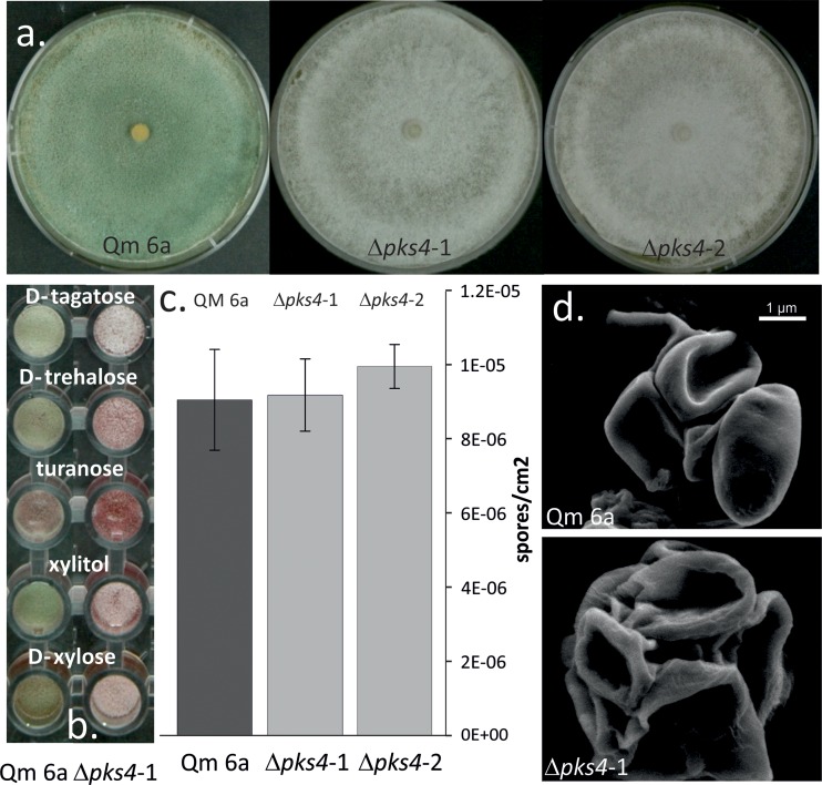
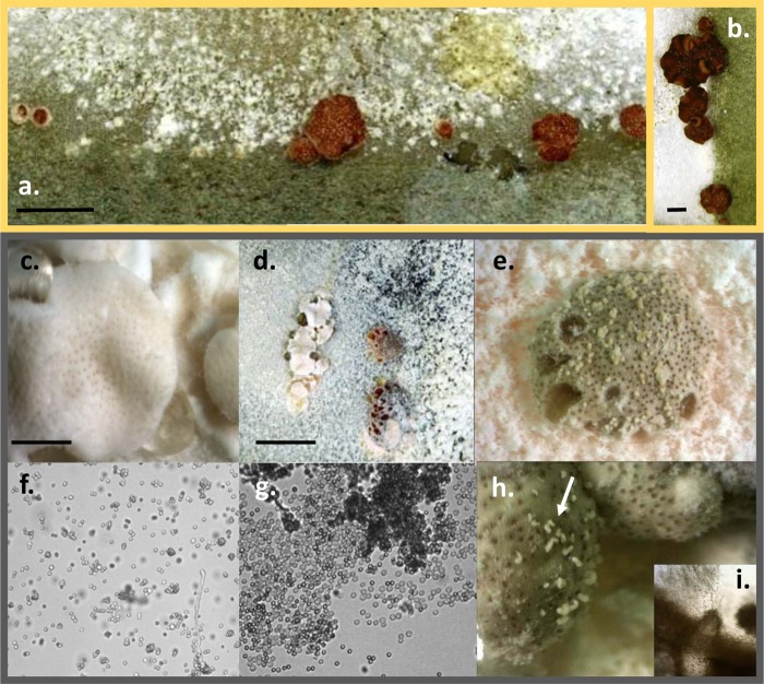
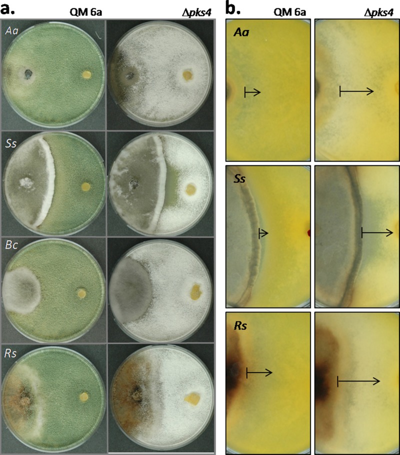
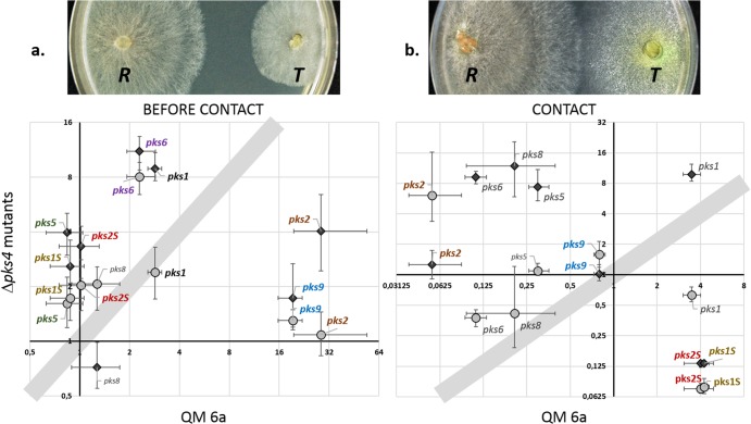
Species of the fungal genus Trichoderma (Hypocreales, Ascomycota) are well-known for their production of various secondary metabolites. Nonribosomal peptides and polyketides represent a major portion of these products. In a recent phylogenomic investigation of Trichoderma polyketide synthase (PKS)-encoding genes, the pks4 from T. reesei was shown to be an orthologue of pigment-forming PKSs involved in synthesis of aurofusarin and bikaverin in Fusarium spp. In this study, we show that deletion of this gene in T. reesei results in loss of green conidial pigmentation and in pigmentation alteration of teleomorph structures. It also has an impact on conidial cell wall stability and the antagonistic abilities of T. reesei against other fungi, including formation of inhibitory metabolites. In addition, deletion of pks4 significantly influences the expression of other PKS-encoding genes of T. reesei. To our knowledge, this is the first indication that a low-molecular-weight pigment-forming PKS is involved in defense, mechanical stability, and stress resistance in fungi.


Figures

Fig 1
Fig 1
Conidiation morphology of QM 6a and the pks4 deletion mutants. (a) Plate macromorphology and conidial pigmentation of QM 6a and Δpks4 cultures. (b) Conidial pigmentation of QM 6a and Δpks4 mutants on selected carbon sources of the Biolog FF microplate phenotype microarrays. (c) Conidial density of QM 6a and Δpks4 mutants' spores produced after 7 days of cultivation PDA. No statistically significant difference in the intensity of conidia production was observed (ANOVA, P > 0.05). (d) Conidial mechanical stability observed under a FEI Quanta 200 FEGSEM after application of vacuum conditions and high pressure, under 25,000× magnification.
Fig 2
Fig 2
Teleomorph morphology of T. reesei QM 6a wild type and Δpks4 mutants. (a) Stromata of wild-type T. reesei strains Qm 6a and CBS 999.97, mated in vitro. (b) Fruiting bodies of strains Δpks4-1 and Qm 6a mated in vitro. (c) Young stromata and mature, partially melanized brownish fruiting bodies of F1 Δpks4 strains mated in vitro. Bar, 2 mm. (d) Partially melanized overmature fruiting structures of F1 Δpks4 strains with dark perithecia openings. Bar, 10 mm. (e) Magnified young white stromata of F1 Δpks4 strains with melanized openings. (f) Conidia of F1 Δpks4 strains. (g and h) Ascospores from the cirri of F1 Δpks4 attached to overmature perithecia, indicated by the arrows in panel h. Magnification (panels f and g), ×200. (i) Pigmented perithecium from the stroma shown in panel h. Magnification, ×200.
Fig 3
Fig 3
Antagonistic potentials of QM 6a and the pks4-1 deletion mutant. Confrontations panels are shown only for Δpks4-1, but both mutants revealed identical mycoparasitic patterns. (a) Front sides of confrontation plates, with plant-pathogenic fungi always on the left side and Trichoderma QM 6a or the mutant always on the right. (b) Back sides of confrontation plates, with plant-pathogenic fungi always on the left side and T. reesei QM 6a or the mutant strain always on the right. Antibiosis zones for Δpks4 strains are indicated by the arrows. Aa, Ss, Bs, and Rs mark Alternaria alternata, Sclerotinia sclerotiorum, Botrytis cinerea, and Rhizoctonia solani, respectively.
Fig 4
Fig 4
Effects of VOCs and WSCs from pks4 deletion mutants and QM 6a on growth of R. solani. (a) Reduction of R. solani growth by the VOCs secreted by T. reesei QM 6a and Δpks4 mutants after 4 days of incubation. Both mutants consistently reduced the growth of R. solani. (b) R. solani growth on PDA medium containing WSCs of T. reesei and the pks4 deletion mutants. (c) Growth of R. solani on medium with WSCs secreted by the pks4 mutants and QM 6a in the presence of VOCs from R. solani during growth of the T. reesei strains.
Fig 5
Fig 5
Expression of pks genes in Δpks4 mutants compared to QM 6a before contact (a) and at contact (b) with Rhizoctonia solani. The gray circles and black diamonds represent data for the Δpks4-1 and Δpks4-2 mutants, respectively. Shadowed areas indicate that there was no difference in regulation of the pks4 deletion mutants compared to QM 6a. Protein numbers of pks genes in the genome of T. reesei are as follows (in parentheses): pks1 (Trire2:65172), pks2 (Trire2:65891), pks5 (Trire2:59482), pks6 (Trire2:60118), pks8 (Trire2:81964), pks9 (Trire2:106272), pks1S (Trire2:73621), pks2S (Trire2:73618). Those genes whose names are shown in color exhibited a consistent expression trend for both Δpks4 mutants. Vertical and horizontal bars indicate standard deviations for the wild type and the mutants, respectively. The images above the plots show the sampling stage of the T. reesei (T) strains under each condition of confrontation with R. solani (R).

References

    1. Druzhinina IS, Seidl-Seiboth V, Herrera-Estrella A, Horwitz BA, Kenerley CM, Monte E, Mukherjee PK, Zeilinger S, Grigoriev IV, Kubicek CP. 2011. Trichoderma: the genomics of opportunistic success. Nat. Rev. Microbiol. 16:749–759 - PubMed
    1. Druzhinina IS, Shelest E, Kubicek CP. 2012. Novel traits of Trichoderma predicted through the analysis of its secretome. FEMS Microbiol. Lett. 337:1–9 - PMC - PubMed
    1. Druzhinina IS, Kubicek CP. 2013. Ecological genomics of Trichoderma. In Martin F. (ed), Ecological genomics of fungi. Wiley-Blackwell, Oxford, United Kingdom
    1. Mukherjee PK, Horwitz BA, Kenerley CM. 2012. Secondary metabolism in Trichoderma: a genomic perspective. Microbiology 158:35–45 - PubMed
    1. Kroken S, Glass NL, Taylor JW, Yoder OC, Turgeon BG. 2003. Phylogenomic analysis of type I polyketide synthase genes in pathogenic and saprobic ascomycetes. Proc. Natl. Acad. Sci. U. S. A. 100:15670–15675 - PMC - PubMed

Publication types