. 2013 Oct;15(10):1197-1205.
doi: 10.1038/ncb2837. Epub 2013 Sep 15.

Cardiolipin externalization to the outer mitochondrial membrane acts as an elimination signal for mitophagy in neuronal cells

Affiliations

Cardiolipin externalization to the outer mitochondrial membrane acts as an elimination signal for mitophagy in neuronal cells

Charleen T Chu et al. Nat Cell Biol. 2013 Oct.

Abstract

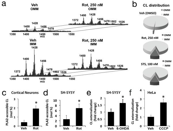
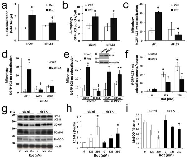
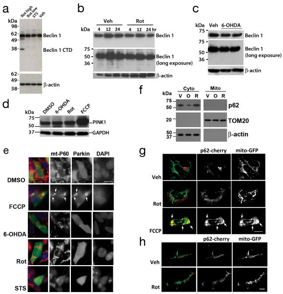
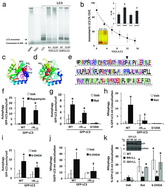
Recognition of injured mitochondria for degradation by macroautophagy is essential for cellular health, but the mechanisms remain poorly understood. Cardiolipin is an inner mitochondrial membrane phospholipid. We found that rotenone, staurosporine, 6-hydroxydopamine and other pro-mitophagy stimuli caused externalization of cardiolipin to the mitochondrial surface in primary cortical neurons and SH-SY5Y cells. RNAi knockdown of cardiolipin synthase or of phospholipid scramblase-3, which transports cardiolipin to the outer mitochondrial membrane, decreased the delivery of mitochondria to autophagosomes. Furthermore, we found that the autophagy protein microtubule-associated-protein-1 light chain 3 (LC3), which mediates both autophagosome formation and cargo recognition, contains cardiolipin-binding sites important for the engulfment of mitochondria by the autophagic system. Mutation of LC3 residues predicted as cardiolipin-interaction sites by computational modelling inhibited its participation in mitophagy. These data indicate that redistribution of cardiolipin serves as an 'eat-me' signal for the elimination of damaged mitochondria from neuronal cells.


Figures

Fig. 1
Fig. 1. Rotenone-induced mitophagy
Rotenone increased GFP-LC3 puncta and colocalization with mitochondria (arrows) in SH-SY5Y cells (a-c, 1 μM) and primary cortical neurons (d, 250 nM), quantified in Fig. 3f and Supplementary Fig. S1b-c. Rotenone increased delivery of MitoTracker Green-stained mitochondria to Lysotracker Red (LTR)-stained lysosomes (e,f), inhibited by siRNA knockdown of Atg7 or LC3 in SH-SY5Y cells. Inset: RNAi knockdown. Rotenone decreased IMM (COXIV), OMM (TOM40) and matrix (MnSOD) protein expression levels in primary neurons (g), reversed by bafilomycin (Baf)-inhibition of autolysosomal degradation, quantified in Supplementary Fig. S1e. Mean +/− s.d. of n=7 independent experiments for b,c, and n=3 independent experiments for f,g (see Statistics Source Data Table); * p < 0.05 vs. vehicle control; † p < 0.05 vs. Rot-siCtrl. Scale = 10 μm. See Supplementary Fig. S7 for uncropped blots.
Fig. 2
Fig. 2. Analysis of mitochondrial CL distribution and externalization
IMM and OMM fractions isolated from primary neurons following the indicated treatments were lipid extracted for LC-MS analysis. MS spectra (a) demonstrated increased CL content of the OMM after rotenone treatment, with diversification of the cluster distribution to the 7 clusters exhibited by the IMM. Pie charts showing the CL distribution between IMM and OMM fractions from toxintreated neurons, normalized to mitochondrial lipid Pi (b). Treatment with rotenone caused significant increases in PLA2-hydrolyzable (surface accessible) CL assessed by LC-MS in primary cortical neurons (c, 250 nM) and SH-SY5Y cells (d, 1 μM). Treatment with 6-OHDA and CCCP increased surface exposure of CL probed with Annexin V in SH-SY5Y (e, 120 μM) and Parkin-expressing-HeLa cells (f, 20 μM), respectively. Mean +/− s.d. of n=3 independent experiments for c-f (see Statistics Source Data Table); * p < 0.05 vs. Control.
Fig. 3
Fig. 3. RNAi knockdown of cardiolipin enzymes attenuate CL exposure and mitophagy
SH-SY5Y cells expressing control (siCtrl) or scramblase-3 siRNA #508 (siPLS3) × 72h were stained with Mitotracker Green FM, a transmembrane potential-independent dye, and treated with rotenone (1 μM). Anionic phospholipids exposed to the outer surface of isolated mitochondria were quantified by flow cytometry for Alexa 647-annexin V binding (a). PLS3 knockdown had no effect on Rot-induced autophagy (b), but decreased mitophagy in SH-SY5Y cells treated with rotenone (c, 1 μM) or 6-OHDA (d, 120 μM). The effects of siPLS3 were recapitulated using a second siRNA #433 (d, Supplementary Fig. S3b, S3g), and reversed by transfection with an RNAi-resistant mouse PLS3 vector (e). Primary neurons transfected with cardiolipin synthase siRNA (siCLS) or scrambled siCtrl were treated with rotenone (250 nM) 72h later and analyzed for autophagy (Supplementary Fig. S1b) or mitophagy by either colocalization analysis (f), or with mitochondrial protein immunoblot (g) and densitometry (h,i; Supplementary Fig. S4c). Knockdown efficiencies are shown in Supplementary Figs. S3 & S4. Mean +/− s.d. of n=4 independent experiments for b,c, and n=3 independent experiments for a, df, h-i (see Statistics Source Data Table); * p < 0.05 vs. vehicle; † p < 0.05 vs. toxin-treated-siCtrl; ** p < 0.05 vs. Rot-siPLS3. See Supplementary Fig. S7 for uncropped blots.
Fig. 4
Fig. 4. Effects of Rot, STS or 6-OHDA on Beclin 1 cleavage and distribution of Parkin and p62
Western blot analysis showed Beclin 1 cleavage products (<49 kDa) were not observed in primary neurons exposed to mitophagy-inducing sublethal rotenone (250 nM × 2h: Rot low) or staurosporine (100 nM × 2h: STS) concentrations (a). However, Beclin-1 was cleaved upon exposure to a lethal dose of rotenone (1 mM × 24h: Rot high). Likewise, SH-SY5Y cells did not show Beclin 1 cleavage upon exposure to rotenone (b) or 6-OHDA (c; duplicate lanes). Data are representative of 2-3 independent experiments per toxin. Cell lysates from stable PINK1-Flag expressing SH-SY5Y cells were treated with 6-OHDA (120 μM), Rot (1 μM), or FCCP (2 μM), and analyzed by Flag immunoblot for PINK1-Flag levels (d). SH-SY5Y cells transfected with HA-Parkin were treated the indicated toxins, immunolabeled for HA (green) and mitochondrial p60 antigen (red) and analyzed for HA-Parkin re-distribution to mitochondria (e). Scale bar: 10μm. FCCP elicited translocation of Parkin to mitochondria (e, arrows), which was not seen with the other mitophagy stimuli. Cytosolic and mitochondrial pellets were prepared from SHSY5Y cells treated with vehicle (V), 6-OHDA (O, 120 μM) or Rot (R, 1 μM). Gels were loaded with cytosolic or mitochondrial proteins, and immunoblotted for p62/SQSMT1, and the indicated fractionation markers (f). Confocal analysis of p62/SQSMT1 distribution in SH-SY5Y cells (g) and in primary cortical neurons (h) that were co-transfected with mCherry-tagged p62/SQSMT1 and mitochondrially targeted GFP. Note recruitment of p62 to large mitochondrial aggregates (g, yellow, arrows) in FCCP-treated cells, which was not observed in control or Rot treated cells. See Supplementary Fig. S7 for uncropped blots.
Fig. 5
Fig. 5. Mitochondrial recognition for mitophagy requires N-terminal amino acids of LC3
Recombinant LC3 incubated with tetraoleoylcardiolipin (TOCL) or dioleoylphosphatidylcholine (DOPC) liposomes at the indicated molar ratios were analyzed by Blue Native PAGE (a); liposome binding impaired the gel entry of proteins. Titration of phospholipids (e.g. TOCL) to LC3 was performed to evaluate the ratio that prevents 50% of the LC3 from entering the gels (IC50) as an index of relative affinity (b). Inset: comparison of IC50 values for TOCL vs. dioleoyl-phosphatidic acid (DOPA) and tetralinoleoyl-CL (TLCL) vs. monolyso-trilinoleoyl-CL (lysoCL). The IC50 for DOPG was >15. * p < 0.05 vs. TOCL; † p < 0.05 s. TLCL. Molecular model of interaction sites for CL binding to LC3 by docking analysis. CL is colored magenta in the top-ranked binding site conformation (c), and cyan in the alternate conformation (d), with the phosphates as spheres (oxygen-red, phosphorusorange). Amino acids interacting with CL are represented as spheres along the LC3 ribbon structure, colored blue>orange from N>C terminus. Thirty-nine LC3 family sequences including isoforms A-C were aligned with ClustalW and displayed using WebLogo, with symbol heights corresponding to relative amino acid frequency (e). Asterisks denote residues predicted to contact CL in the favored (magenta) and alternate conformations (cyan). Arrowhead shows position of N-terminal truncation used to create GFP-LC3 deletion mutants, which were analyzed for GFP-LC3 puncta formation (f,g,i) and participation in mitophagy (h.j) in response to the indicated stimuli. Dual point mutations were prepared based on predicted CL contact residues (e), and analyzed for participation in mitophagy elicited by Rot or 6-OHDA (k). Inset: expression of GFP-LC3 plasmids (Uncropped blots in Supplementary Fig. S7). Mean +/− s.d. of n=3 independent experiments for f-h, and n=4 independent experiments for i,j (see Statistics Source Data Table); * p < 0.05 vs. respective Veh. † p < 0.05 vs. toxin-treated-WT.
Fig. 6
Fig. 6. Schematic diagram summarizing the proposed role of CL in mitochondrial autophagy
Mitochondrial injuries induce a PLS3-dependent externalization of CL to the outer surface of mitochondria. Cargo targeting into autophagosomes are facilitated by interactions of CL with the N-terminal domain of LC3. Clearance of CL-exposed mitochondria by mitophagy (green arrows) would be predicted to prevent CL oxidation and accumulation of pro-apoptotic signals (red arrow).

References

    1. Kim I, Lemasters JJ. Mitophagy selectively degrades individual damaged mitochondria after photoirradiation. Antioxidants & redox signaling. 2011;14:1919–1928. - PMC - PubMed
    1. Kanki T, Wang K, Cao Y, Baba M, Klionsky DJ. Atg32 is a mitochondrial protein that confers selectivity during mitophagy. Dev Cell. 2009;17:98–109. - PMC - PubMed
    1. Narendra D, Tanaka A, Suen DF, Youle RJ. Parkin is recruited selectively to impaired mitochondria and promotes their autophagy. The Journal of cell biology. 2008;183:795–803. - PMC - PubMed
    1. Liu L, et al. Mitochondrial outer-membrane protein FUNDC1 mediates hypoxiainduced mitophagy in mammalian cells. Nature cell biology. 2012;14:177–185. - PubMed
    1. Schlame M. Cardiolipin synthesis for the assembly of bacterial and mitochondrial membranes. Journal of lipid research. 2008;49:1607–1620. - PMC - PubMed

Publication types

MeSH terms