. 2013 Dec;27(12):5059-71.
doi: 10.1096/fj.13-232371. Epub 2013 Sep 13.

Structural characterization of a fusion glycoprotein from a retrovirus that undergoes a hybrid 2-step entry mechanism

Affiliations

Structural characterization of a fusion glycoprotein from a retrovirus that undergoes a hybrid 2-step entry mechanism

Halil Aydin et al. FASEB J. 2013 Dec.

Abstract

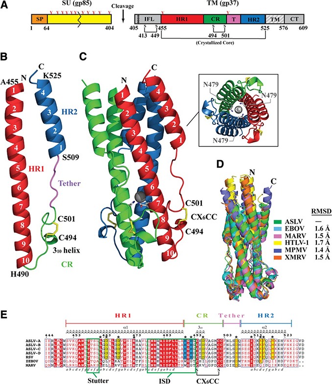
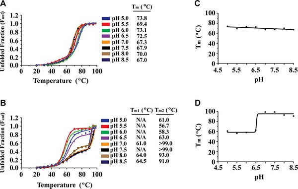
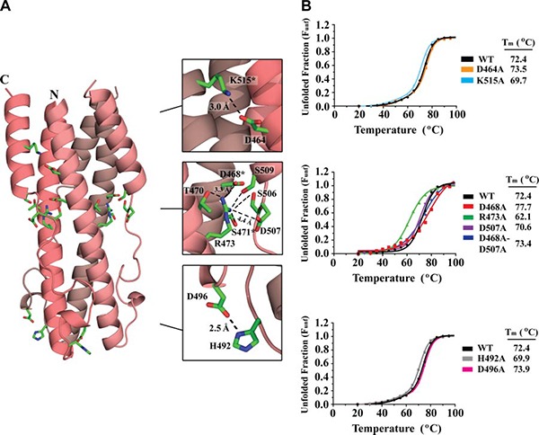
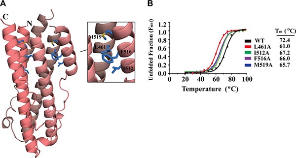
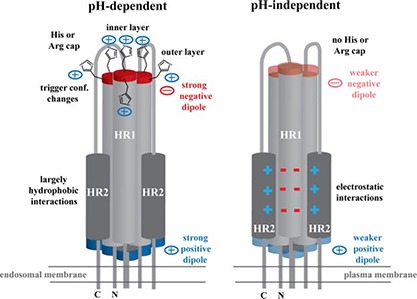
Entry of enveloped viruses into host cells is mediated by their surface envelope glycoproteins (Env). On the surface of the virus, Env is in a metastable, prefusion state, primed to catalyze the fusion of the viral and host membranes. An external trigger is needed to promote the drastic conformational changes necessary for the fusion subunit to fold into the low-energy, 6-helix bundle. These triggers typically facilitate pH-independent entry at the plasma membrane or pH-dependent entry in a low-pH endosomal compartment. The α-retrovirus avian sarcoma leukosis virus (ASLV) has a rare, 2-step entry mechanism with both pH-dependent and pH-independent features. Here, we present the 2.0-Å-resolution crystal structure of the ASLV transmembrane (TM) fusion protein. Our structural and biophysical studies indicated that unlike other pH-dependent or pH-independent viral TMs, the ASLV fusion subunit is stable irrespective of pH. Two histidine residues (His490 and His492) in the chain reversal region confer stability at low pH. A structural comparison of class I viral fusion proteins suggests that the presence of a positive charge, either a histidine or arginine amino acid, stabilizes a helical dipole moment and is a signature of fusion proteins active at low pH. The structure now reveals key residues and features that explain its 2-step mechanism, and we discuss the implications of the ASLV TM structure in the context of general mechanisms required for membrane fusion.

Keywords: ASLV; HTLV-1; TM; helix dipole moment.


Figures

Figure 1
Figure 1
Structure of ASLV TM. A) Schematic of the ASLV Env. CR, chain reversal region; CT, cytoplasmic tail; HR1, heptad repeat 1 region; HR2, heptad repeat 2 region; IFL, internal fusion loop; SP, signal peptide; SU, surface attachment subunit; TM, transmembrane domain. Colored regions correspond to the ASLV TM core that was crystallized. Red Y‐shaped symbols denote N‐linked glycans. TM fusion subunit contains 3 disulfide linkages: one within the hydrophobic internal fusion loop, one in the chain‐reversal region, and an intermolecular covalent linkage between the SU and TM. B) Monomer of the ASLV TM. Features in the TM are color coded to the regions shown in panel A. C) Trimeric ASLV TM postfusion peplomer. The 3 ASLV TM monomers are shown in red, blue, and green. Inset: view of ASLV TM down the 3‐fold axis, showing the chloride ion bound by 3 aspargine residues within the inner HR1 core. D) Structural superimposition of ASLV TM and other retroviral and filoviral fusion subunits. PDB coordinate files used are as follows: ASLV, 4JPR; EBOV, 2EBO; MARV, 4G2K; HTLV‐1, 1MG1; MPMV, 4JF3; XMRV, 4JGS. E) Primary sequence alignment of ASLV TM subtypes A–D, Rous sarcoma virus (RSV) TM, EBOV GP2, and MARV GP2. The 3–4 periodicity of the HRs is shown below the alignment. Stutter region, immunosuppressive domain (ISD), and CX6CC motif are highlighted within labeled green boxes.
Figure 2
Figure 2
ASLV TM stability as a function of pH. A, B) CD thermal denaturation profiles of ASLV TM (A) and HTLV‐1 gp21 (B) at pH values between 5.0 and 8.5. CD signal was normalized between 0 (folded) and 1 (unfolded). C, D) Plot of Tm vs. pH for ASLV TM (C) and HTLV‐1 gp21 (D). ASLV TM is stable from pH 5.0 to pH 8.5, whereas HTLV‐1 gp21 is highly stable at pH > 7.0.
Figure 3
Figure 3
ASLV TM electrostatic interactions. A) Ribbon diagram of ASLV TM with ion‐pair interactions shown as green sticks. Each TM monomer is shown in a different shade of pink. Inset: zoomed view of electrostatic interactions between the HR1‐HR2 and HR1‐HR1 interfaces and the CR region. Distances between residues are shown in angstroms; asterisks indicate residues contributed by a neighboring molecule. B) CD thermal denaturation profiles of wild‐type and salt bridge ASLV TM mutants. CD signals were normalized between 0 (folded) and 1 (unfolded); melting temperatures are shown at right.
Figure 4
Figure 4
ASLV TM hydrophobic interactions. A) Ribbon diagram of ASLV TM with hydrophobic interactions between the HR1‐HR2 regions. Each TM monomer is shown in a different shade of pink. Inset: zoomed view of the hydrophobic interactions (blue sticks). B) CD thermal denaturation profiles of wild‐type and hydrophobic ASLV TM mutants. CD signals were all normalized between 0 (folded) and 1 (unfolded); melting temperatures are shown at right.
Figure 5
Figure 5
ASLV TM CR region histidine residues are important for stability at low pH. A) Left panels: ribbon diagrams of ASLV TM, HTLV‐1 gp21, IAV HA2, and EBOV GP2. Molecules are viewed down the 3‐fold axis with the CR region facing toward the viewer. Right panels: electrostatic potential mapped onto the molecular surface. Red and blue regions denote negative and positive charges, respectively. Positively charged residues that stabilize the negative helix dipole moment are highlighted as green sticks. B) CD thermal denaturation profiles of ASLV TM His490Ala, His490Arg, His492Ala, His492Glu, and HTLV‐1 gp21 Lys394His at pH 8.0, 7.5, 6.5, and 5.0. CD signal was normalized between 0 (folded) and 1 (unfolded).
Figure 6
Figure 6
Summary of pH‐dependent and pH‐independent fusion protein properties. Cartoon shows the viral fusion subunit in the postfusion conformation. Viruses that require low pH for entry, such as ASLV, IAV, and LCMV, fuse at the endosomal membrane and have strong positive and negative charges at their ends due to its helix dipole moment. Negatively charged helix dipole is stabilized by two layers of positively charged histidine or arginine capping residues (only histidine is shown). In addition, the outer layer of histidine residues may be involved in triggering conformational changes in the prefusion viral glycoprotein on entering a low‐pH environment. HR1‐HR2 interface is in general mediated by largely hydrophobic interactions. Viruses that undergo a pH‐independent entry process, such as retroviruses, fuse at the plasma membrane. The helix dipole moment is decreased, and no histidine or arginine residues are located in the CR region to stabilize the negative helix dipole. Moreover, the HR1‐HR2 interface is stabilized with a larger number of electrostatic interactions rather than hydrophobic forces.

References

    1. Harrison, S. C. (2008) Viral membrane fusion. Nat. Struct. Mol. Biol. 15, 690–698 - PMC - PubMed
    1. Weissenhorn, W. , Dessen, A. , Calder, L. J. , Harrison, S. C. , Skehel, J. J. , and Wiley, D. C. (1999) Structural basis for membrane fusion by enveloped viruses. Mol. Membr. Biol. 16, 3–9 - PubMed
    1. Plemper, R. K. (2011) Cell entry of enveloped viruses. Curr. Opin. Virol. 1, 92–100 - PMC - PubMed
    1. Dimitrov, D. S. (2004) Virus entry: molecular mechanisms and biomedical applications. Nat. Rev. Microbiol. 2, 109–122 - PMC - PubMed
    1. White, J. M. , Delos, S. E. , Brecher, M. , and Schornberg, K. (2008) Structures and mechanisms of viral membrane fusion proteins. Crit. Rev. Biochem. Mol. Biol. 43, 189–219 - PMC - PubMed

Publication types

Associated data

LinkOut - more resources