. 2013 Oct;16(10):1417-25.
doi: 10.1038/nn.3516. Epub 2013 Sep 15.

Synapse maturation by activity-dependent ectodomain shedding of SIRPα

Affiliations

Synapse maturation by activity-dependent ectodomain shedding of SIRPα

Anna B Toth et al. Nat Neurosci. 2013 Oct.

Abstract

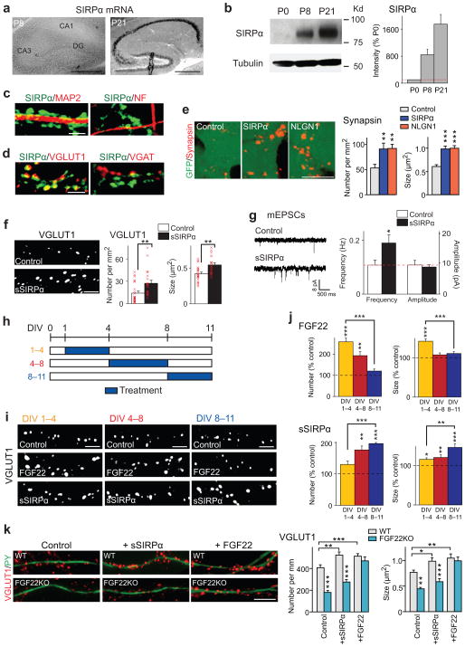
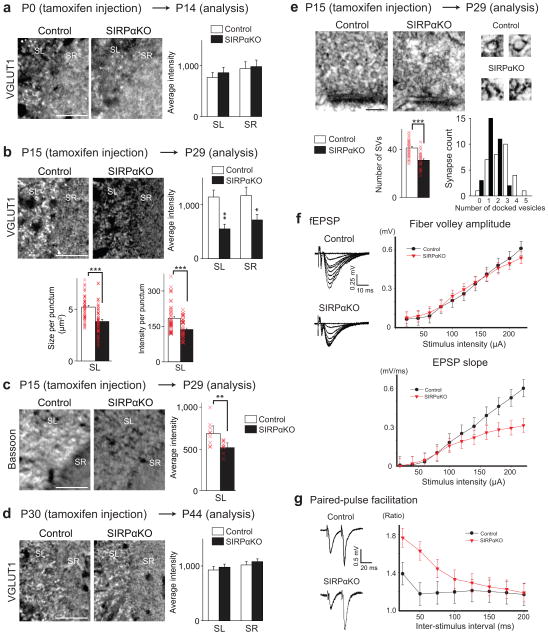
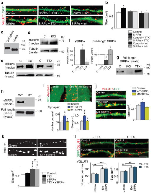
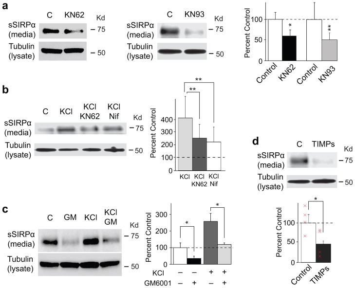
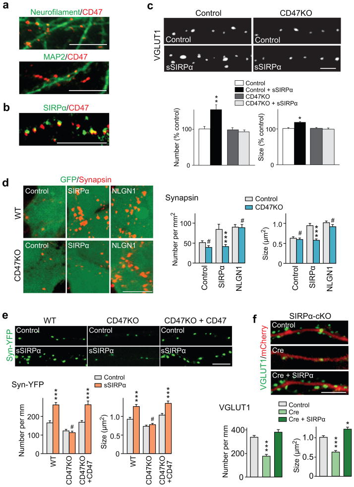
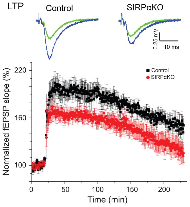
Formation of appropriate synaptic connections is critical for proper functioning of the brain. After initial synaptic differentiation, active synapses are stabilized by neural activity-dependent signals to establish functional synaptic connections. However, the molecular mechanisms underlying activity-dependent synapse maturation remain to be elucidated. Here we show that activity-dependent ectodomain shedding of signal regulatory protein-α (SIRPα) mediates presynaptic maturation. Two target-derived molecules, fibroblast growth factor 22 and SIRPα, sequentially organize the glutamatergic presynaptic terminals during the initial synaptic differentiation and synapse maturation stages, respectively, in the mouse hippocampus. SIRPα drives presynaptic maturation in an activity-dependent fashion. Remarkably, neural activity cleaves the extracellular domain of SIRPα, and the shed ectodomain in turn promotes the maturation of the presynaptic terminal. This process involves calcium/calmodulin-dependent protein kinase, matrix metalloproteinases and the presynaptic receptor CD47. Finally, SIRPα-dependent synapse maturation has an impact on synaptic function and plasticity. Thus, ectodomain shedding of SIRPα is an activity-dependent trans-synaptic mechanism for the maturation of functional synapses.


Conflict of interest statement

COMPETING FINANCIAL INTERESTS

The authors declare no competing financial interests.

Figures

Figure 1
Figure 1. FGF22 and SIRPα promote the early or late stage of glutamatergic presynaptic differentiation
(a) In situ hybridization for Sirpα in the hippocampus during synapse formation (positive signals in black). Sirpα mRNA is highly expressed at P21, the time for synapse maturation, but not at P8, the time for initial synapse differentiation. Reproduced three times. (b) Western blotting for the SIRPα protein (tubulin as control) in the hippocampus. The amount of SIRPα significantly increases from P8 to P21. Full-length blots are presented in Supplementary Figure 10. (c,d) Hippocampal cultures at DIV11 were stained with the antibodies indicated. (c) SIRPα proteins are abundant on MAP2-positive dendrites but not on neurofilament (NF)-positive axons.(d) SIRPα is concentrated at VGLUT1-postive glutamatergic synapses but not at VGAT-positive GABAergic synapses. Reproduced five times. (e) HEK cells expressing SIRPα, neuroligin1 (NLGN1), or control HEK cells (labeled with GFP) were co-cultured with hippocampal neurons for 2 days and stained for synapsin. The synapsin puncta formed on HEK cells expressing SIRPα are significantly more dense and larger than those formed on control HEK cells and are comparable to the ones on HEK cells expressing NLGN1. Data are from 24/23/22 fields from 5 cultures. (f,g) Recombinant soluble SIRPα (sSIRPα) was applied to hippocampal cultures from DIV1–11.(f) sSIRPα treatment significantly increases the number (x 1,000 puncta per mm2) and size of VGLUT1 puncta as compared to PBS control (n = 57 fields from 5 cultures). (g) Representative traces and summary data of whole-cell recordings of mEPSCs from control and sSIRPα-treated hippocampal neurons. mEPSC frequency, but not amplitude, increases by sSIRPα treatment. (n = 57/63 cells from 5 cultures). (h) Schematic timeline of the experiment shown in (i,j). Cultured hippocampal cells were treated with FGF22 or sSIRPα from DIV1–4 (beginning of synapse formation), DIV4–8 (middle of synapse formation), or DIV8–11 (ending of synapse formation). All cultures were fixed on DIV11. (i) Staining of hippocampal cultures for VGLUT1 after treatment with FGF22 or sSIRPα as shown in (h). (j) Numbers and sizes of VGLUT1 puncta after treatment on DIV1–4, DIV4–8, or DIV8–11. FGF22 treatment is most effective at increasing VGLUT1 clustering when incubated from DIV1–4, while sSIRPα is most effective when incubated from DIV8–11. Data are shown as percentage of PBS control and from 32/43/40/34/27/26 fields from 5 cultures. (k) sSIRPα or FGF22 was applied to hippocampal cultures prepared from WT or FGF22KO mice, and the cultures were stained for VGLUT1 and Py, which labels dendrites of CA3 pyramidal neurons. Fewer and smaller VGLUT1 puncta were on CA3 neurons in FGF22KO cultures relative to WT cultures; the defects were rescued by the application of FGF22, but not by sSIRPα. n = 19/23/25/25/17/23 neurites from 3 cultures. Bars in the graphs are mean ± SEM. *p < 0.05, **p < 0.01, ***p < 0.0001 (here and in subsequent figures) by Student’s t-test (f,g) or by ANOVA followed by Tukey test (e,j,k). Scale bars, 500 μm (a), 10 μm (e,k), and 5 μm (others).
Figure 2
Figure 2. SIRPα is required for the maturation, but not induction or maintenance, of excitatory presynaptic terminals in the hippocampus in vivo
(a–d) SIRPα was inactivated around the time of initial synapse differentiation (P0–P14; a), synapse maturation (P15–P29; b,c), or synapse maintenance (P30–P44; d) by tamoxifen injections into conditional SIRPαKO mice. Control animals also received tamoxifen. Hippocampal sections from the control and SIRPαKO mice were stained for VGLUT1 (a,b,d) or bassoon (c). Images are from CA3 (positive signals in white). SL, stratum lucidum; SR, stratum radiatum. Graphs show the measurements of the relative intensity of VGLUT1 or bassoon staining (% control). (a) Mice were injected with tamoxifen at P0 and analyzed at P14. No significant difference in the VGLUT1 staining intensity between control and SIRPαKO at this time period. (b,c) Mice were injected with tamoxifen at P15 and analyzed at P29. The VGLUT1 (b) and bassoon (c) staining intensities are significantly decreased in SIRPαKO mice relative to control. Quantifications of the average size and intensity of VGLUT1 puncta are also shown in (b). (d) Mice were injected with tamoxifen at P30 and analyzed at P44. No significant difference between control and SIRPαKO at this time period. Staining data are from 12/20 sections from 3/5 mice. (e) Electron microscopic analysis of asymmetric (excitatory) synapses in CA3 of P29 mice injected with tamoxifen at P15. High magnification views of synaptic vesicles (SVs) are shown on the right. The numbers of SVs within 400 nm of the active zone and the numbers of docked vesicles are quantified. Significantly fewer SVs and docked vesicles are found in asymmetric synapses in SIRPαKO mice relative to control. Data are from 30 synapses from 3 mice. Student’s t-test (a–e). Scale bars, 50 μm (a) and 100 nm (e). (f) Evoked fEPSPs were recorded in acute slices from CA1 of SIRPαKO mice and control littermates (tamoxifen injections at P15, analyses at P29). Sample traces of fEPSP recordings are shown. In input-output curves (right), fEPSP slope, but not fiber volley amplitude, is significantly decreased in SIRPαKO mice relative to control littermates (p < 0.001 by two-way ANOVA; n = 9/13 cells from 4 mice). (g) Paired-pulse facilitation (PPF) across a range of inter-stimulus intervals for evoked EPSCs from SIRPαKO mice and control littermates. PPF is significantly enhanced in SIRPαKO mice (p < 0.001 by two-way ANOVA; n = 11/15 cells from 4 mice).
Figure 3
Figure 3. Cleavage of the extracellular domain of SIRPα is activity-dependent and is necessary for SIRPα-dependent presynaptic maturation
(a,b) Hippocampal neurons were transfected with a GFP plasmid (Control) or SIRPα and GFP plasmids (SIRPα) at DIV4. TTX or a cocktail of inhibitors for neurotransmitter receptors (APV, CNQX, bicuculline) was applied from DIV5. Cultures were stained for VGLUT1 on DIV13. VGLUT1 clustering on the dendrites of SIRPα-transfected neurons is increased in size as compared to control, but addition of TTX or the inhibitor cocktail prevents this increase. n = 10 neurites from 3 cultures. (c) Hippocampal neurons were cultured for 11 days. Media and cell lysates were assayed for SIRPα. Reproduced five times. (d–g) Hippocampal neurons were cultured for 10–12 days and then incubated with KCl, bicuculline, or TTX for 1–3 days. Media was collected and assayed for the amount of secreted SIRPα (sSIRPα) by immunoprecipitation followed by Western blotting (d,e). Cell lysates were also prepared and assayed for tubulin (d,e) and full-length SIRPα remaining on the cell (g) by Western blotting. In all conditions tested, tubulin levels are similar among the cell lysates. (d) Addition of KCl to activate neurons increases the amount of secreted SIRPα in the media as compared to control. (e) Bicuculline increases the amount of sSIRPα in the media, while TTX decreases it. (f) Quantification of the band intensity from results such as those shown in (d,e,g). Intensities were normalized against the intensity of the control band. n = 5/3 blots from 5/3 cultures. (g) In the cell lysate, the amount of full-length SIRPα is decreased in KCl-treated cultures and increased in TTX-treated cultures. (h) Verification of cleavage-resistant mutant SIRPα. HEK cells were transfected with the wild-type SIRPα or cleavage-resistant mutant SIRPα (MT) plasmid for 2 days. Media and cell lysates were assayed for sSIRPα or full-length SIRPα. MT-SIRPα shows no release of sSIRPα. Reproduced three times. (i) HEK cells expressing WT- or MT-SIRPα and control HEK cells (labeled with GFP) were co-cultured with hippocampal neurons for 2 days and stained for synapsin. MT-SIRPα does not increase the number and size of synapsin puncta. Data are from 24/24/33 fields from at 5 cultures. (j) Hippocampal neurons were transfected with the GFP plasmid (Control) or the WT- or MT-SIRPα plasmid together with the GFP plasmid at DIV4 and stained for VGLUT1 at DIV13. VGLUT1 puncta on the dendrites of WT-SIRPα-transfected neurons, but not on those of MT-SIRPα-transfected neurons, are increased in size as compared to control. n = 23 neurites from 3 cultures. (k) Presynaptic maturation induced by soluble SIRPα is not dependent on neural activity. Hippocampal neurons were treated with a bath application of sSIRPα and/or TTX from DIV1, and stained for VGLUT1 at DIV13. sSIRPα increases the size of VGLUT1 puncta, and adding TTX has no effect on the VGLUT1 clustering induced by sSIRPα. n = 13 fields from 3 cultures. (l) Hippocampal neurons were transfected with the GFP plasmid (Control) or the Ext-SIRPα and GFP plasmids (Ext-SIRPα = secretable SIRPα) at DIV4. TTX was applied from DIV5. Cultures were stained for VGLUT1 on DIV13. VGLUT1 clustering on the dendrites of Ext-SIRPα-transfected neurons is increased in number and size as compared to control, and addition of TTX has no effect on this increase. n = 26/23/21/24 neurites from 3 cultures. Student’s t-test (b,f) or ANOVA followed by Tukey test (i–l). #: not significant. Scale bars, 10 μm (i,l), 5 μm (a,j,k).
Figure 4
Figure 4. SIRPα-dependent presynaptic maturation involves calcium channel, CaMK, and MMP
(a–d) Hippocampal neurons were cultured for 10–12 days and then incubated with a calcium channel blocker - nifedipine, CaMK inhibitors - KN62 or KN93, or MMP inhibitors - GM6001 or TIMPs, in the presence or absence of KCl for 1–3 days. Media was collected and assayed for the amount of secreted SIRPα. Cell lysates were assayed for tubulin. Quantification of the sSIRPα band intensity is shown in the graphs (normalized against control). Nifedipine, KN62, KN93, GM6001, and TIMPs effectively decrease the amount of sSIRPα found in the media (Student’s t-test). n = 4 blots from 4 cultures.
Figure 5
Figure 5. CD47 is the presynaptic receptor for SIRPα-mediated presynaptic maturation
(a,b) Hippocampal neurons were stained for CD47 together with neurofilament, MAP2 or SIRPα. CD47 puncta are abundant in neurofilament-positive axons and not in MAP2-positve dendrites (a) and co-localized with SIRPα (b). Reproduced five times. (c) Hippocampal neurons prepared from CD47KO mice and control littermates were treated with sSIRPα and stained for VGLUT1. sSIRPα does not increase the number and size of VGLUT1 puncta in CD47KO neurons. n = 80/93/102/108 fields from 4 cultures. (d) HEK cells expressing SIRPα, neuroligin1, or control HEK cells (labeled with GFP) were co-cultured with hippocampal neurons prepared from WT or CD47KO mice for 2 days and stained for synapsin. HEK cells expressing SIRPα induce presynaptic differentiation in co-cultured WT hippocampal neurons, but fail to do so in co-cultured CD47KO neurons. HEK cells expressing neuroligin1 induce presynaptic differentiation in both WT and CD47KO neurons. Graphs show quantification of synapsin puncta number and size. Data are from 38/34/34/35/42/40 fields from 5 cultures. (e) Presynaptic expression of CD47 restores responsiveness to sSIRPα in CD47KO neurons. Hippocampal neurons prepared from WT or CD47KO mice were transfected with the synaptophysin-YFP plasmid or with the synaptophysin-YFP and CD47 plasmids. Cultures were treated with sSIRPα, and presynaptic differentiation of transfected neurons was assessed by synaptophysin-YFP clustering. sSIRPα does not increase the number and size of synaptophysin-YFP puncta in CD47KO neurons, but introduction of CD47 restores responsiveness to sSIRPα. n = 33/37/33/35/37/36 neurites from 3 cultures. (f) Postsynaptic expression of SIRPα rescues presynaptic defects in SIRPαKO neurons. Hippocampal neurons prepared from conditional SIRPαKO mice were transfected with an mCherry plasmid (control), Cre and mCherry plasmids (knockout) or Cre, mCherry and SIRPα plasmids (rescue) and stained for VGLUT1. Both the number and size of VGLUT1 puncta on mCherry-expressing dendrites were decreased in SIRPαKO relative to control, but postsynaptic expression of SIRPα rescues the defects. n = 41/33/36 neurites from 3 cultures. Student’s t-test (c) or ANOVA followed by Tukey test (d–f). Scale bar, 5 μm (c), 10 μm (all others).
Figure 6
Figure 6. Impact of SIRPα-dependent presynaptic maturation on synaptic plasticity
Hippocampal slices were prepared from SIRPαKO mice and control littermates and fEPSPs recorded at CA3–CA1 synapses. LTP was induced by high frequency stimulation (four trains of 1s, 100 Hz stimulations spaced by 30s intervals). LTP is significantly impaired in SIRPαKO mice (p < 0.05 by Student’s t-test at 1 hour after the LTP induction). n = 20/10 slices from 11/7 mice.

Comment in

References

    1. Lipska BK, Halim ND, Segal PN, Weinberger DR. Effects of reversible inactivation of the neonatal ventral hippocampus on behavior in the adult rat. J Neurosci. 2002;22:2835–2842. - PMC - PubMed
    1. Pfeiffer BE, et al. Fragile X mental retardation protein is required for synapse elimination by the activity-dependent transcription factor MEF2. Neuron. 2010;66:191–197. - PMC - PubMed
    1. Kasai H, Fukuda M, Watanabe S, Hayashi-Takagi A, Noguchi J. Structural dynamics of dendritic spines in memory and cognition. Trends Neurosci. 2010;33:121–129. - PubMed
    1. Sanes JR, Lichtman JW. Development of the vertebrate neuromuscular junction. Annu Rev Neurosci. 1999;22:389–442. - PubMed
    1. Waites CL, Craig AM, Garner CC. Mechanisms of vertebrate synaptogenesis. Annu Rev Neurosci. 2005;28:251–274. - PubMed

Publication types

Substances

LinkOut - more resources