. 2013 Oct;19(10):1318-24.
doi: 10.1038/nm.3270. Epub 2013 Sep 15.

Endogenous factor VIII synthesis from the intron 22-inverted F8 locus may modulate the immunogenicity of replacement therapy for hemophilia A

Collaborators, Affiliations

Endogenous factor VIII synthesis from the intron 22-inverted F8 locus may modulate the immunogenicity of replacement therapy for hemophilia A

Gouri Shankar Pandey et al. Nat Med. 2013 Oct.

Abstract

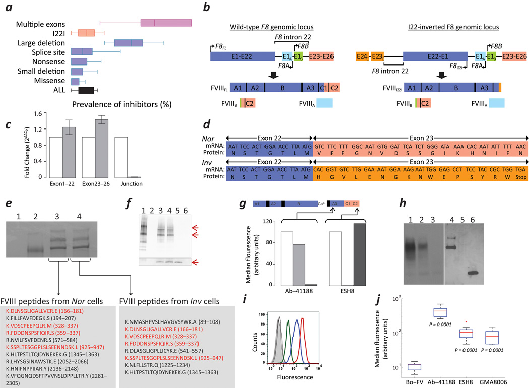
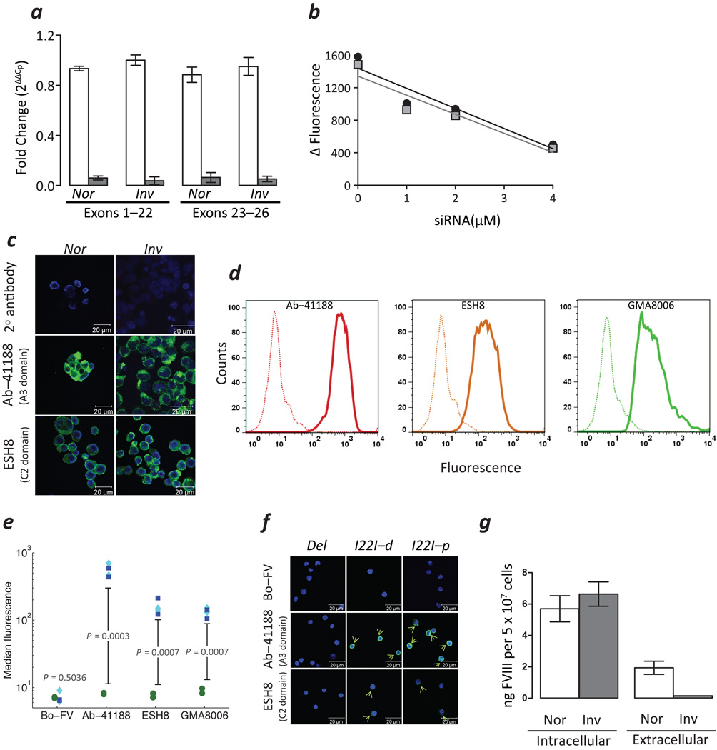
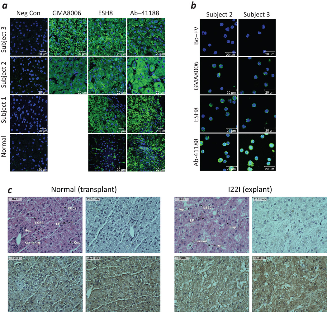
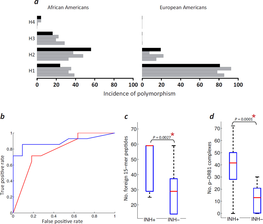
Neutralizing antibodies (inhibitors) to replacement factor VIII (FVIII, either plasma derived or recombinant) impair the effective management of hemophilia A. Individuals with hemophilia A due to major deletions of the FVIII gene (F8) lack antigenically cross-reactive material in their plasma ("CRM-negative"), and the prevalence of inhibitors in these individuals may be as high as 90%. Conversely, individuals with hemophilia A caused by F8 missense mutations are CRM-positive, and their overall prevalence of inhibitors is <10% (ref. 2). Individuals with the F8 intron 22 inversion (found in ∼50% of individuals with severe hemophilia A) have been grouped with the former on the basis of their genetic defect and CRM-negative status. However, only ∼20% of these individuals develop inhibitors. Here we demonstrate that the levels of F8 mRNA and intracellular FVIII protein in B lymphoblastoid cells and liver biopsies from individuals with the intron 22 inversion are comparable to those in healthy controls. These results support the hypothesis that most individuals with the intron 22 inversion are tolerized to FVIII and thus do not develop inhibitors. Furthermore, we developed a new pharmacogenetic algorithm that permits the stratification of inhibitor risk for individuals and subpopulations by predicting the immunogenicity of replacement FVIII using, as input, the number of putative T cell epitopes in the infused protein and the competence of major histocompatibility complex class II molecules to present such epitopes. This algorithm showed statistically significant accuracy in predicting the presence of inhibitors in 25 unrelated individuals with the intron 22 inversion.


Figures

Figure 1
Figure 1
Expression of FVIII in cells derived from subjects with HA. (a) Prevalence of inhibitors in individuals with HA due to various types of F8 gene defects. (b) Structure of the wild-type (left) and I22-inverted F8 (right) and the predicted protein products. (c) mRNA expression of exons 1–22, exons 23–26, and the junction between exons 22 and 23 in the Nor (empty bars) and Inv (filled bars) cells (mean ± SD; n=3). (d) Sequence of cDNA obtained from Nor and Inv cells at the junction of exons 22 and 23. (e) Commassie Blue stained SDS-PAGE gel following immuno-precipitation of the following: (1) Negative control, immuno-precipitation in the absence of rFVIII or cell lysates. (2) ~100 ng purified FVIII. (3) lysate of > 50 × 106 Nor cells. (4) lysate of > 50 × 106 Inv cells. Human-FVIII specific peptides identified by mass spectrometric analysis of bands co-migrating with the purified rFVIII are shown in the lower panels. Peptides in red are those identified in both Nor and Inv cells. (f) Immunoblot following immuno-precipitation using a cocktail of mAbs to FVIII followed by immuno-blotting using a sheep FVIII-specific pAb: (1) ~100 ng purified FVIIIFL. (2) Blank lane. (3) Lysate of > 50 × 10 6Nor cells. (4) Lysate of >50 × 106 Inv cells. (5) ~100 ng of the purified C2 domain of FVIII. (6) Negative control, immuno-precipitation in the absence of rFVIII or cell lysates. The lower panel depicts the lower molecular weight bands overexposed using a more sensitive chemiluminescent substrate. (g) The MFI of NIH-3T3 cells transfected with F8FL (unfilled), F8I22I (light grey) and F8B (dark grey) stained with either Ab-41188 or ESH8. Arrows indicate site specificity of mAbs to FVIII molecule. (h) Immunoblots of cell lysates of HuH-7 cells transiently transfected with F8FL (Lanes 1 & 4), F8I22I (Lanes 2 & 5) and F8B (Lanes 3 & 6); domain-specific mAbs to FVIII, Ab-41188 (Lanes 1–3) and GMA8006 (Lanes 4–6) were used to probe a Western Blot of cell lysates. (i) Intracellular expression of FVIII in permeabilized Inv cells detected by flow cytometry using isotype controls IgG1 and IgG2a (grey filled and grey solid line respectively), Bo-FV (black, dotted line) and the mAbs to FVIII; Ab-41188 (red, solid), ESH8 (blue, solid) or GMA8006 (green, solid). (j) Bar and whisker plots showing the MFI of PBMCs obtained from individuals with the HA due to the I22I and labelled with the indicated Abs.
Figure 2
Figure 2
FVIII expression in subjects with HA. (a) Expression of indicated mRNAs (mean ± SD; n = 3) in Nor and Inv cells treated with Smart Pool siRNA specific to FVIII (F8-siRNA, unfilled bars) or scrambled non-specific siRNA (NT-siRNA, filled gray bars). (b) Relative expression of FVIII estimated in Inv cells treated with indicated concentrations of F8-siRNA by flow cytometry using mAbs, Ab-41188 (grey squares) and ESH8 (black circles). (c) Confocal images of permeabilized Nor (left panels) and Inv cells (right panels) labelled with the indicated mAbs. Areas stained by the Abs to FVIII are depicted in green and the DAPI stained nuclei in blue. (d) Detection of intracellular FVIII in a flow cytometry assay using the indicated mAbs in PBMCs obtained from individuals with HA due to either a deletion in the F8 gene (dotted lines) or the I22I (solid lines). (e) The flow cytometry assay depicted in Fig. 2d was carried out on samples from multiple subjects. The MFI of cells obtained from subjects with the I22I (blue squares, proximal; cyan diamonds, distal) were compared with those obtained from subjects with the large deletion (green circles) when detected with the indicated mAbs. (f) Confocal images of permeabilized PBMCs from subjects with a large deletion and I22I (both distal (I22I-d) and proximal (I22I-p)) labelled with the indicated mAbs. Areas stained by the Abs to FVIII are depicted in green and the DAPI stained nuclei in blue. Green arrows show cells stained by FVIII-specific mAbs. (g) FVIII levels in the conditioned medium (extracellular) and lysates (intracellular) of Nor and Inv cells estimated by ELISA (mean ± SD; n = 3).
Figure 3
Figure 3
FVIII expression in liver tissue obtained from individuals with HA (a) Confocal images of histologic sections obtained from the explanted liver of the individual with the I22I whose cells were used to generate the Inv cells (Sample 1), biopsies of his transplanted liver (Normal), and explanted livers from two additional subjects with the I22I (Subjects 2 & 3) immuostained with the indicated mAbs. The areas stained by the Abs specific to FVIII are depicted in green and the DAPI stained nuclei in blue. (b) Confocal images of PBMCs which were frozen at the time of transplant for Subjects 2 and 3 (Fig. 3a) stained with the indicated mAbs. Areas stained by the Abs specific to FVIII are depicted in green and the DAPI stained nuclei in blue. (c) Immunohistochemistry stained sections from a biopsy of the normal liver after its transplantation into the individual with the I22I (left) and the explanted liver from the same individual (right). Clockwise from upper left: (i) H&E stained sections; (ii) negative control sections stained with only the HRP-conjugated goat anti-mouse-IgG-Fc-secondary antibody; (iii) positive Ab-41188 stained sections and (iv) positive ESH8 stained sections.
Figure 4
Figure 4
A pharmacogenomic algorithm for personalized assessment of inhibitor risk in subjects with HA. (a) Incidence of four common FVIII variants (H1, wild-type; H2, D1241E; H3, D1241E & M2238V; H4, R484H & D1241E) in African American (left panel) and European American (right panel) individuals with HA who were evaluated in three previous studies (grey bars) and this study (black bars). (b) ROC curves comparing the number of foreign peptides (red line) and number of binders, i.e. peptide-MHC-II affinity ≤500 nM (blue line), as predictors of the development of inhibitors in individual subjects with HA. (c) The number of foreign 15-mer peptides each HA subject was exposed to in the inhibitor positive (INH+) and inhibitor negative (INH−) groups are depicted as box plots where the central red mark is the median and the edges represent the 25th and 75th percentiles. (d) Box plots (see c) depicting the number of peptide-DRB1 (p-DRB1) complexes in the INH+ and INH− groups.

References

    1. Lillicrap D. Improvements in factor concentrates. Curr. Opin. Hematol. 2010;17:393–397. - PubMed
    1. Wight J, Paisley S. The epidemiology of inhibitors in haemophilia A: a systematic review. Haemophilia. 2003;9:418–435. - PubMed
    1. Gouw SC, et al. F8 gene mutation type and inhibitor development in patients with severe hemophilia A: systematic review and meta-analysis. Blood. 2012;119:2922–2934. - PubMed
    1. Jenkins PV, et al. Analysis of intron 22 inversions of the factor VIII gene in severe hemophilia A: implications for genetic counseling. Blood. 1994;84:2197–2201. - PubMed
    1. Egler C, et al. Kinetic parameters of monoclonal antibodies ESH2, ESH4, ESH5, and ESH8 on coagulation factor VIII and their influence on factor VIII activity. J. Mol. Recognit. 2009;22:301–306. - PubMed

Publication types