

Optical coherence tomography findings in spinocerebellar ataxia-3
- PMID: 24037234
- PMCID: PMC3869502
- DOI: 10.1038/eye.2013.201
Optical coherence tomography findings in spinocerebellar ataxia-3
Abstract
Purpose: To report optical coherence tomography (OCT) findings in order to detect subclinical alterations of the afferent visual pathways in spinocerebellar ataxia 3 (SCA-3).
Patients and methods: Nine genetically confirmed patients (18 eyes) were evaluated with a complete ophthalmologic examination including visual acuity, colour vision, visual field test, and retinal nerve fibre layer (RNFL) and macular thickness with OCT Cirrus HD. A neurological examination was performed and the Scale for the Assessment and Rating of Ataxia (SARA score) was determined in all patients.
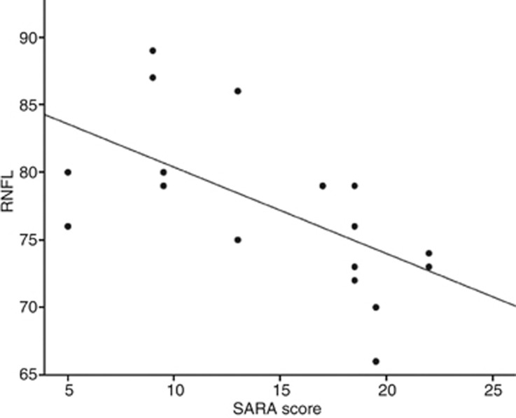
Results: The mean RNFL thickness was 77.39 microns, standard deviation (SD) was ± 5.93. In 15 eyes (83.33%), the mean RNFL thickness was lower than the population average considering age and sex. In 10 cases, there was a reduction of the RNFL thickness in the superior sector, eight in the inferior and four in the nasal. Temporal sector RNFL thickness was preserved in all eyes. RNFL thickness was inversely correlated to SARA score (r=-0.64, P=0.012). The mean macular thickness was 252.61 microns, SD ± 22.80, being inferior respecting average population in only two eyes (11.11%). In four patients, (eight eyes) OCT studies were performed during a mean follow-up of 14.25 months, and in five eyes (62.50%) there was a mild trend to a RNFL thickness decrease in this period.
Conclusion: A mild and progressive decrease in RNFL thickness can be observed in SCA-3 patients. A negative correlation exists between an anatomic marker (RNFL thickness) and a clinical severity scale (SARA score); thus, RNFL thickness could be considered as a promising biomarker of the disease.
Figures
References
-
- Miller RC, Tewari A, Miller JA, Garbern J, Van Stavern GP. Neuro-ophthalmologic features of spinocerebellar ataxia type 7. J Neuro-Ophthalmol. 2009;29 (3:180–186. - PubMed
-
- Fortuna F, Barboni P, Liguori R, Valentino ML, Savini G, Gellera C, et al. Visual system involvement in patients with Friedreichs ataxia. Brain. 2009;132:116–123. - PubMed
-
- Moschos MM, Tagaris G, Markopoulos I, Margetis I, Tsapakis S, Kanakis M, et al. Morphologic changes and retinal impairment in patients with Parkinson disease without visual loss. Eur J Ophthalmol. 2010;21:24–29. - PubMed
-
- Lu Y, Li Z, Zhang X, Ming B, Jia J, Wang R, et al. Retinal nerve fibre layer structure abnormalities in early Alzheimers disease: evidence in optical coherence tomography. Neurosci Lett. 2010;480:69–72. - PubMed
MeSH terms
LinkOut - more resources
Full Text Sources
Other Literature Sources
Medical
