. 2013 Dec;34(5-6):369-78.
doi: 10.1007/s10974-013-9359-4. Epub 2013 Sep 14.

Localisation of AMPK γ subunits in cardiac and skeletal muscles

Affiliations

Localisation of AMPK γ subunits in cardiac and skeletal muscles

Katalin Pinter et al. J Muscle Res Cell Motil. 2013 Dec.

Abstract

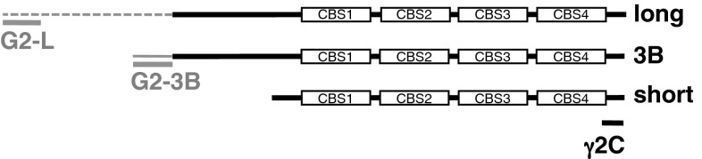
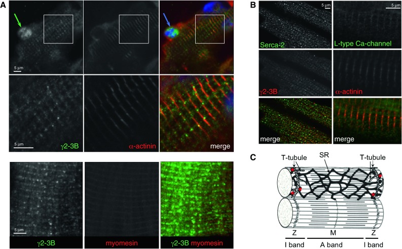
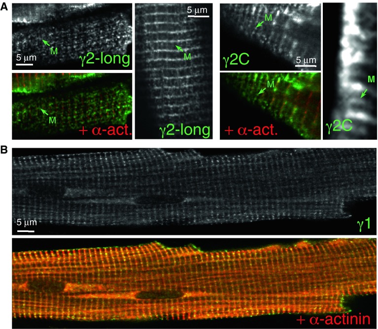
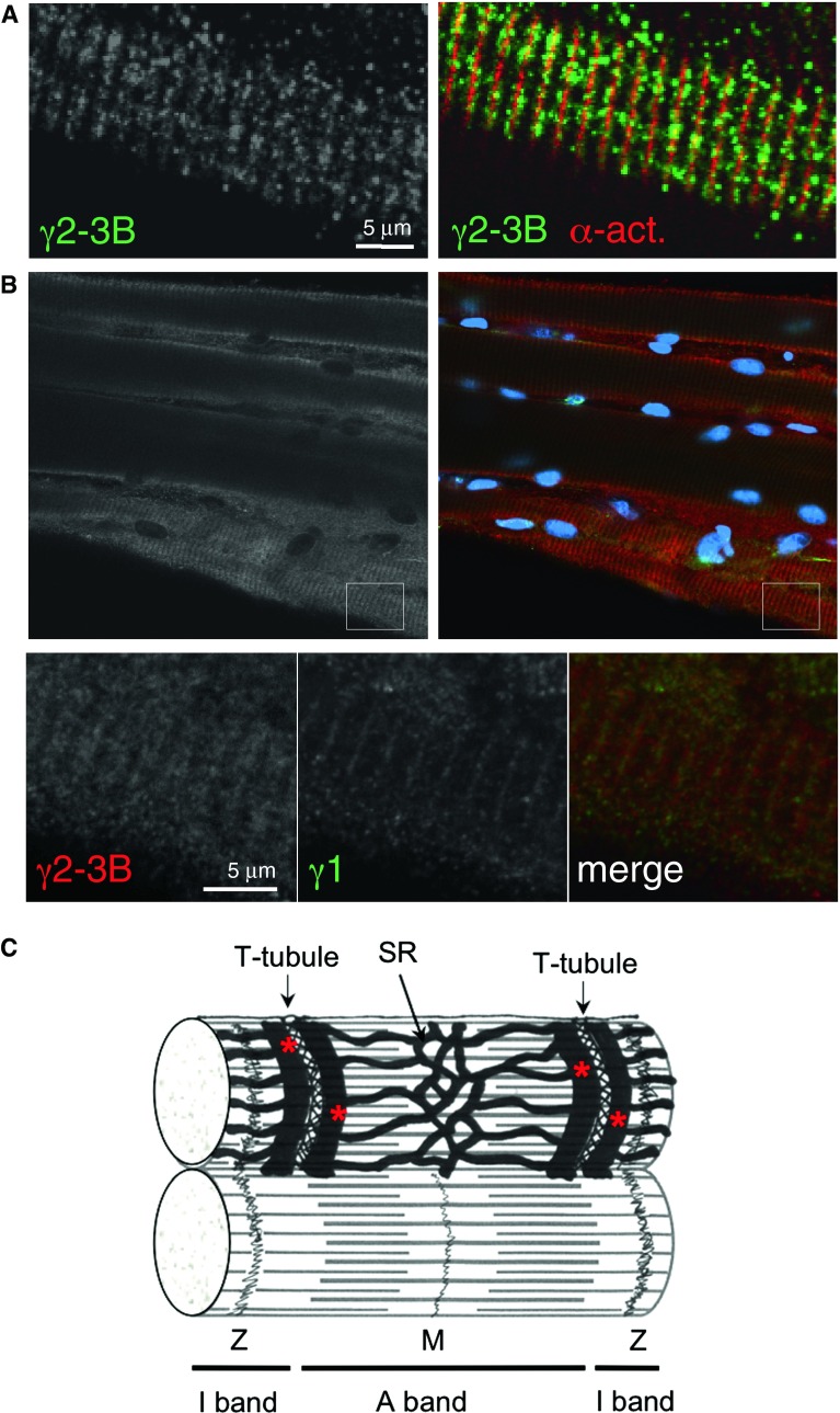
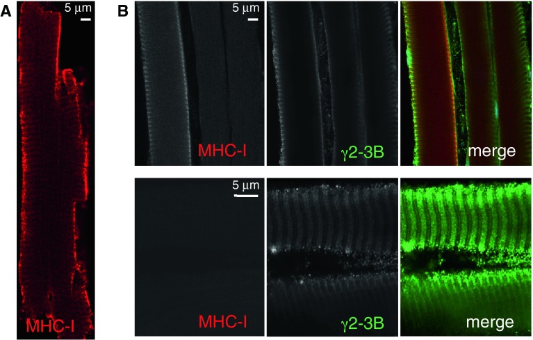
The trimeric protein AMP-activated protein kinase (AMPK) is an important sensor of energetic status and cellular stress, and mutations in genes encoding two of the regulatory γ subunits cause inherited disorders of either cardiac or skeletal muscle. AMPKγ2 mutations cause hypertrophic cardiomyopathy with glycogen deposition and conduction abnormalities; mutations in AMPKγ3 result in increased skeletal muscle glycogen. In order to gain further insight into the roles of the different γ subunits in muscle and into possible disease mechanisms, we localised the γ2 and γ3 subunits, along with the more abundant γ1 subunit, by immunofluorescence in cardiomyocytes and skeletal muscle fibres. The predominant cardiac γ2 variant, γ2-3B, gave a striated pattern in cardiomyocytes, aligning with the Z-disk but with punctate staining similar to T-tubule (L-type Ca(2+) channel) and sarcoplasmic reticulum (SERCA2) markers. In skeletal muscle fibres AMPKγ3 localises to the I band, presenting a uniform staining that flanks the Z-disk, also coinciding with the position of Ca(2+) influx in these muscles. The localisation of γ2-3B- and γ3-containing AMPK suggests that these trimers may have similar functions in the different muscles. AMPK containing γ2-3B was detected in oxidative skeletal muscles which had low expression of γ3, confirming that these two regulatory subunits may be co-ordinately regulated in response to metabolic requirements. Compartmentalisation of AMPK complexes is most likely dependent on the regulatory γ subunit and this differential localisation may direct substrate selection and specify particular functional roles.


Figures

Fig. 1
Fig. 1
Mapping the γ2 immunogen sequences on the γ2 variants. Domain diagrams of the three γ2 variants showing the unique sequences of γ2-long and γ2-3B in grey dotted and solid lines respectively, along with the location of the immunogen sequences
Fig. 2
Fig. 2
Subcellular localisation of AMPK γ2-3B in mouse ventricular cardiomyocytes. (A) Localisation of AMPK-γ2-3B—G2-3B antibody. Enlarged sections (boxed areas) are shown in the second row of images. (B) Staining pattern of the T-tubules and SR. Z-disk marker is α-actinin; M-line marker is myomesin. (C) Ultrastructure of cardiomyocytes—a schematic diagram showing the sarcomere and the T-tubule/SR system (based on Katz 1975); the terminal cisternae of SR are highlighted by the red stars
Fig. 3
Fig. 3
Subcellular localisation of γ2 and γ1 in mouse ventricular cardiomyocytes. (A) Staining patterns produced by antibodies to γ2-long (G2-L) and by a pan-γ2 antibody (γ2C). The strongest staining is around the Z-disk with these antibodies; antibodies G2-L and γ2C detect γ2 protein in the M-line. (B) Localisation of AMPK γ1 in the Z-disk by co-staining the cells with the Z-disk marker α-actinin antibody
Fig. 4
Fig. 4
Immunofluorescence staining of mouse skeletal muscle fibres. (A) Localisation of the AMPK γ subunits in skeletal muscle fibres. AMPK with γ1 is in the Z-disk; the γ3 staining is more punctate around the Z-disk. There is γ3 staining in the nucleus. (B) The staining pattern for γ3 appears to be a very regular doublet flanking the Z-disk (enlargement of the boxed area, double green arrows). Nuclear staining with the γ3 antibody is very prominent. Marker for the Z-disk is γ1, and myomesin for the M-line. (C) Staining pattern of the T-tubules/SR system is similar to the γ3 staining. (A,C) EDL muscle fibres; (B) White quadriceps fibres
Fig. 5
Fig. 5
Detection of γ2-3B in skeletal muscle fibres. (A) EDL fibre stained with G2-3B and α-actinin antibodies. (B) Soleus fibres; the Z-disk marked by γ1 staining. The boxed area is enlarged and showing the regular γ-3B staining pattern that is around the Z-disk (second row of images). (C) Ultrastructure of skeletal muscle fibre—a schematic diagram (based on Eisenberg et al. 1974); the terminal cisternae of SR are highlighted by the red stars
Fig. 6
Fig. 6
Expression of γ2-3B and slow myosin heavy chain in cardiac and skeletal muscle. (A) Cardiomyocyte stained with anti-slow myosin heavy chain (MHC-I); (B) A selection of soleus muscle fibres stained with both G2-3B and MHC-I antibodies. The weak M-line staining with G2-3B may depict the M-line part of the SR (see diagram in Fig. 5C)

References

    1. Alzamora R, Gong F, Rondanino C, Lee JK, Smolak C, Pastor-Soler NM, Hallows KR. AMP-activated protein kinase inhibits KCNQ1 channels through regulation of the ubiquitin ligase Nedd4-2 in renal epithelial cells. Am J Physiol Renal Physiol. 2010;299(6):F1308–F1319. doi: 10.1152/ajprenal.00423.2010. - DOI - PMC - PubMed
    1. Andersen MN, Rasmussen HB. AMPK: a regulator of ion channels. Commun Integr Biol. 2012;5(5):480–484. doi: 10.4161/cib.21200. - DOI - PMC - PubMed
    1. Andersson L. Identification and characterization of AMPK gamma 3 mutations in the pig. Biochem Soc Trans. 2003;31(Pt 1):232–235. - PubMed
    1. Arad M, Benson DW, Perez-Atayde AR, McKenna WJ, Sparks EA, Kanter RJ, McGarry K, Seidman JG, Seidman CE. Constitutively active AMP kinase mutations cause glycogen storage disease mimicking hypertrophic cardiomyopathy. J Clin Invest. 2002;109(3):357–362. - PMC - PubMed
    1. Banerjee SK, McGaffin KR, Pastor-Soler NM, Ahmad F (2009) SGLT1 is a novel cardiac glucose transporter that is perturbed in disease states. Cardiovasc Res 84(1):111–118 - PMC - PubMed

Publication types

Substances

LinkOut - more resources