

Apoptosis-related gene expression in glioblastoma (LN-18) and medulloblastoma (Daoy) cell lines
- PMID: 24037645
- PMCID: PMC3844829
- DOI: 10.1007/s13577-011-0029-9
Apoptosis-related gene expression in glioblastoma (LN-18) and medulloblastoma (Daoy) cell lines
Abstract
The expression of apoptosis genes in a commercial pre-designed low-density array from Applied Biosystems was evaluated in two human brain cancer cell models, LN-18 and Daoy (HTB-186™) in comparison to the reference human primary endothelial cells under basic conditions. Analysis of the gene expression in the cancer cell lines compared to the normal control revealed features reflecting anti-apoptotic and inflammatory characteristics of the former. There was an overall downregulation of apoptosis-stimulating genes in both cancer cell lines, along with an upregulation of certain apoptosis inhibitors. A number of genes demonstrated statistically significant changes in their expressions, including BAX (BCL2-associated X protein); the CARD4/NLR family, CARD domain containing 4; CASP10 (caspase 10, apoptosis-related cysteine peptidase); DAP1 (death-associated protein kinase 1), and BIRC5 (baculoviral IAP repeat-containing 5). Anti-apoptotic potential in both cell lines was demonstrated by changes in the Bax:Bcl-2 ratio and downregulation of the APAF1 gene in LN18 cells. There was also significant downregulation of extrinsic signals and the TNF/FADD/inflammatory cascade, and upregulation of caspase inhibitors (IAPs). These results provided a novel molecular characterization of important human cancer cell lines, which might provide a useful research tool for investigating the experimental model of the CNS cell.
Figures
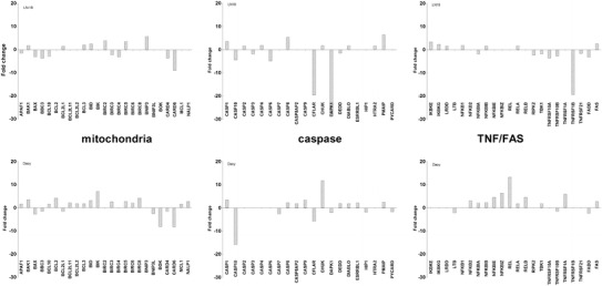
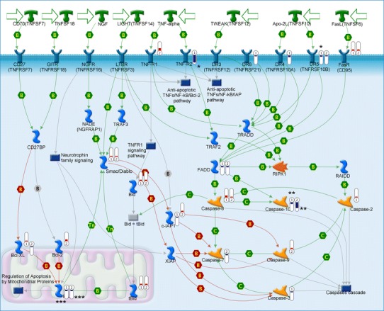
 means decreased gene expression.
  means increased gene expression; height of bars corresponds to the change value. Numbers in symbols indicate: 1 LN18, 2 Daoy cell lines; number of asterisks reflects the statistical significance of the p value (* p < 0.05, ** p < 0.01, *** p < 0.001)
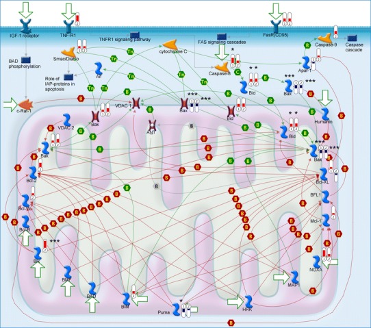 means decreased gene expression. means increased gene expression; height of bars corresponds to the change value. Numbers in symbols indicate: 1 LN18, 2 Daoy cell lines; number of asterisks reflects the statistical significance of the p value (* p < 0.05, ** p < 0.01, *** p < 0.001)
 means decreased gene expression. means increased gene expression; height of bars corresponds to the change value. Numbers in symbols indicate: 1 LN18, 2 Daoy cell lines; number of asterisks reflects the statistical significance of the p value (* p < 0.05, ** p < 0.01, *** p < 0.001)
References
-
- Giangaspero F, Bigner SH, Kleihues P, Pietsch T, Trojanowski JQ. Medulloblastoma. In: Kleihues P, Cavenee WK, editors. Tumours of the nervous system. Lyon: IARC; 2000. p. 129–39.
-
- ATCC. ATCC corporate website. http://www.atcc.org/.
Publication types
MeSH terms
Substances
LinkOut - more resources
Full Text Sources
Other Literature Sources
Research Materials
Miscellaneous
