. 2014 Feb;59(2):555-66.
doi: 10.1002/hep.26712. Epub 2013 Dec 20.

Reciprocal regulation of microRNA-122 and c-Myc in hepatocellular cancer: role of E2F1 and transcription factor dimerization partner 2

Affiliations

Reciprocal regulation of microRNA-122 and c-Myc in hepatocellular cancer: role of E2F1 and transcription factor dimerization partner 2

Bo Wang et al. Hepatology. 2014 Feb.

Abstract

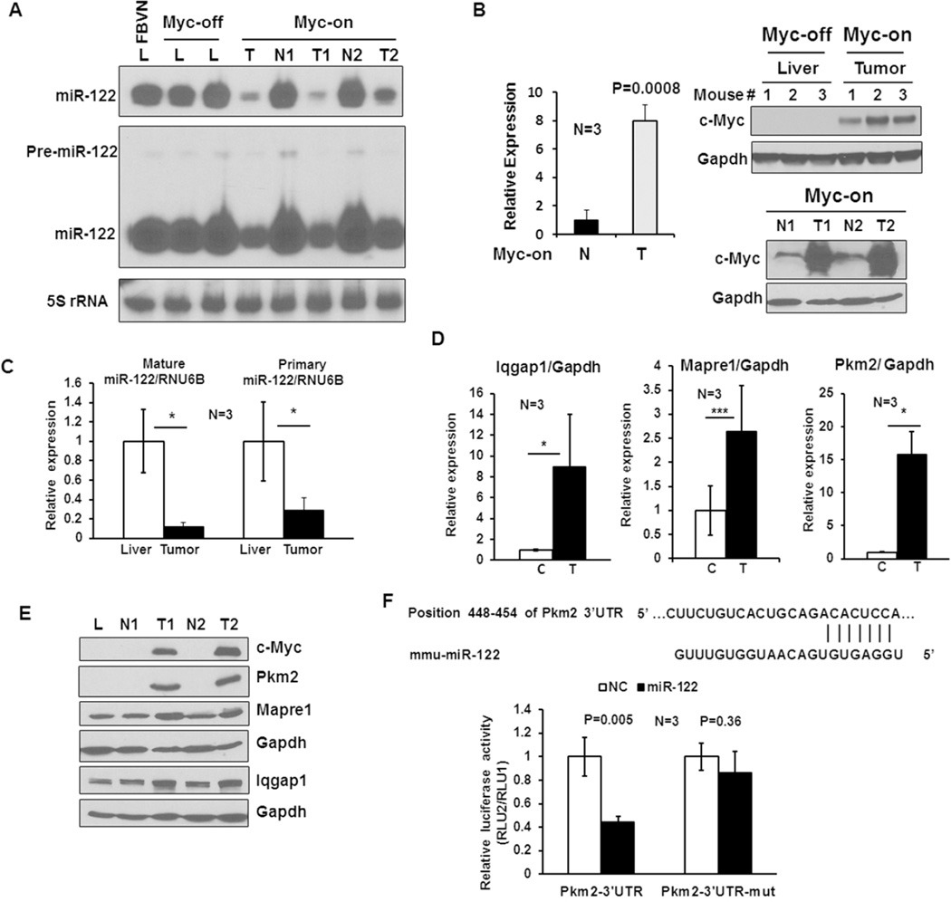
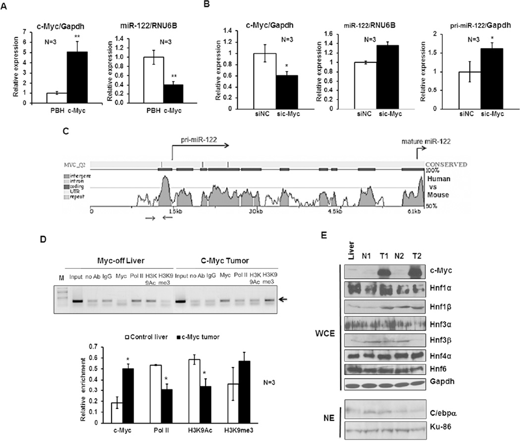
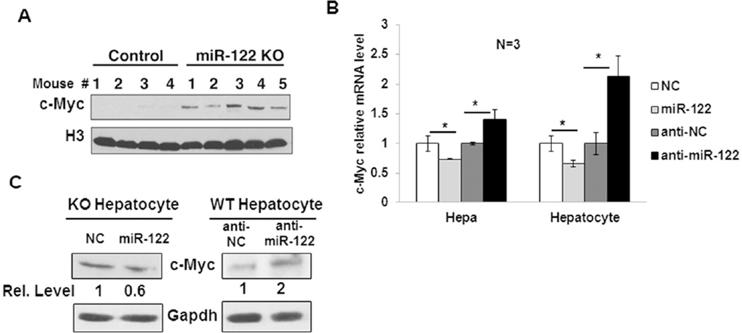
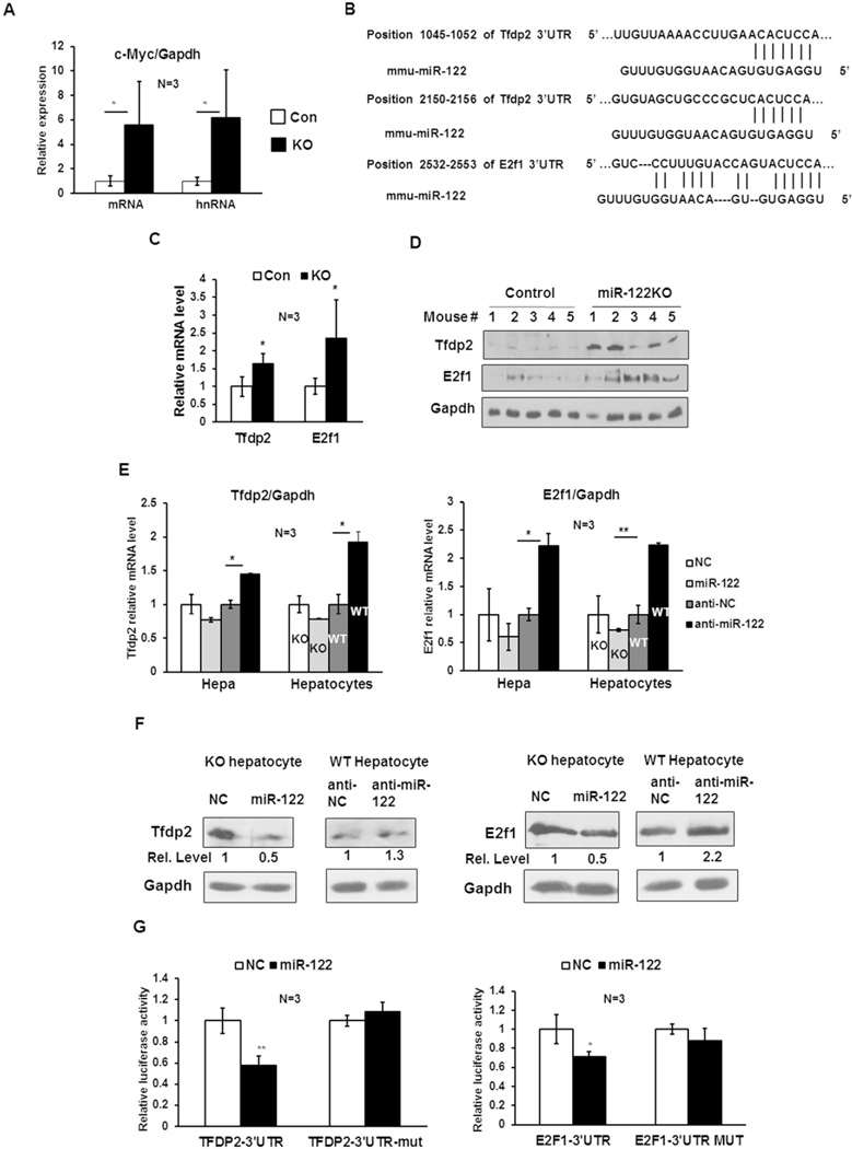
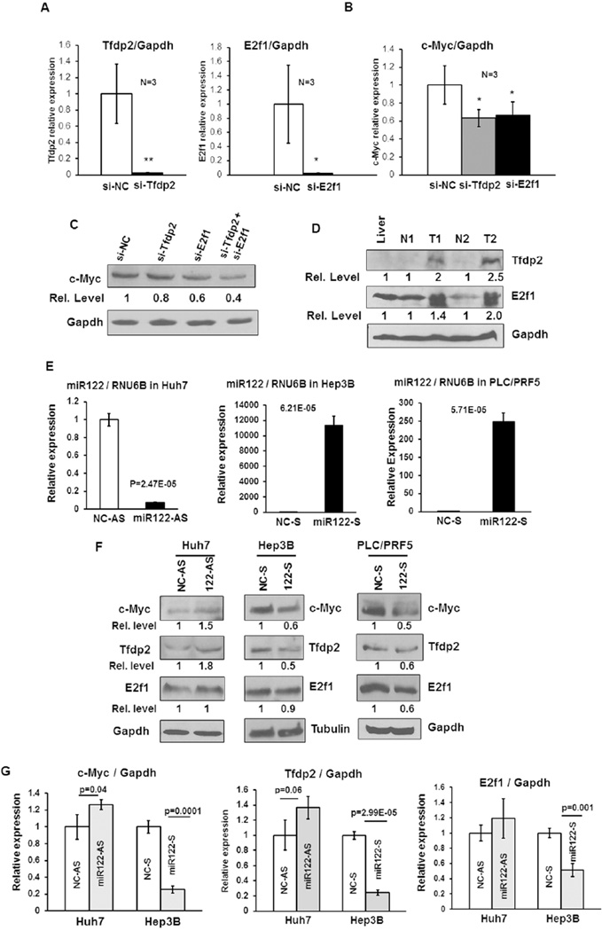
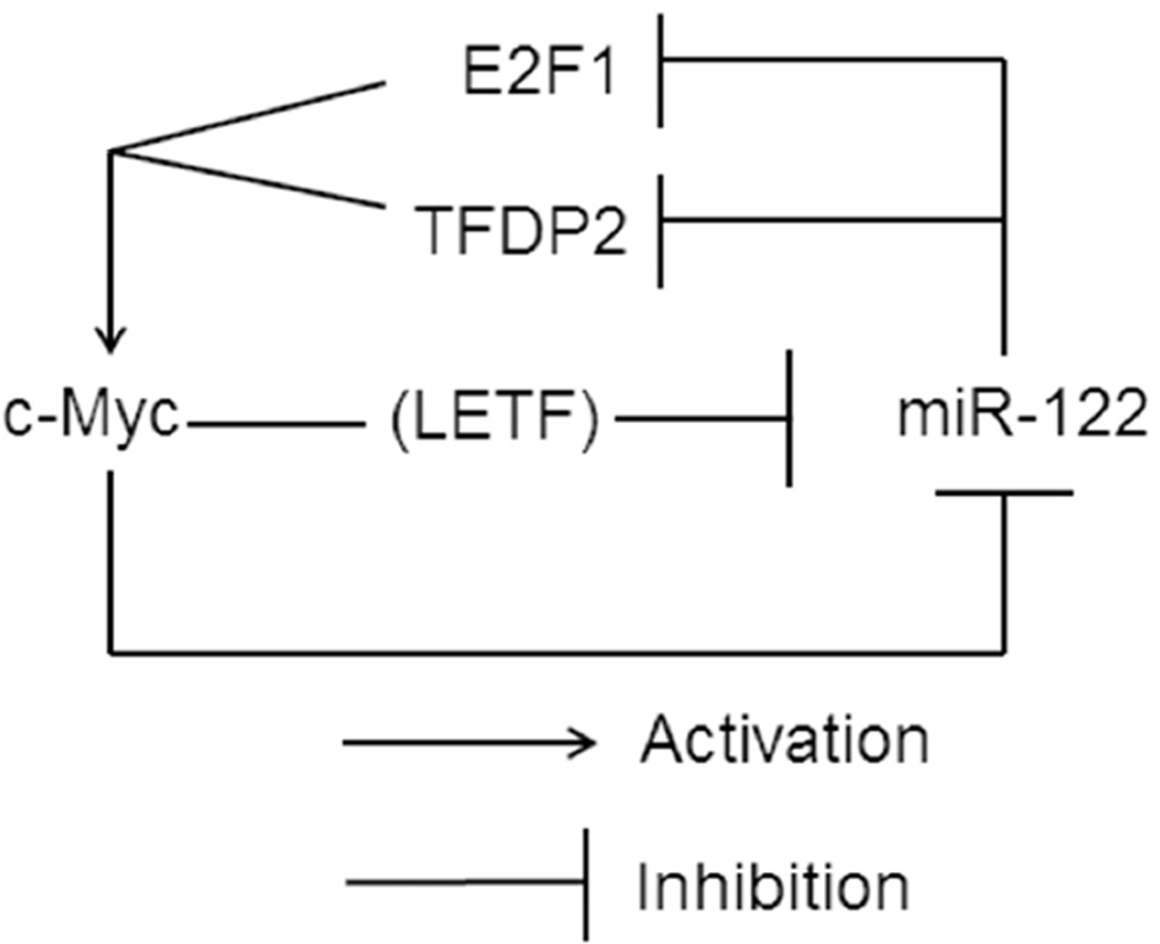
c-Myc is a well-known oncogene frequently up-regulated in different malignancies, whereas liver-specific microRNA (miR)-122, a bona fide tumor suppressor, is down-regulated in hepatocellular cancer (HCC). Here we explored the underlying mechanism of reciprocal regulation of these two genes. Real-time reverse-transcription polymerase chain reaction (RT-PCR) and northern blot analysis demonstrated reduced expression of the primary, precursor, and mature miR-122 in c-MYC-induced HCCs compared to the benign livers, indicating transcriptional suppression of miR-122 upon MYC overexpression. Indeed, chromatin immunoprecipitation (ChIP) assay showed significantly reduced association of RNA polymerase II and histone H3K9Ac, markers of active chromatin, with the miR-122 promoter in tumors relative to the c-MYC-uninduced livers, indicating transcriptional repression of miR-122 in c-MYC-overexpressing tumors. The ChIP assay also demonstrated a significant increase in c-Myc association with the miR-122 promoter region that harbors a conserved noncanonical c-Myc binding site in tumors compared to the livers. Ectopic expression and knockdown studies showed that c-Myc indeed suppresses expression of primary and mature miR-122 in hepatic cells. Additionally, Hnf-3β, a liver enriched transcription factor that activates miR-122 gene, was suppressed in c-MYC-induced tumors. Notably, miR-122 also repressed c-Myc transcription by targeting transcriptional activator E2f1 and coactivator Tfdp2, as evident from ectopic expression and knockdown studies and luciferase reporter assays in mouse and human hepatic cells.

Conclusion: c-Myc represses miR-122 gene expression by associating with its promoter and by down-regulating Hnf-3β expression, whereas miR-122 indirectly inhibits c-Myc transcription by targeting Tfdp2 and E2f1. In essence, these results suggest a double-negative feedback loop between a tumor suppressor (miR-122) and an oncogene (c-Myc).


Figures

Figure 1
Figure 1. miR-122 is downregulated in c-Myc-induced liver tumors
A. Northern blot analysis of precursor and mature miR-122 levels in the normal liver (L) from FBVN and (Myc-off) mice, as well as benign liver (N) and tumor (T) from the same (Myc-on) mouse. Total RNA (25 µg) from each sample was separated by denaturing PAGE (15% acrylamide), transferred onto Nylon membrane, cross-linked and subjected to Northern blot analysis with 32P-labeled anti-miR-122 deoxyoligonucleotide. Membrane was exposed to 4hr and 72hr at −80°C to detect mature and precursor miR-122, respectively. The blot was hybridized to anti-sense 5S rRNA probe to demonstrate equal loading of RNA. B. qRT-PCR and Western blot analysis of c-Myc expression in the liver (Myc-off), Myc-on benign liver (N) and tumor (T) tissues in the presence and absence of doxycycline, respectively. Whole tissue extracts (100 µg protein) were immunoblotted with an antibody specific for human c-Myc and reprobed with Gapdh antibody. C. qRT-PCR analysis of primary and mature miR-122 in Myc-off livers and Myc-on tumors using Taqman kit. Data was normalized to RNU6B. D. qRT-PCR analysis of mRNA levels of specified miR-122 targets in livers and tumors. C: control liver, T: tumor. E. Western blot analysis of selected miR-122 targets in liver and tumor whole tissue extracts (100 µg). L: FBVN liver, N: benign liver and T: Tumor. F. Luciferase reporter assay demonstrating Pkm2 is a target of miR-122. Upper panel: miR-122 cognate site in 3’-UTR of Pkm2 gene. Lower panel: relative luciferase activity in Hepa cells transfected with the psiCHECK2 harboring wild type (Pkm2-3’-UTR) or mutant (Pkm2-3’-UTR-mut) site along with miR-122 mimic (miR-122) or negative control RNA (NC).
Figure 2
Figure 2. c-Myc represses miR-122 expression in vitro
A. Relative c-Myc and mature miR-122 expression in Hepa cells transfected with MYC expression vector (pBH-c-Myc) or pBH (vector control). Hepa cells were transfected with specified plasmids and RNAs isolated 36–48 hr post-transfection were subjected to qRT-PCR analysis. The data was normalized to Gapdh or RNU6B. Relative level in vector-transfected cells was assigned a value of 1. B. Relative c-Myc, mature and primary miR-122 expression in Hepa cells transfected with c-Myc siRNA (sic-Myc) or negative control siRNA (siNC). Cellular RNAs isolated after 36–48 hr were analyzed by qRT-PCR. C. Conserved miR-122 promoter region upstream of +1 site harbors a candidate noncanonical c-Myc binding site predicted by rVista program. Sequence conservation of miR-122 gene between mouse and human as analyzed by rVista. Arrows denote the chromatin immunoprecipitation (ChIP) analysis primers spanning c-Myc site. D. ChIP analysis showed increased binding of c-Myc but reduced association of RNA polymerase II (Pol II) and histone H3K9Ac (a marker of active gene) with miR-122 promoter region in c-Myc induced tumors compared to Myc-off livers. Equal amounts of chromatin (DNA) from livers and tumors were subjected to ChIP assay with specific antibodies. Rabbit IgG and protein G beads alone were used as negative controls. 1:100 dilution of input was used for amplification of miR-122 promoter. Upper panel: A representative photograph of ethidium bromide stained agarose gel containing PCR products obtained with specified antibodies in ChIP assay. Lower panel: Quantitative analysis of ChIP data from 3 different livers and tumors. E. Western blot analysis of c-Myc, LETFs and C/ebpα expression in whole cell extracts (WCE) or nuclear extracts (NE) of Myc-off liver, as well as benign liver (N) and tumor (T) from Myc-on.
Figure 3
Figure 3. c-Myc is upregulated in miR-122KO mouse livers
A. Western blot analysis of c-Myc protein levels in the control and miR-122KO liver nuclear extracts using an antibody that detects both mouse and human Myc proteins. Histone H3 was used as a loading control. B. qRT-PCR analysis of endogenous c-Myc mRNA expression in Hepa and KO hepatocytes transfected with miR-122 mimic (miR122) or negative control mimic (NC) as well as in Hepa cells and wild-type (WT) hepatocytes transfected with miR-122 inhibitor (anti-miR122) or negative control inhibitor (anti-NC). C. Western blot analysis of c-Myc in KO and WT hepatocytes transfected with miR-122 mimic and inhibitor, respectively along with respective negative controls as described in 3B.
Figure 4
Figure 4. Tfdp2 and E2f1, targets of miR-122, are involved in the regulation of c-Myc expression by miR-122
A. qRT-PCR analysis of c-Myc mRNA and hnRNA (primary transcript) levels in control (floxed) and miR-122KO (KO) mouse livers demonstrated comparable increase in both in KO livers. B. Potential miR-122 seed match in Tfdp2 and E2f1 3’UTRs predicted by TargetScan and RNA22 programs, respectively. C. qRT-PCR analysis of Tfdp2 and E2f1 mRNA levels in control and KO mouse livers. D. Western blot analysis of Tfdp2, E2f1 and Gapdh in the whole liver extracts. E. qRT-PCR analysis of Tfdp2 and E2f1 in Hepa cells and KO hepatocytes transfected with miR-122 mimic (miR-122), negative control mimic (NC) as well as in Hepa cells and WT hepatocytes transfected with miR-122 inhibitor (anti-miR-122) or negative control inhibitor (anti-NC). F. Western blot analysis of Tfdp2 and E2f1 in KO and WT hepatocytes transfected with miR-122 mimic or inhibitors as described in 4E. G. Tfdp2 and E2f1 3’UTR driven luciferase assays were performed as described in 1F.
Figure 5
Figure 5. Tfdp2 and E2f1 are involved in the regulation of c-Myc expression in miR-122KO mouse livers
A. qRT-PCR analysis of Tfdp2 and E2f1 in respective siRNA or negative control siRNA (siNC) transfected KO hepatocytes. B. qRT-PCR analysis of c-Myc in KO hepatocytes transfected with siRNA specific for Tfdp2, E2f1 or negative control siRNA (siNC). C. Western blot analysis of c-Myc in Tfdp2 and E2f1 siRNA transfected KO hepatocytes. D. Western blot analysis of E2f1 and Tfdp2 in c-Myc-induced tumors (T), benign (N) and control livers. E, F. qRT-PCR analysis of miR-122 (E) and western blot analysis of c-MYC, E2F1 and TFDP2 (F) in Huh7 cells transfected with 50 nM of anti-miR-122 (122-AS) or anti-sense NC-RNA (NC-AS) and Hep3B and PLC/PRF5 cells transfected with 50nM miR-122 mimic (122-S) or sense NC-RNA (NC-S). G. qRT-PCR analysis of c-MYC, E2F1 and TFDP2 in Huh7 and Hep3B cells.
Figure 6
Figure 6. A model depicting reciprocal regulation between c-Myc and miR-122 based on the observations in miR-122KO and c-MYC transgenic mouse models

References

    1. Bartel DP. MicroRNAs: target recognition and regulatory functions. Cell. 2009;136:215–233. - PMC - PubMed
    1. Pasquinelli AE. MicroRNAs and their targets: recognition, regulation and an emerging reciprocal relationship. Nat Rev Genet. 2012;13:271–282. - PubMed
    1. Hafner M, Landthaler M, Burger L, Khorshid M, Hausser J, Berninger P, Rothballer A, et al. Transcriptome-wide identification of RNA-binding protein and microRNA target sites by PAR-CLIP. Cell. 2010;141:129–141. - PMC - PubMed
    1. Ebert MS, Sharp PA. Roles for microRNAs in conferring robustness to biological processes. Cell. 2012;149:515–524. - PMC - PubMed
    1. Mendell JT, Olson EN. MicroRNAs in stress signaling and human disease. Cell. 2012;148:1172–1187. - PMC - PubMed

Publication types

MeSH terms