Review
. 2013 Oct;74(4):506-16.
doi: 10.1002/ana.24014.

Energy failure: does it contribute to neurodegeneration?

Affiliations
Review

Energy failure: does it contribute to neurodegeneration?

Divya Pathak et al. Ann Neurol. 2013 Oct.

Abstract

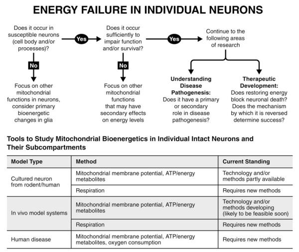
Energy failure from mitochondrial dysfunction is proposed to be a central mechanism leading to neuronal death in a range of neurodegenerative diseases. However, energy failure has never been directly demonstrated in affected neurons in these diseases, nor has it been proved to produce degeneration in disease models. Therefore, despite considerable indirect evidence, it is not known whether energy failure truly occurs in susceptible neurons, and whether this failure is responsible for their death. This limited understanding results primarily from a lack of sensitivity and resolution of available tools and assays and the inherent limitations of in vitro model systems. Major advances in these methodologies and approaches should greatly enhance our understanding of the relationship between energy failure, neuronal dysfunction, and death, and help us to determine whether boosting bioenergetic function would be an effective therapeutic approach. Here we review the current evidence that energy failure occurs in and contributes to neurodegenerative disease, and consider new approaches that may allow us to better address this central issue.


Figures

FIGURE 1
FIGURE 1
Potential mitochondria-based mechanisms by which Parkinson disease proteins may produce energy failure and neuronal death. The schematic illustrates the known effects of loss of parkin or PINK1 (mitochondrial biogenesis, mitochondrial transport, mitochondrial turnover,, dynamics,, and respiration,,) or increased synuclein (mitochondrial turnover, dynamics,, and respiration,). These primary changes may result in disruptions of the normal mitochondrial distribution and/or function, and this in turn could lead to energy failure and neuronal death. Other mitochondrial functions, such as reactive oxygen species (ROS) production, calcium buffering, and roles in apoptotic pathways could also be altered and contribute to cell-death pathways. DJ-1 and LRRK2 can also affect mitochondrial function, but are not depicted in this schematic.
FIGURE 2
FIGURE 2
Regulation of mitochondrial bioenergetics in axons. In most non-neural cells, the capacity of mitochondria to produce energy depends on the mitochondrial mass and function, but they may be less dependent on subcellular distribution due to adenosine triphosphate (ATP) diffusion. In axons, however, a normal distribution of mitochondria may also be required to minimize energy gradients. This is illustrated here by the hypothetical gradations in color in the neuron, in which lighter blue reflects higher ATP levels, whereas darker reflects lower levels. Therefore, factors that disrupt the normal distribution of mitochondria, such as mitochondrial dynamics (the balance between mitochondrial fusion and fission) and mitochondrial motility may have more prominent effects on energy levels in axons. Notably, mitochondria in axons are also smaller and more mobile and may have different lifespans than those in the cell body, suggesting that the intrinsic function of mitochondria in the cell body versus axons may also differ.
FIGURE 3
FIGURE 3
Energy failure in individual neurons. The schematic illustrates an algorithm of critical but challenging questions to determine whether energy failure occurs in individual neurons, including their processes, whether and how it contributes to degeneration, and how it might be targeted therapeutically. The table summarizes the availability of tools and methods to assess the bioenergetic function of mitochondria in individual neurons in model systems and human disease. ATP = adenosine triphosphate.

References

    1. Zheng B, Liao Z, Locascio JJ, et al. PGC-1alpha, a potential therapeutic target for early intervention in Parkinson’s disease. Sci Transl Med. 2010;2:52ra73. - PMC - PubMed
    1. Parihar MS, Brewer GJ. Mitoenergetic failure in Alzheimer disease. Am J Physiol Cell Physiol. 2007;292:C8–C23. - PubMed
    1. Kapogiannis D, Mattson MP. Disrupted energy metabolism and neuronal circuit dysfunction in cognitive impairment and Alzheimer’s disease. Lancet Neurol. 2011;10:187–198. - PMC - PubMed
    1. Mochel F, Haller RG. Energy deficit in Huntington disease: why it matters. J Clin Invest. 2011;121:493–499. - PMC - PubMed
    1. Nunnari J, Suomalainen A. Mitochondria: in sickness and in health. Cell. 2012;148:1145–1159. - PMC - PubMed

Publication types

MeSH terms