Review
. 2014 Feb;243(2):201-15.
doi: 10.1002/dvdy.24036. Epub 2013 Oct 24.

Mechanisms of muscle growth and atrophy in mammals and Drosophila

Affiliations
Review

Mechanisms of muscle growth and atrophy in mammals and Drosophila

Rosanna Piccirillo et al. Dev Dyn. 2014 Feb.

Abstract

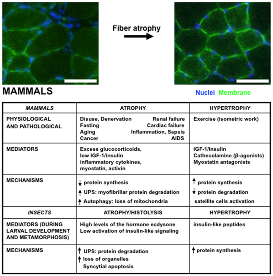
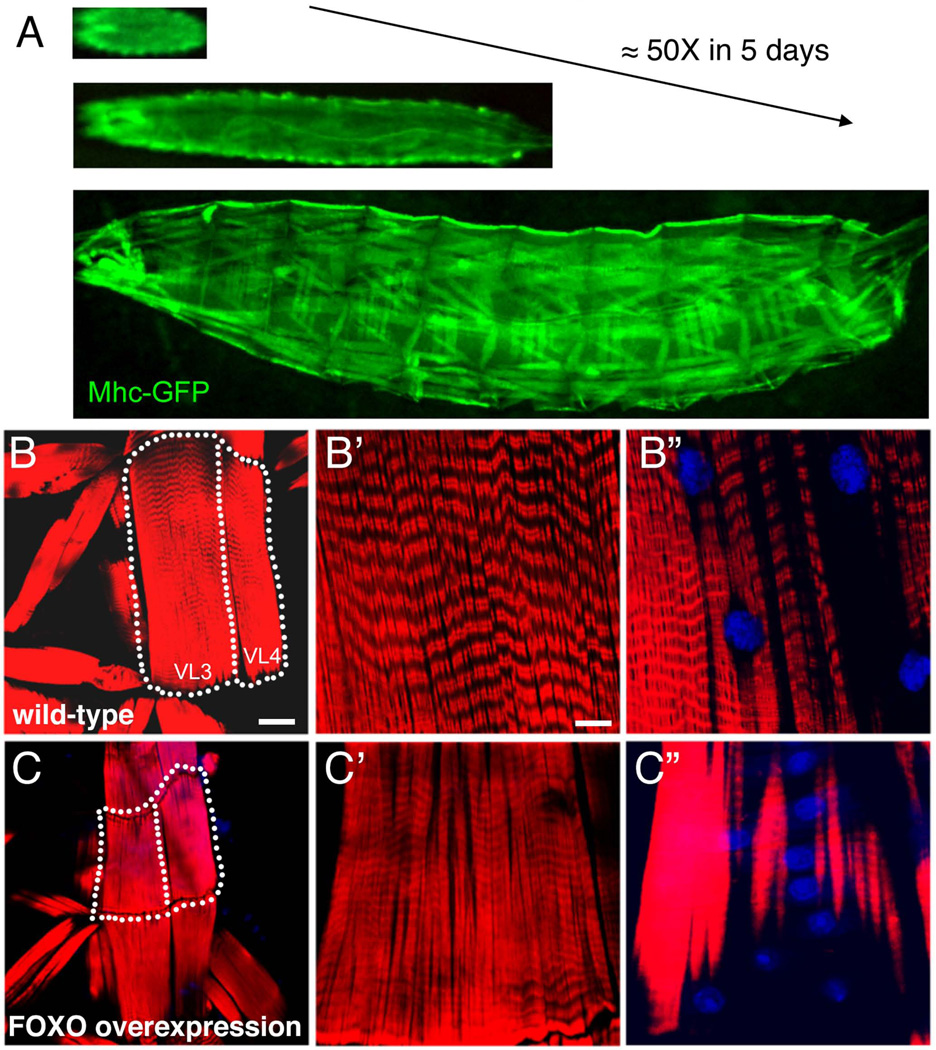
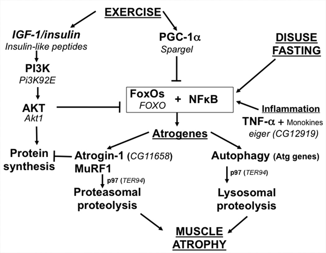
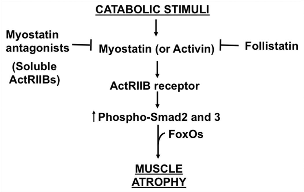
Background: The loss of skeletal muscle mass (atrophy) that accompanies disuse and systemic diseases is highly debilitating. Although the pathogenesis of this condition has been primarily studied in mammals, Drosophila is emerging as an attractive system to investigate some of the mechanisms involved in muscle growth and atrophy.

Results: In this review, we highlight the outstanding unsolved questions that may benefit from a combination of studies in both flies and mammals. In particular, we discuss how different environmental stimuli and signaling pathways influence muscle mass and strength and how a variety of disease states can cause muscle wasting.

Conclusions: Studies in Drosophila and mammals should help identify molecular targets for the treatment of muscle wasting in humans.

Keywords: animal models of muscle wasting; muscle atrophy; proteostasis; skeletal muscle growth.


Figures

Figure 1
Figure 1
Environmental stimuli and signaling pathways increasing proteolysis and depressing protein synthesis and leading to muscle atrophy. Representative fields of a transverse section of muscle fibers from fed mice or weight-matched mice deprived of food for 48h are shown. Nuclei and membranes are indicated in blue (Hoechst) and green, respectively. Scale bar is 50 µm.
Figure 2
Figure 2
Developmental growth of skeletal muscles in Drosophila larvae is inhibited by FOXO overexpression. A) Skeletal muscle size dramatically increases by 50-fold in the larval stage of development, which lasts 5 days. Muscle growth results from enhanced protein synthesis without any addition of muscle nuclei. Larvae expressing a GFP-tagged Myosin Heavy Chain (Mhc) protein in body wall muscles are shown. B) The size increase of body wall muscles is inhibited by overexpressing the transcription factor FOXO in muscles (C, Dmef2-Gal4 UAS-foxo versus Dmef2-Gal4 in B). Ventral Longitudinal muscles 3 and 4 (VL3 and VL4) from animals at the end of larval development are outlined for comparison in B and C. The dramatic increase in muscle mass observed during Drosophila larval development provides a sensitive setup for the identification of evolutionarily conserved genes regulating muscle mass. Micrographs in B’-B” and C’-C” outline sarcomeres and nuclei within muscle fibers (F-actin, red; Nuclei, blue). Scale bar is 40 µm in B-C and 10 µm in B’-B” and C’-C”. See Demontis and Perrimon, 2009, for more information.
Figure 3
Figure 3
Signaling pathways increasing proteolysis, depressing protein synthesis and leading to muscle atrophy. A comparison of the molecular players in Drosophila (indicated in Italics) and mammals is shown.
Figure 4
Figure 4
Myostatin signaling pathway as a basis to counteract muscle wasting. Molecules to inhibit this pathway are currently in human clinical trials to cure the muscle wasting associated with Duchenne Muscular Dystrophy.

References

    1. Acharyya S, Ladner KJ, Nelsen LL, Damrauer J, Reiser PJ, Swoap S, Guttridge DC. Cancer cachexia is regulated by selective targeting of skeletal muscle gene products. J Clin Invest. 2004;114:370–378. - PMC - PubMed
    1. Askanas V, Engel WK. Inclusion-body myositis and myopathies: different etiologies, possibly similar pathogenic mechanisms. Curr Opin Neurol. 2002;15:525–531. - PubMed
    1. Baehrecke EH. Autophagic programmed cell death in Drosophila. Cell Death Differ. 2003;10:940–945. - PubMed
    1. Baracos VE, DeVivo C, Hoyle DH, Goldberg AL. Activation of the ATP-ubiquitin-proteasome pathway in skeletal muscle of cachectic rats bearing a hepatoma. Am J Physiol. 1995;268:E996–E1006. - PubMed
    1. Bayline RJ, Duch C, Levine RB. Nerve-muscle interactions regulate motor terminal growth and myoblast distribution during muscle development. Dev Biol. 2001;231:348–363. - PubMed

Publication types

LinkOut - more resources