. 2013 Nov;34(11):1558-67.
doi: 10.1002/humu.22429.

Position of glycine substitutions in the triple helix of COL6A1, COL6A2, and COL6A3 is correlated with severity and mode of inheritance in collagen VI myopathies

Affiliations

Position of glycine substitutions in the triple helix of COL6A1, COL6A2, and COL6A3 is correlated with severity and mode of inheritance in collagen VI myopathies

Russell J Butterfield et al. Hum Mutat. 2013 Nov.

Abstract

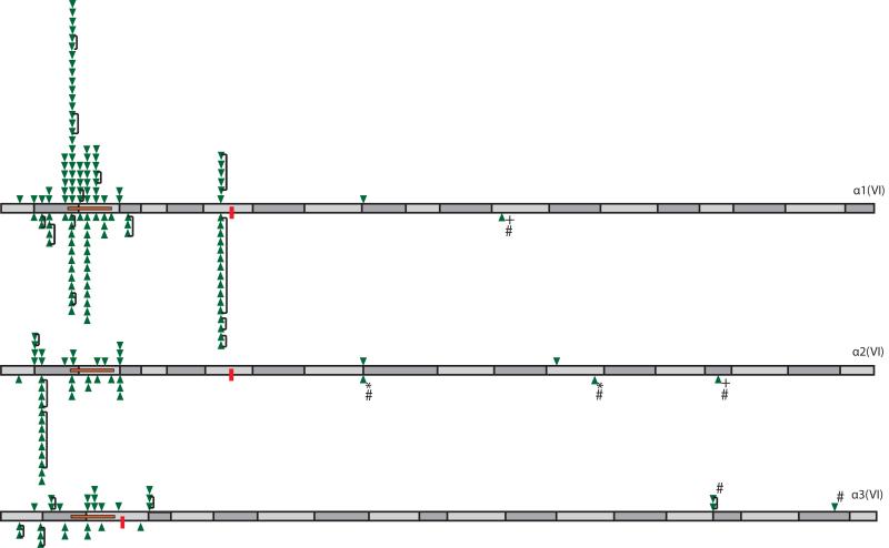
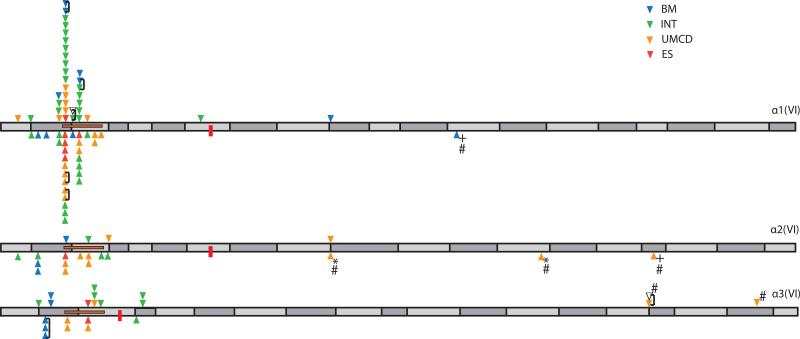
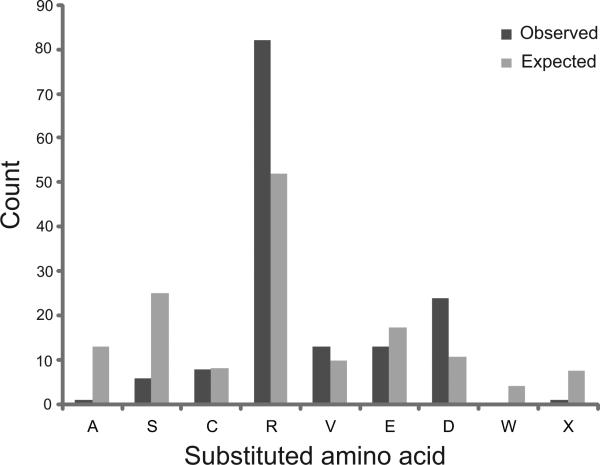
Glycine substitutions in the conserved Gly-X-Y motif in the triple helical (TH) domain of collagen VI are the most commonly identified mutations in the collagen VI myopathies including Ullrich congenital muscular dystrophy, Bethlem myopathy, and intermediate (INT) phenotypes. We describe clinical and genetic characteristics of 97 individuals with glycine substitutions in the TH domain of COL6A1, COL6A2, or COL6A3 and add a review of 97 published cases, for a total of 194 cases. Clinical findings include severe, INT, and mild phenotypes even from patients with identical mutations. INT phenotypes were most common, accounting for almost half of patients, emphasizing the importance of INT phenotypes to the overall phenotypic spectrum. Glycine substitutions in the TH domain are heavily clustered in a short segment N-terminal to the 17th Gly-X-Y triplet, where they are acting as dominants. The most severe cases are clustered in an even smaller region including Gly-X-Y triplets 10-15, accounting for only 5% of the TH domain. Our findings suggest that clustering of glycine substitutions in the N-terminal region of collagen VI is not based on features of the primary sequence. We hypothesize that this region may represent a functional domain within the triple helix.

Keywords: Bethlem myopathy; Ullrich congenital muscular dystrophy; collagen VI; genotype-phenotype correlation.


Figures

Figure 1
Figure 1. Domain structure and assembly of collagen VI
(A) Domain structure of collagen VI α1(VI), α2(VI), and α3(VI) chains. N-terminal von Willebrand factor A domains are light gray and C-terminal von Willebrand factor A domains are dark gray. Cysteine residues in the 30th Gly-X-Y triplet of COL6A1 and COL6A2 and 17th triplet of COL6A3 important for higher order assembly are labeled ‘S’. The 17th Gly-X-Y triplet (delineated with a dashed line) is an important landmark with 89% of glycine substitutions N-terminal this site. A cysteine residue at this site in the α3(VI) chain in this is important for disulfide binding of tetramers. (B) Assembly of collagen VI highlighting, first, association of α1(VI), α2(VI), and α3(VI) chains to form the monomer subunit, second, antiparallel association of monomers stabilized by disulfide bonds between cysteine residues in the 30th Gly-X-Y triplet of α1(VI) and α2(VI) chains with the C-terminal globular domains of the adjacent monomer, and third, parallel association of dimers stabilized by disulfide bonds formed between cysteine residues in the 17th triplet of adjacent α3(VI) chains. Final assembly into beaded microfibrils occurs by extracellular association of tetramers in end-to-end fashion (Chu, et al., 1990b; Pace, et al., 2008). Size of the globular N- and C-terminal domains and the length of the TH domain are proportional to size estimations proposed by Beecher et al.(Beecher, et al., 2011)
Figure 2
Figure 2. Summary of known cases with glycine substitutions in the TH domain of α1(VI), α2(VI), and α3(VI) chains (194 cases)
Triangles above the axis represent glycine substitutions identified in our cohort and below the axis represent glycine substitutions identified in published cases. Related individuals are linked by a bracket. Position of cysteine residues important for dimerization (α1(VI), α2(VI)), and tetramerization (α3(VI)) are marked with a red box. The critical region important for assembly proposed by Pace et al. is marked with a horizontal orange box. *indicates both alleles in one patient with two different glycine substitutions. + represents patients homozygous glycine substitutions. # indicates alleles known/suspected to be acting in recessive fashion. TH domain exons are indicated by alternating dark and light grey rectangles.
Figure 3
Figure 3. Clinical phenotype and position of mutation for patients with glycine substitutions in the TH domain (103 cases)
Triangles above the axis represent glycine substitutions identified in our cohort and below the axis represent glycine substitutions identified in published cases. Related individuals are linked by a bracket. Position of cysteine residues important for dimerization (α1(VI), α2(VI)), and tetramerization (α3(VI)) are marked with a red box. The critical region important for assembly proposed by Pace et al. is marked with a horizontal orange box. + represent patients homozygous glycine substitutions. * denotes both alleles in one patient with two different glycine substitutions, # indicates alleles known or suspected to be acting in recessive fashion. TH domain exons are indicated by alternating dark and light grey rectangles.
Figure 4
Figure 4. Distribution of glycine and non-glycine substitutions on the α1(VI), α2(VI), and α3(VI) chain
Position of cysteine residues important for dimerization (α1(VI), α2(VI)), and tetramerization (α3(VI)) are marked with a small vertical box. The critical region important for assembly proposed by Pace et al. is marked with a horizontal box. (Pace, et al., 2008) (A) Clustering of glycine substitutions in TH domain of α1(VI), α2(VI), and α3(VI) chains. Tic marks above the axis represent alleles identified in our cohort. Tic marks below the axis represent alleles identified in published cases. The position of CpG motifs with potential to cause a glycine substitution are marked with diamonds. The three most commonly observed substitutions (p.G284R, p.G290R, and p.G293R) are all in the COL6A1 gene and are all in the context of CpG motifs. Glycine substitutions in the context of CpG elsewhere in the TH domain were only seen at 2 of the possible 43 CpG sites (COL6A2: c.1450G>A, p.G484R; COL6A3: c.6175G>T p.G2059C). Neither of these has more than 1 independent observation among the 148 unrelated cases. (B) Distribution of intermediate frequency variants (0.05%-5% minor allele frequency, full height tic), and low frequency variants (<0.05% minor allele frequency, half height tic) in the TH domain of α1(VI), α2(VI), and α3(VI) chains. Non-glycine missense variants observed in the TH domain in our clinical sequencing effort (tic marks above the axis) and in dbSNP135 (tic marks below the axis) are not clustered on COL6A1, nor are they clustered within the TH domain.
Figure 5
Figure 5. Distribution of observed and expected glycine substitutions in α1(VI), α2(VI), and α3(VI) chains in 148 unrelated cases based on neighbor dependent mutation rates
Order of substituted amino acids on the X-axis is based on the increasing severity of disruption to the stability of the triple helix.
Figure 6
Figure 6. Immunofluorescent staining of muscle (A) and cultured fibroblasts (B) from a patient with c.812G>A; p.G271D mutation in COL6A2
Dual labeling of collagen VI (green) and collagen IV (red) in muscle shows presence of collagen VI in the ECM, but poor co-localization with the basement membrane (yellow). Scale bar is 75 μm. Inset shows higher magnification for patient muscle (A,b). Staining for collagen VI in cultured fibroblasts from the same patient shows decreased deposition and speckled appearance of collage VI in the ECM of patient fibroblasts compared to control (B;a,b). Collagen VI staining in the presence of TritonX-100 to permeablize the cell membrane demonstrates intracellular retention of collagen VI in patient fibroblasts (B: c,d).

References

    1. Baker NL, Morgelin M, Pace RA, Peat RA, Adams NE, Gardner RJ, Rowland LP, Miller G, De Jonghe P, Ceulemans B. Molecular consequences of dominant Bethlem myopathy collagen VI mutations. Ann Neurol. 2007;62(4):390–405. others. - PubMed
    1. Baker NL, Morgelin M, Peat R, Goemans N, North KN, Bateman JF, Lamande SR. Dominant collagen VI mutations are a common cause of Ullrich congenital muscular dystrophy. Hum Mol Genet. 2005;14(2):279–93. - PubMed
    1. Baldock C, Sherratt MJ, Shuttleworth CA, Kielty CM. The supramolecular organization of collagen VI microfibrils. J Mol Biol. 2003;330(2):297–307. - PubMed
    1. Beecher N, Roseman AM, Jowitt TA, Berry R, Troilo H, Kammerer RA, Shuttleworth CA, Kielty CM, Baldock C. Collagen VI, conformation of A-domain arrays and microfibril architecture. J Biol Chem. 2011;286(46):40266–75. - PMC - PubMed
    1. Brinas L, Richard P, Quijano-Roy S, Gartioux C, Ledeuil C, Lacene E, Makri S, Ferreiro A, Maugenre S, Topaloglu H. Early onset collagen VI myopathies: Genetic and clinical correlations. Ann Neurol. 2010;68(4):511–20. others. - PubMed

Publication types

MeSH terms