

Protective effect of naringenin on acetic acid-induced ulcerative colitis in rats
- PMID: 24039355
- PMCID: PMC3769899
- DOI: 10.3748/wjg.v19.i34.5633
Protective effect of naringenin on acetic acid-induced ulcerative colitis in rats
Abstract
Aim: To evaluate the ameliorative effect of naringenin (NG) during ulcerative colitis (UC) in rats.
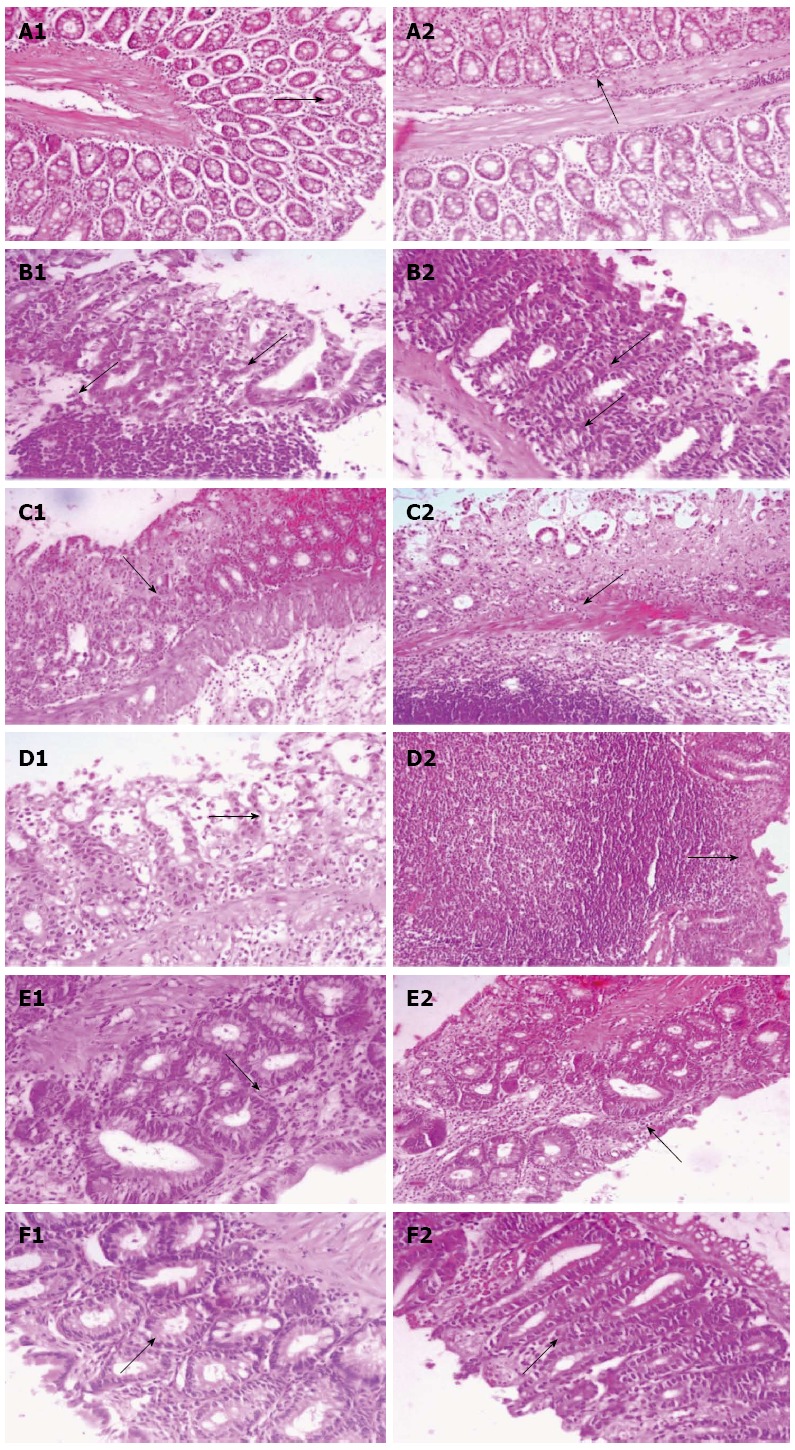
Methods: Rats were treated with three different doses (25, 50 and 100 mg/kg per day) of NG and a single dose of mesalazine (MES, 300 mg/kg per day) for seven days prior to ulcerative colitis induction by 4% acetic acid (AA). Twenty four hours after AA rectal administration, animals were scarified and the colonic tissues were dissected. Colonic mucus content was estimated using Alcian blue dye binding technique. In colon tissues, levels of total glutathione sulphadryls (T-GSH), non-protein sulphadryls (NP-SH) and thiobarbituric acid reactive substances (TBARS) were evaluated. The activities of the antioxidant enzymes, catalase (CAT) and superoxide dismutase (SOD) were measured. Concentrations of nucleic acids (DNA and RNA) and total protein were also estimated in colon tissues. Colonic levels of tumor necrosis factor-α (TNF-α), interleukin-1β (IL-1β), interleukin-6 (IL-6), prostaglandin E2 (PGE2) and nitric oxide (NO) were estimated. In cross section of colitis tissue the histopathological changes were observed.
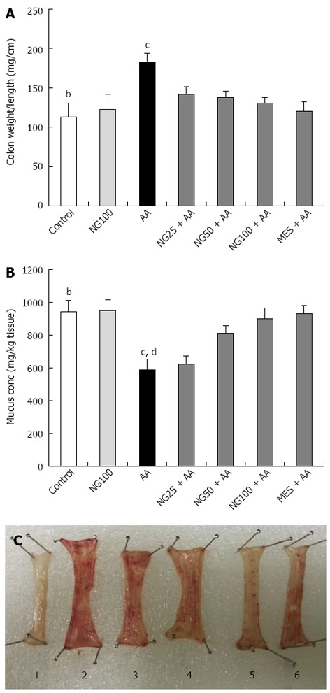
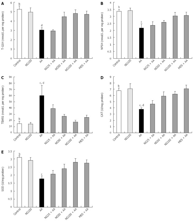
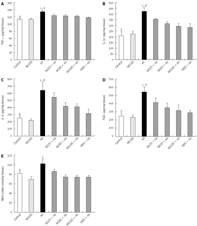
Results: Colonic mucus content was decreased in AA compared to controls (587.09 ± 65.59 mg/kg vs 941.78 ± 68.41 mg/kg, P < 0.001). AA administration markedly reduced T-GSH (5.25 ± 0.37 nmol/L vs 3.04 ± 0.24 nmol/L, P < 0.01), NP-SH (3.16 ± 0.04 nmol/L vs 2.16 ± 0.30 nmol/L, P < 0.01), CAT (6.77 ± 0.40 U/mg vs 3.04 ± 0.2 U/mg, P < 0.01) and SOD (3.10 ± 0.11 U/mg vs 1.77 ± 0.18 U/mg, P < 0.01) while TBARS, TNF-α, IL-1β, IL-6, PGE2 and NO levels (15.09 ± 3.84 nmol/L vs 59.90 ± 16.34 nmol/L, P < 0.01; 113.56 ± 1.91 pg/mg vs 134.24 ± 4.77 pg/mg, P < 0.01; 209.20 ± 36.38 pg/mg vs 422.19 ± 31.47 pg/mg, P < 0.01; 250.83 ± 25.09 pg/mg vs 638.58 ± 115.9 pg/mg, P < 0.01; 248.19 ± 36.98 pg/mg vs 541.74 ± 58.34 pg/mg, P < 0.01 and 81.26 ± 2.98 mmol/g vs 101.90 ± 10.73 mmol/g, P < 0.001) were increased in colon of rats with UC compared controls respectively.Naringenin supplementation, significantly and dose dependently increased the colonic mucus content. The elevated TBARS levels were significantly decreased (39.35 ± 5.86 nmol/L, P < 0.05; 26.74 ± 3.17 nmol/L, P < 0.01 nmol/L and 17.74 ± 2.69 nmol/L, P < 0.01) compared to AA (59.90 ± 16.34 nmol/L) group while the decreased levels of T-GSH and NP-SH and activities of CAT and SOD found increased by NG treatments in dose dependent manner. The decreased values of nucleic acids and total protein in AA group were also significantly (P < 0.01) increased in all three NG supplemented groups respectively. NG pretreatment inhibited the TNF-α levels (123.76 ± 3.76 pg/mg, 122.62 ± 3.41 pg/mg and 121.51 ± 2.61 pg/mg vs 134.24 ± 4.78 pg/mg, P < 0.05) compared to AA group, respectively. Interleukins, IL-1β and IL-6 levels were also decreased in NG50 + AA (314.37 ± 16.31 pg/mg and 292.58 ± 23.68 pg/mg, P < 0.05) and NG100 + AA (416.72 ± 49.62 pg/mg and 407.96 ± 43.87 pg/mg, P < 0.05) when compared to AA (352.46 ± 8.58 pg/mg and 638.58 ± 115.98 pg/mg) group. Similar decrease (P < 0.05) was seen in PGE2 and NO values when compared to AA group. The group pretreated with MES, as a reference drug, showed significant (P < 0.01) protection against the changes induced in colon tissue by AA administration respectively.
Conclusion: In present study, NG produced antioxidant and anti-inflammatory effects demonstrating protective effect in inflammatory bowel disease.
Keywords: Inflammatory bowel disease; Naringenin; Oxidative stress; Ulcerative colitis.
Figures
References
-
- Selling JH, Pasricha PJ. Pharmacotherapy of inflammatory bowel disease. In: Gilman’s G, editor. The Pharmacological Basis of Therapeutics. New York: McGraw-Hill Publications; 2006. p. 1009.
-
- Parkes M, Jewell D. Ulcerative colitis and Crohns disease: molecular genetics and clinical implications. Expert Rev Mol Med. 2001;2001:1–18. - PubMed
-
- Halpin SJ, Ford AC. Prevalence of symptoms meeting criteria for irritable bowel syndrome in inflammatory bowel disease: systematic review and meta-analysis. Am J Gastroenterol. 2012;107:1474–1482. - PubMed
-
- Lih-Brody L, Powell SR, Collier KP, Reddy GM, Cerchia R, Kahn E, Weissman GS, Katz S, Floyd RA, McKinley MJ, et al. Increased oxidative stress and decreased antioxidant defenses in mucosa of inflammatory bowel disease. Dig Dis Sci. 1996;41:2078–2086. - PubMed
Publication types
MeSH terms
Substances
LinkOut - more resources
Full Text Sources
Other Literature Sources
Medical
Miscellaneous
