Cerebral and splenic infarctions after injection of N-butyl-2-cyanoacrylate in esophageal variceal bleeding
- PMID: 24039373
- PMCID: PMC3769917
- DOI: 10.3748/wjg.v19.i34.5759
Cerebral and splenic infarctions after injection of N-butyl-2-cyanoacrylate in esophageal variceal bleeding
Abstract
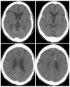
Variceal bleeding is the most serious complication of portal hypertension, and it accounts for approximately one fifth to one third of all deaths in liver cirrhosis patients. Currently, endoscopic treatment remains the predominant method for the prevention and treatment of variceal bleeding. Endoscopic treatments include band ligation and injection sclerotherapy. Injection sclerotherapy with N-butyl-2-cyanoacrylate has been successfully used to treat variceal bleeding. Although injection sclerotherapy with N-butyl-2-cyanoacrylate provides effective treatment for variceal bleeding, injection of N-butyl-2-cyanoacrylate is associated with a variety of complications, including systemic embolization. Herein, we report a case of cerebral and splenic infarctions after the injection of N-butyl-2-cyanoacrylate to treat esophageal variceal bleeding.
Keywords: Cerebrum; Esophageal varix; Infarction; N-butyl-2-cyanoacrylate; Spleen.
Figures


References
-
- Schuman BM, Beckman JW, Tedesco FJ, Griffin JW, Assad RT. Complications of endoscopic injection sclerotherapy: a review. Am J Gastroenterol. 1987;82:823–830. - PubMed
-
- Helmy A, Hayes PC. Review article: current endoscopic therapeutic options in the management of variceal bleeding. Aliment Pharmacol Ther. 2001;15:575–594. - PubMed
-
- Habib SF, Muhammad R, Koulaouzidis A, Gasem J. Pulmonary embolism after sclerotherapy treatment of bleeding varices. Ann Hepatol. 2008;7:91–93. - PubMed
-
- Habib A, Sanyal AJ. Acute variceal hemorrhage. Gastrointest Endosc Clin N Am. 2007;17:223–252, v. - PubMed
-
- Ryan BM, Stockbrugger RW, Ryan JM. A pathophysiologic, gastroenterologic, and radiologic approach to the management of gastric varices. Gastroenterology. 2004;126:1175–1189. - PubMed
Publication types
MeSH terms
Substances
LinkOut - more resources
Full Text Sources
Other Literature Sources
Medical

