. 2013;9(9):e1003585.
doi: 10.1371/journal.ppat.1003585. Epub 2013 Sep 5.

Viral membrane fusion and nucleocapsid delivery into the cytoplasm are distinct events in some flaviviruses

Affiliations

Viral membrane fusion and nucleocapsid delivery into the cytoplasm are distinct events in some flaviviruses

Adel M Nour et al. PLoS Pathog. 2013.

Abstract

Flaviviruses deliver their genome into the cell by fusing the viral lipid membrane to an endosomal membrane. The sequence and kinetics of the steps required for nucleocapsid delivery into the cytoplasm remain unclear. Here we dissect the cell entry pathway of virions and virus-like particles from two flaviviruses using single-particle tracking in live cells, a biochemical membrane fusion assay and virus infectivity assays. We show that the virus particles fuse with a small endosomal compartment in which the nucleocapsid remains trapped for several minutes. Endosomal maturation inhibitors inhibit infectivity but not membrane fusion. We propose a flavivirus cell entry mechanism in which the virus particles fuse preferentially with small endosomal carrier vesicles and depend on back-fusion of the vesicles with the late endosomal membrane to deliver the nucleocapsid into the cytoplasm. Virus entry modulates intracellular calcium release and phosphatidylinositol-3-phosphate kinase signaling. Moreover, the broadly cross-reactive therapeutic antibody scFv11 binds to virus-like particles and inhibits fusion.


Conflict of interest statement

The authors have declared that no competing interests exist.

Figures
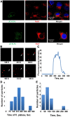
Figure 1
Figure 1. Endocytosis, live-cell tracking and membrane fusion kinetics of JE-VLPs.
(A) 15 min after treatment of Vero cells with 200 µl JE-VLPs (at 17 pM or 50 ng/ml E protein), JEV E protein (green, detected with anti-West Nile primary and fluorescein-labeled secondary antibodies) colocalizes with endocytic markers Rab5 and Rab7 (red). The Pearson colocalization coefficient was 0.19 and 0.34 for E-Rab5 and E-Rab7, respectively. (B) Snapshots of JE-VLPs labeled with self-quenching concentrations of rhodamine C18 (R18) infecting live Vero cells. VLPs were tracked for 20 min after infection. Individual particles were identified and tracked with ImageJ. (C) Fluorescence intensity of the tracked particle marked with an arrow in B. Early in the endocytic pathway, approximately 5 min after infection, fusion of the viral and cellular membranes causes dilution and dequenching of the red R18 dye. R18 fluorescence remains unexpectedly stable for approximately 4 min before rapid decay due to diffusion of the dye into endosomal membranes, suggesting that the dye is transiently trapped in a small endocytic compartment. (D) Duration of R18 fluorescence, from half-maximal dequenching to half-maximal decay, for 14 tracked JE-VLPs. On average the dye remained trapped in a small endocytic compartment (with fluorescence remaining constant) for 251±97 s prior to dilution of the dye and fluorescence decay. (E) Half-time of R18 fluorescence decay (time from maximal to half-maximal fluorescence) for 14 tracked particles. The average decay halftime was 94±64 s. See also Figures S1 and S2 and Movie S1.
Figure 2
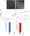
Figure 2. Effects on chloroquine on membrane fusion of JE-VLPs and YFV in Vero cells.
(A) 0.1 g/l chloroquine (194 µM) had no notable toxic effect on the cells as indicated by differential interference contrast (DIC) microscopy of untreated cells (left) and chloroquine-treated cells (right). Cells were treated with 0.1 g/l chloroquine and infected with R18-labeled JE-VLPs (200 µl at 17 pM), (B), or with YFV (MOI = 1), (C). Chloroquine blocked VLP membrane fusion, as judged by the lack of R18 fluorescence dequenching in a field of treated cells (red curve) relative to the normal dequenching of R18 in untreated cells (blue curve). (D) Relative qRT-PCR of viral RNA in untreated (mock) and chloroquine-treated Vero cells 1 h post-infection with YFV (MOI = 1). Endosomal and cytosolic fractions were separated and total RNA was extracted from the cytosolic fraction as described in the Materials and Methods. RT-PCR was used to quantify the 3′-UTR of YFV genomic RNA. Error bars represent the standard error of the mean (SEM) of three experiments. Treatment with chloroquine reduced RNA release into the cytoplasm by >95%. (E) Plaque assay with BHK cells infect with YFV (MOI = 0.1) in presence and in absence of chloroquine. BHK cells were infected with MOI 0.1 of YFV in presence and in absence of 0.1 g/l chloroquine. Chloroquine treatment completely inhibited YFV replication. See also Figure S3.
Figure 3
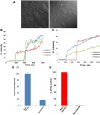
Figure 3. Microtubules are required for RNA delivery into the cytoplasm and viral infectivity but not membrane fusion.
Cells were pretreated with 20 µM nocodazole (a microtubule polymerization inhibitor) for 1 h before treatment with R18-labeled JE-VLPs or YFV. (A) DIC micrograph of the treated cells showing that nocodazole has no toxic effect on Vero cells although the cell morphology is altered. (B) JE-VLPs (200 µl at 17 pM) and (C) YFV (MOI = 0.1–1) fused normally in Vero cells in the presence of 20 µM nocodazole, as indicated by the R18 fluorescence dequenching profiles of three representative tracked particles. (D) qRT-PCR nucleocapsid delivery assay showing that nocodazole reduced YFV nucleocapsid delivery into the cytoplasm of Vero cells by approximately 80% relative to untreated cells. Error bars represent the standard error of the mean (SEM) of three experiments. (E) Plaque assay showing that nocodazole treatment inhibited YFV replication, reducing the number of viral plaques by >97%. See also Movie S2.
Figure 4
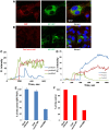
Figure 4. BMP, a lipid specific to late endosomes, is required for nucleocapsid delivery into the cytoplasm and viral infectivity but not membrane fusion.
Cells were cultured overnight in media containing 50 µg/ml anti-BMP antibody. Cells were washed and the medium was changed before immunostaining, treatment with R18-labeled JE-VLPs (200 µl at 17 pM) or with YFV (MOI = 0.1–1). (A) Immunostaining of BMP in BHK cells. Left: BMP staining (Texas Red) was mainly perinuclear. Center: NS1-GFP (green) expression in BHK cells to transcomplement the ΔNS1-YFV genome with NS1 for viral production as described in Ref. . (B) BHK cells treated with total mouse IgG instead of anti-BMP antibody. No antibody staining is detectable in the cells (left panel). JE-VLPs, (C), or YFV, (D), fused normally in Vero cells pretreated with anti-BMP antibody, as indicated by the R18 fluorescence dequenching profiles of three representative tracked particles. Error bars represent the standard error of the mean (SEM) of three experiments. (E) qRT-PCR assay showing that pretreatment of Vero cells with anti-BMP antibody reduced the delivery of YFV nucleocapsid into the cytoplasm by approximately 70% relative to untreated cells or cell treated with total mouse IgG. (F) Plaque assay showing that pretreatment of BHK cells with the anti-BMP antibody partially inhibited YFV replication, reducing the number of viral plaques by approximately 65% relative to the controls.
Figure 5
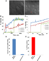
Figure 5. PI(3) kinase activity is required for RNA delivery into the cytoplasm and viral infectivity but not membrane fusion.
Cells were pretreated with 0.1 µM wortmannin for 1 h prior to treatment with JE-VLPs (200 µl at 17 pM) or infection with YFVs (MOI = 1). (A) DIC micrograph of the treated cells showing that wortmannin had no toxic effects although treated cells showed a characteristic increase in the number of intracellular vacuoles (red arrows). JE-VLPs, (B), or YFV, (C), fused normally in Vero cells pretreated with wortmannin, as indicated by the R18 fluorescence dequenching profiles of three representative tracked particles. (D) qRT-PCR assay showing that pretreatment of Vero cells with wortmannin reduced the delivery of YFV nucleocapsid into the cytoplasm by 95% relative to untreated cells. Error bars represent the standard error of the mean (SEM) of three experiments. (E) Plaque assay showing that pretreatment of BHK cells with wortmannin reduced YFV replication and viral plaque formation to background levels. See also Movie S3.
Figure 6
Figure 6. JE-VLPs and YFV bind to phosphatidylserine and fuse with liposomes with a lipid composition similar to early endosomes and ECVs.
(A) Liposomes were produced from an ECV-like lipid mixture (3∶1∶1∶1∶4, Chol∶PE∶PI(3)P∶PS∶PC) as described in the Materials and Methods. JE-VLPs fused efficiently with the liposomes at pH 5.5 but not at pH 8.4. (B) YFV fused normally with the synthetic liposomes at pH 5.5 but pretreatment of the virus with 2 mM DEPC for 30 min blocked fusion at pH 5.5. Error bars represent the standard error of the mean (SEM) of three experiments with fluorescence measured in triplicate for each experiment. (C) Beads coated with the anionic lipid PS pulled down JE-VLPs and YFV. Beads coated with heparan sulfate (HS) were used as a positive control and uncoated beads were used as a negative control. (D) Beads coated with PI(3)P did not bind to either JE-VLPs or YFV. HS-beads and uncoated beads were used as positive and negative controls, respectively.
Figure 7
Figure 7. A single-chain variable region fragment of neutralizing anti-West Nile virus E antibody 11 (scFv11) binds JE-VLPs but not YFV in vivo and in vitro.
(A) ELISA of JE-VLPs and YFVs treated with scFv11. The antibody fragment bound to JE-VLPs but not YFV. Error bars represent the standard error of the mean (SEM) of three experiments. (B) Size-exclusion chromatography was used to confirm the binding of scFv11 to JE-VLPs in solution. VLPs and scFv11-VLP complexes eluted in the void volume (8 ml); free or excess scFv11 eluted at 16 ml. (C) The R18 fluorescence intensity of a field of cells treated with purified scFv11-JE-VLP complexes was tracked by confocal microscopy. No dequenching was observed until free JE-VLPs were added (red arrow at 200 s). (D) Pretreatment of R18-labeled JE-VLPs with 1 µM scFv11 reduced the membrane fusion activity of the VLPs with synthetic ECV-like liposomes (see Figure 6) at pH 5.5 by approximately 50%. (E) Isothermal titration calorimetry (ITC) was used to determine the equilibrium dissociation constant of scFv11 from JE-VLPs, (K d = 152±21 nM, and the stoichiometry of scFv11 binding to JE-VLPs, 0.648±0.013 scFv11 molecules per E protein. See also Figure S8.
Figure 8
Figure 8. Proposed model of flavivirus cell entry.
After binding to their receptors, flaviviruses enter the clathrin-mediated endocytic pathway and are directed to early endosomes (EEs). Virus entry causes an increase in cytoplasmic calcium levels, which may cause a redistribution of phospholipids in the cellular membranes. When the pH of the early endosomal compartments is reduced to approximately 6.5, about 5 min after cellular attachment, virus particles fuse preferentially with ECVs, due to the large excess of ECV membranes over the limiting endosomal membrane. The enrichment of specific anionic lipids in ECVs such as PS may further promote fusion with ECVs over fusion with the limiting endosomal membrane. The nucleocapsid remains trapped in the ECV lumen for several minutes, until the ECV fuses back with the limiting membrane of the late endosome (LE). This back-fusion event requires microtubule transport, PI(3)P-dependent signaling, the late endosomal anionic lipid BMP, and cellular fusion proteins. The PI(3)P kinase inhibitor wortmannin inhibits the formation of ECVs. In the absence of ECVs, flaviviruses either fail to fuse completely with the EE membrane (e.g. fusion proceeds only as far as hemifusion of the proximal lipid layers) or they fuse with as yet unidentified endosomal compartments in which the nucleocapsid remains trapped.

References

    1. Morita E, Sundquist WI (2004) Retrovirus budding. Annu Rev Cell Dev Biol 20: 395–425. - PubMed
    1. Salonen A, Ahola T, Kaariainen L (2005) Viral RNA replication in association with cellular membranes. Curr Top Microbiol Immunol 285: 139–173. - PMC - PubMed
    1. Gruenberg J (2001) The endocytic pathway: a mosaic of domains. Nature reviews 2: 721–730. - PubMed
    1. Lemichez E, Bomsel M, Devilliers G, vanderSpek J, Murphy JR, et al. (1997) Membrane translocation of diphtheria toxin fragment A exploits early to late endosome trafficking machinery. Molecular microbiology 23: 445–457. - PubMed
    1. Le Blanc I, Luyet PP, Pons V, Ferguson C, Emans N, et al. (2005) Endosome-to-cytosol transport of viral nucleocapsids. Nat Cell Biol 7: 653–664. - PMC - PubMed

Publication types

MeSH terms