. 2013;9(9):e1003608.
doi: 10.1371/journal.ppat.1003608. Epub 2013 Sep 5.

Multicellular bacteria deploy the type VI secretion system to preemptively strike neighboring cells

Affiliations

Multicellular bacteria deploy the type VI secretion system to preemptively strike neighboring cells

Christopher J Alteri et al. PLoS Pathog. 2013.

Abstract

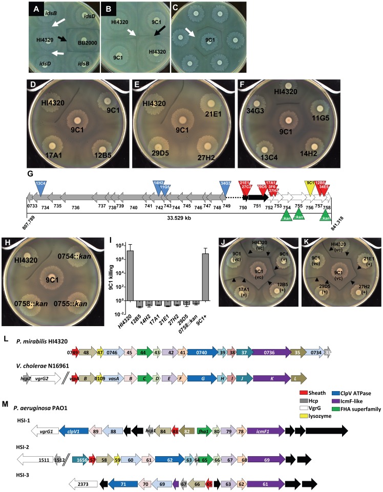
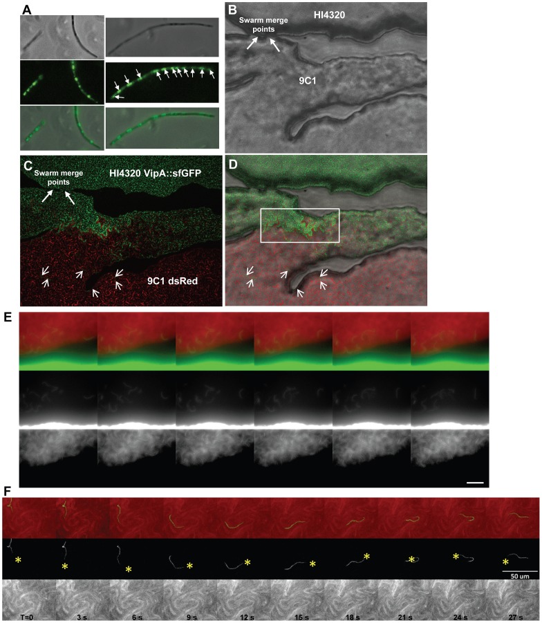
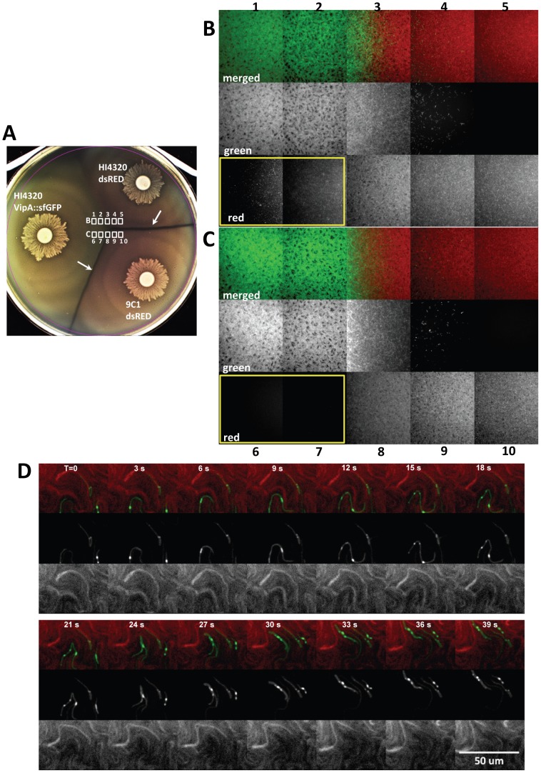
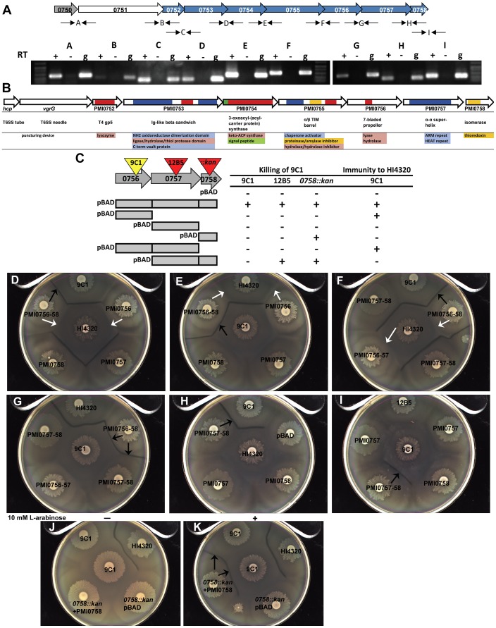
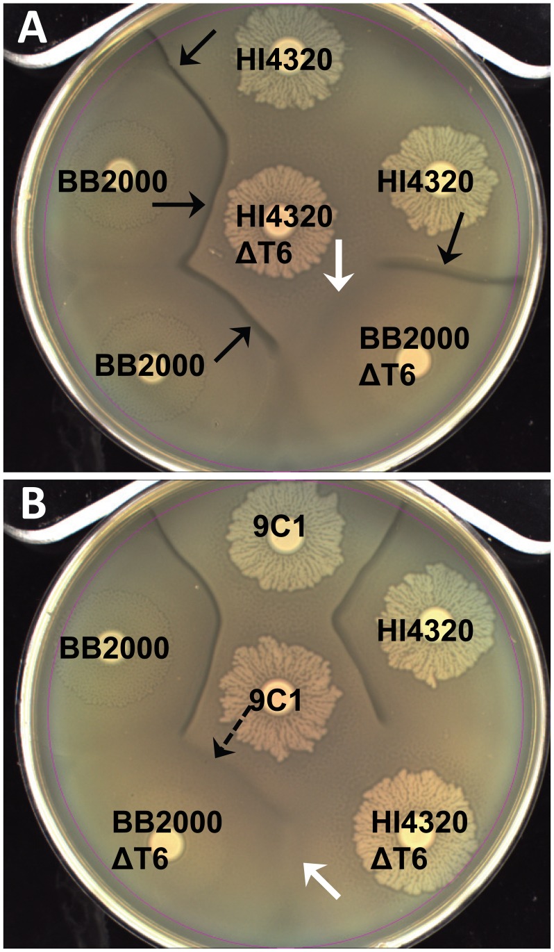
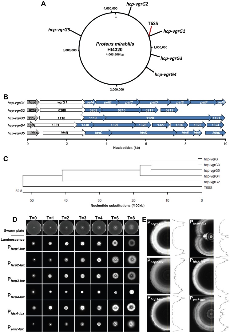
The Type VI Secretion System (T6SS) functions in bacteria as a contractile nanomachine that punctures and delivers lethal effectors to a target cell. Virtually nothing is known about the lifestyle or physiology that dictates when bacteria normally produce their T6SS, which prevents a clear understanding of how bacteria benefit from its action in their natural habitat. Proteus mirabilis undergoes a characteristic developmental process to coordinate a multicellular swarming behavior and will discriminate itself from another Proteus isolate during swarming, resulting in a visible boundary termed a Dienes line. Using transposon mutagenesis, we discovered that this recognition phenomenon requires the lethal action of the T6SS. All mutants identified in the genetic screen had insertions within a single 33.5-kb region that encodes a T6SS and cognate Hcp-VrgG-linked effectors. The identified T6SS and primary effector operons were characterized by killing assays, by construction of additional mutants, by complementation, and by examining the activity of the type VI secretion system in real-time using live-cell microscopy on opposing swarms. We show that lethal T6SS-dependent activity occurs when a dominant strain infiltrates deeply beyond the boundary of the two swarms. Using this multicellular model, we found that social recognition in bacteria, underlying killing, and immunity to killing all require cell-cell contact, can be assigned to specific genes, and are dependent on the T6SS. The ability to survive a lethal T6SS attack equates to "recognition". In contrast to the current model of T6SS being an offensive or defensive weapon our findings support a preemptive mechanism by which an entire population indiscriminately uses the T6SS for contact-dependent delivery of effectors during its cooperative mode of growth.


Conflict of interest statement

The authors have declared that no competing interests exist.

Figures

Figure 1
Figure 1. Social recognition and Dienes line formation results from bacterial killing, dependent on the T6SS and divergent hcp-vgrG effector operon.
(A) Non-identical P. mirabilis strains, BB2000 and HI4320, form a Dienes line (black arrow). Mutations within idsB and idsD of HI4320 do not form a Dienes line with their parental strain (white arrows). (B) One of 1,920 HI4320 Tn5 insertion mutants, identified as 9C1, formed a Dienes line (black arrow) with its parental strain. HI4320 merges with itself at the center of the plate (white arrow). (C) Transposon mutants were rescreened against 9C1 to find mutants that lost the ability to form a Dienes line (arrow). (D, E) Mutant 9C1 merged with itself and insertion mutants 12B5, 17A1, 21E1, 27H2, and 29D5. (F) 9C1 merges with T6SS mutants 11G5, 14H2, 13C4, and 34G3. (G) Transposon insertions (red triangles) in genes located within the same operon as the 9C1 mutant (yellow triangle). The three genes not identified in the genetic screen were disrupted by targeted insertion of antibiotic resistance gene (green triangles). hcp and vgrG homologs (black arrows) and additional genes predicted to encode effectors delivered by the T6SS (white arrows) are indicated. Additional mutants (13C4, 14H2, 11G5, and 34G3) have transposon insertions (blue triangles) in genes encoding conserved components of the T6SS (grey arrows). The black dotted line represents intergenic sequence. (H) Mutants PMI0754::kan, 0755::kan, and 0758::kan merge with 9C1. (I) Killing assays confirming 9C1 killing are reported as [(CFU of test strain/CFU of 9C1)output]/[(CFU of test strain/CFU of 9C1)input]. Strain HI4230 kills mutant 9C1 by at least 7-logs. Mutant 9C1 complemented with the primary hcp-vgrG effector operon (9C1+) kills mutant 9C1. (J, K) Dienes line formation (arrows) was restored when the indicated mutants are complemented with the entire primary hcp-vgrG effector operon, (+); empty vector, (vc). (L) The 17 genes that encode the HI4320 T6SS are highly conserved with the well characterized T6SS genes from V. cholerae N16961 and have identical gene order in their respective chromosome and (M) comparison to the three known T6SSs (HSI-1, HSI-2, and HSI-3) encoded within the genome of P. aeruginosa PA01. The arrows are color-coded based upon known or predicted gene function. Black arrows represent genes that are not found in either P. mirabilis HI4320 or V. cholerae N16961 T6SS gene locus. See also Table S1.
Figure 2
Figure 2. Visualization of the T6SS activity during multicellular infiltration and killing.
(A) Actively swarming P. mirabilis HI4230 cells expressing VipA::sfGFP viewed under phase contrast and by fluorescence microscopy. Punctate green staining represents sites of T6SS assembly (arrows). (B) HI4320 and mutant 9C1 are observed merging at their swarm fronts on an agar surface (arrows) under phase contrast. (C) HI4320 VipA::sfGFP is observed infiltrating deep into the opposing 9C1 swarm expressing dsRED. Short arrows indicate individual HI4320 swarm cells within the 9C1 swarm. (D) Merged image of (B) and (C). The boxed region encapsulates intense green straining representing increased sfGFP-signal due to increased assembly of the T6SS sheath (VipA::sfGFP) by the front swarm edge of wild-type HI4320. (E) Visualization of individual HI4320 wild-type swarmer cells (green) that have infiltrated and are demonstrating multicellular swarming within the susceptible 9C1 swarm (red). (F) The forward movement of one individual HI4320 swarm cell over 30 seconds is indicated by yellow asterisk. Elapsed time (T) in seconds is indicated. Each frame in (E, F) represents 3 seconds elapsed time and the white bar is 50 µM. See also Figure S1, S2, and Movie S1 and S2.
Figure 3
Figure 3. Infiltration of resistant and sensitive opposing swarms by P. mirabilis HI4320 expressing VipA::sfGFP.
(A) Agar plate inoculated with P. mirabilis HI4320 VipA::sfGFP opposing strains HI4320 or mutant 9C1 expressing dsRED. Dienes lines (white arrows) formed between HI4320 VipA::sfGFP or HI4320 dsRED and 9C1 dsRED but not between HI4320 VipA::sfGFP and HI4320 dsRED. The fields examined by fluorescence microscopy (white boxes) are indicated at the intersection of the swarms. Active infiltration of (B) HI4320 dsRED and (C) mutant 9C1 dsRED by HI4320 expressing VipA::sfGFP. The numbered panels in (B) and (C) correspond to the numbered areas indicated in (A) and were viewed directly on the agar plate at the time of intersection. For each field, the individual green and red channels from the merged image are shown to maximize visualization of the infiltrating swarm. In (B) and (C) the panels boxed with a yellow border show dsRED-expressing bacteria infiltrating into the HI4320 VipA::sfGFP swarm are only observable with HI4320 dsRED (B1 and B2 red); the susceptible 9C1 dsRED is undetectable (C6 and C7 red). (D) Infiltrating HI4320 expressing VipA::sfGFP demonstrate numerous areas of T6SS activity (green) when in direct contact with target 9C1 cells expressing dsRED. Elapsed time (T) is indicated in seconds. See also Movie S3, S4, S5.
Figure 4
Figure 4. The primary hcp-vgrG T6SS effector operon encodes both killing and immunity functions.
(A) Transcription of the primary effector operon based upon RT-PCR. Intergenic primer pairs (thin black arrows) flanking the open reading frames of PMI0750-PMI0759 are represented by letters. For each reaction (+) cDNA made from RNA using reverse transcriptase, (−) no RT enzyme, or (g) genomic DNA purified from HI4320 were used as template for PCR. (B) Predicted structural homology and potential functions for the effectors and other proteins encoded within the primary hcp-vgrG operon. Functional domains are color coded beneath each gene. (C) Wild-type HI4320, transposon mutants 9C1 and 12B5, and mutant 0758::kan expressing pBAD empty vector or pBAD containing the indicated genes were inoculated onto agar containing 10 mM L-arabinose opposing mutant 9C1 containing empty vector. The presence of a Dienes line (+) indicates killing of 9C1. Mutant 9C1 expressing the same constructs were also examined against wild-type HI4320 for complementation (+) of the 9C1 immunity defect as indicated by absence of a Dienes line (immunity to HI4320). (D–G) P. mirabilis HI4320 and mutant 9C1 (center) were assessed for Dienes line formation against 9C1 with pBAD containing the indicated genes. PMI0756 is necessary and sufficient to restore 9C1 immunity against parental HI4320 (white arrows), while all three genes encoded by PMI0756, PMI0757, and PMI0758 are necessary and sufficient to restore both 9C1 immunity against HI4320 and 9C1 killing of 9C1 lacking PMI0756 (black arrows). (H, I) Mutant 12B5 (peripheral swarms) contains a transposon insertion in PMI0757 and is unable to kill mutant 9C1 unless 12B5 is complemented with pBAD containing PMI0757 and PMI0758. Parental HI4320 does not form a Dienes line with mutant 12B5 (pBAD). Complementation of 12B5 and the resulting Dienes line formation with 9C1 are indicated with black arrows in (H) and (I). All plates in (D–I) contain 10 mM L-arabinose and swarms labeled HI4320, 9C1, and 12B5 contain pBAD empty vector. (J) HI4320 lacking PMI0758 (0758::kan) carrying an uninduced clone of PMI0758 on pBAD does not form a Dienes line with mutant 9C1 in the absence of arabinose (−). (K) The ability to form a Dienes line with mutant 9C1 is fully restored by arabinose induction of the pBAD promoter (+) to express PMI0758 (0758::kan +PMI0758). See also Figure S3 and S4.
Figure 5
Figure 5. Contact-dependent preemptive antagonism is dependent on the T6SS.
(A) A Dienes line (black arrows) forms between two different wild-type isolates, HI4320 and BB2000 (strain A and B kill each other). Loss of the T6SS (ΔT6) in either isolate by disruption of PMI0742 does not affect the discriminatory Dienes line (strain A kills strain B or strain B kills strain A). Loss of the T6SS in both isolates allows non-identical swarms to merge and the lack of T6SS-dependent killing appears as recognition (white arrow). (B) Mutant 9C1 maintains the ability to form a Dienes line with non-identical BB2000 lacking a T6SS (dashed arrow). No line appears when HI4320 and BB2000 both lack the T6SS (white arrow). See also Figure S5.
Figure 6
Figure 6. The P. mirabilis HI4320 genome contains one primary and four orphan hcp-vgrG effector operons that are expressed during swarming.
(A) A circular representation of the P. mirabils HI4320 genome depicting the location of the primary hcp-vgrG effector operon (hcp-vgrG1), divergent T6SS, and the four orphan hcp-vgrG effector operons (hcp-vgrG2-5). (B) PMI0750–PMI0758 encode the primary hcp-vgrG1 effector operon (pef) adjacent to the T6SS operon (see Figure 2); PMI0207–PMI0212 encode the hcp-vgrG2 effector operon; PMI1117–PMI1121 encode the hcp-vgrG3 effector operon; PMI1332-PMI1324 encode the hcp-vgrG4 effector operon; and PMI2990–PMI2996 is the ids operon (hcp-vgrG5). Genes with homology to hcp (grey), vgrG (white), and predicted T6SS effectors (blue) are shown. (C) Alignment of nucleotide sequences of the putative promoter regions upstream of the hcp genes and PMI0749 (vipA) the first gene of the T6SS operon. (D) P. mirabilis HI4320 expressing hcp promoter::luxCDABE transcription fusions were observed over time during active swarming. Individual promoter fusions are indicated to the left of the swarm panels and the time (T) in h is indicated. A representative time course of swarming is shown in the top row for reference. (E) A separate agar plate was inoculated to capture the 18 h time point before the swarm front reached the edge of the plate for each hcp promoter-luciferase transcriptional fusion. Density traces (black line) are shown to visualize changes in luminescence throughout the entire multicellular swarm population. See also Figure S6, S7, S8, S9.

References

    1. Mougous JD, Cuff ME, Raunser S, Shen A, Zhou M, et al. (2006) A virulence locus of Pseudomonas aeruginosa encodes a protein secretion apparatus. Science 312: 1526–1530. - PMC - PubMed
    1. Pukatzki S, Ma AT, Sturtevant D, Krastins B, Sarracino D, et al. (2006) Identification of a conserved bacterial protein secretion system in Vibrio cholerae using the Dictyostelium host model system. Proc Natl Acad Sci U S A 103: 1528–1533. - PMC - PubMed
    1. Pukatzki S, Ma AT, Revel AT, Sturtevant D, Mekalanos JJ (2007) Type VI secretion system translocates a phage tail spike-like protein into target cells where it cross-links actin. Proc Natl Acad Sci U S A 104: 15508–15513. - PMC - PubMed
    1. Ma AT, Mekalanos JJ (2010) In vivo actin cross-linking induced by Vibrio cholerae type VI secretion system is associated with intestinal inflammation. Proc Natl Acad Sci U S A 107: 4365–4370. - PMC - PubMed
    1. Hood RD, Singh P, Hsu F, Guvener T, Carl MA, et al. (2010) A type VI secretion system of Pseudomonas aeruginosa targets a toxin to bacteria. Cell Host Microbe 7: 25–37. - PMC - PubMed

Publication types

MeSH terms

Substances