. 2013;9(9):e1003744.
doi: 10.1371/journal.pgen.1003744. Epub 2013 Sep 5.

Bi-modal distribution of the second messenger c-di-GMP controls cell fate and asymmetry during the caulobacter cell cycle

Affiliations

Bi-modal distribution of the second messenger c-di-GMP controls cell fate and asymmetry during the caulobacter cell cycle

Sören Abel et al. PLoS Genet. 2013.

Abstract

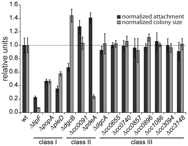
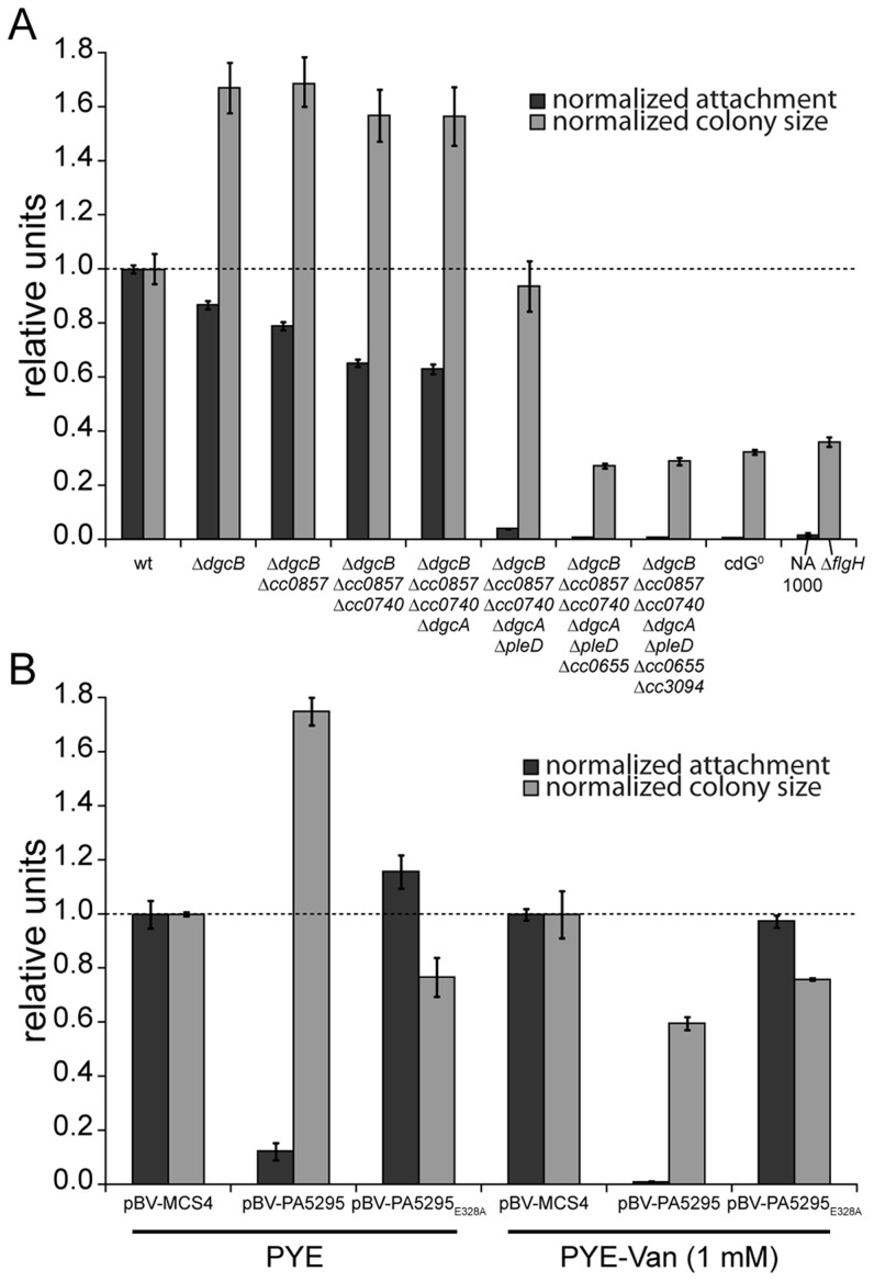
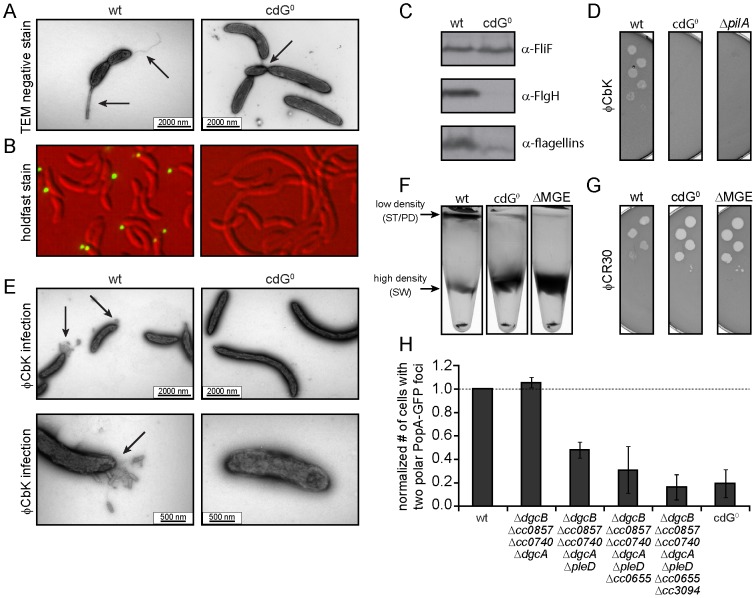
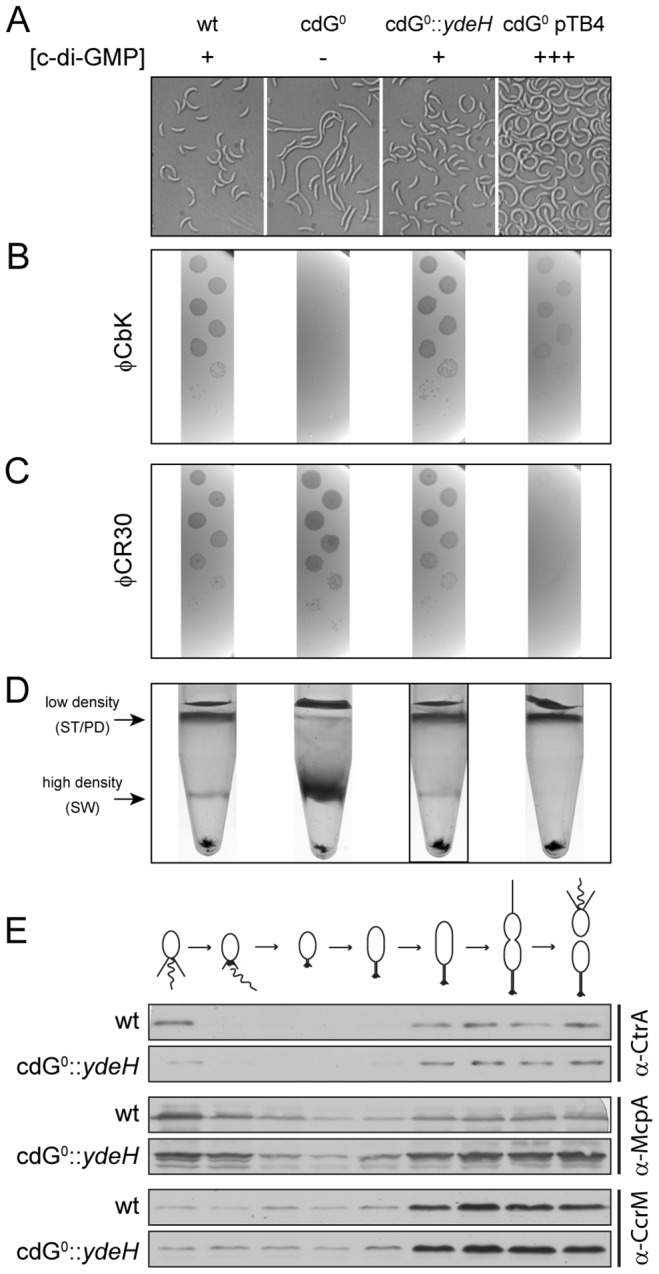
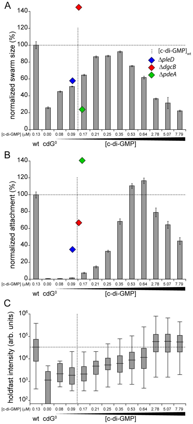
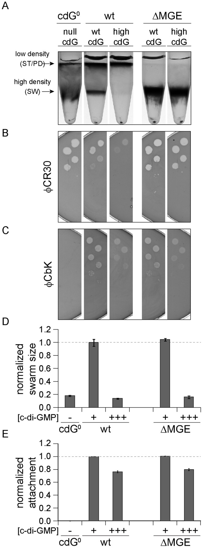
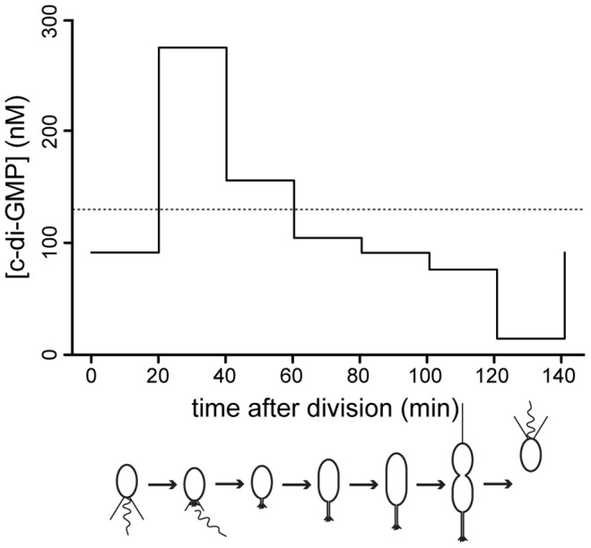
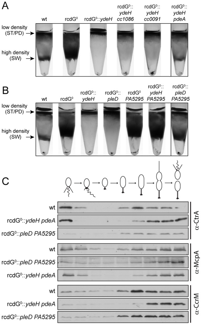
Many bacteria mediate important life-style decisions by varying levels of the second messenger c-di-GMP. Behavioral transitions result from the coordination of complex cellular processes such as motility, surface adherence or the production of virulence factors and toxins. While the regulatory mechanisms responsible for these processes have been elucidated in some cases, the global pleiotropic effects of c-di-GMP are poorly understood, primarily because c-di-GMP networks are inherently complex in most bacteria. Moreover, the quantitative relationships between cellular c-di-GMP levels and c-di-GMP dependent phenotypes are largely unknown. Here, we dissect the c-di-GMP network of Caulobacter crescentus to establish a global and quantitative view of c-di-GMP dependent processes in this organism. A genetic approach that gradually reduced the number of diguanylate cyclases identified novel c-di-GMP dependent cellular processes and unraveled c-di-GMP as an essential component of C. crescentus cell polarity and its bimodal life cycle. By varying cellular c-di-GMP concentrations, we determined dose response curves for individual c-di-GMP-dependent processes. Relating these values to c-di-GMP levels modeled for single cells progressing through the cell cycle sets a quantitative frame for the successive activation of c-di-GMP dependent processes during the C. crescentus life cycle. By reconstructing a simplified c-di-GMP network in a strain devoid of c-di-GMP we defined the minimal requirements for the oscillation of c-di-GMP levels during the C. crescentus cell cycle. Finally, we show that although all c-di-GMP dependent cellular processes were qualitatively restored by artificially adjusting c-di-GMP levels with a heterologous diguanylate cyclase, much higher levels of the second messenger are required under these conditions as compared to the contribution of homologous c-di-GMP metabolizing enzymes. These experiments suggest that a common c-di-GMP pool cannot fully explain spatiotemporal regulation by c-di-GMP in C. crescentus and that individual enzymes preferentially regulate specific phenotypes during the cell cycle.


Conflict of interest statement

The authors have declared that no competing interests exist.

Figures

Figure 1
Figure 1. Motility and attachment behavior of C. crescentus is modulated by several GGDEF- and EAL-domain proteins.
Surface attachment (black bars) and colony size on motility agar plates (grey bars) of mutants lacking individual GGDEF/EAL domain proteins are indicated relative to the wild type. Each bar represents the mean of seven independent experiments; the error bars represent the standard deviation; the dotted line indicates the wild-type behavior. The lines under the gene names outline the phenotypic classes. Class I: non-canonical behavior, class II: canonical behavior, class III: no phenotype (see main text for detailed information).
Figure 2
Figure 2. C-di-GMP is essential for motility and attachment in C. crescentus.
A) A strain devoid of all potential diguanylate cyclases (cdG0 strain; CB15 Δcc0655 Δcc0740 Δcc0857 Δcc0896 Δcc3094 ΔdgcA ΔdgcB ΔpleD) was generated by cumulative deletions of genes that code for GGDEF domain proteins. Both surface attachment (black bars) and colony size on semi-solid agar plates as measure for motility (grey bars) of all mutant intermediates are shown normalized to the corresponding wild-type phenotype. Strain NA1000 and a ΔflgH mutant are shown as non-attaching and non-motile controls, respectively. The mean of eight experiments is given. B) Motility and attachment scores of wild-type C. crescentus strains carrying a plasmid expressing a heterologous phosphodiesterase (PA5295) under control of the inducible vanillate promoter. Each phenotype was normalized to cells carrying the empty plasmid backbone and compared to a strain expressing an active site mutant of the PDE, both under conditions with residual promoter activity (PYE) or full promoter activity (PYE-Van (1 mM)). The bars indicate the mean of six experiments; error bars represent the standard deviation; the dotted line indicates the wild-type behavior.
Figure 3
Figure 3. Depletion of c-di-GMP leads to severe deficiencies in development and cell morphology.
A) Flagellum and stalk biogenesis: Representative transmission electron micrographs of wild-type (left panel) and cdG0 cells (right panel). Arrows highlight the flagellum, the stalk or a misplaced division septum, respectively. B) Holdfast biogenesis: Representative fluorescent micrographs of wild type (left panel) and cdG0 cells (right panel) after staining with fluorescently labeled wheat germ agglutinin. The holdfast specific lectin stain is shown in green and overlaid with a DIC image (red). C) Expression of late flagellar genes: The expression of representative flagellar proteins belonging to class II (FliF), class III (FlgH) and class IV (flagellins) of the flagellar hierarchy are analyzed in wild type (left) and the cdG0 strain (right) by immunoblots with specific antibodies. D) Pili-specific phage φCbK sensitivity: Plaque formation of a 1∶10 serial dilution of phage φCbK was assessed on a lawn of wild type (left), cdG0 strain (middle) and a pilA mutant (right) lacking the major pili subunit. E) Pili-specific phage φCbK sensitivity: Representative transmission electron micrographs of negatively stained wild type (left) and cdG0 strain (right) after brief exposure to the pili specific phage φCbK. Phage particles attached to the cell poles are highlighted by arrows. F) Cell type-specific cell density: Cells of the wild type (left), the cdG0 strain (middle) and a mutant lacking a mobile genetic element (MGE) were separated by density gradient centrifugation. Arrows indicate the low- and high-density bands. The wild type low-density band contains a mixture of stalked (ST) and predivisonal (PD) cells while the high-density band consists of a homogenous population of swarmer (SW) cells. G) Protection from phage φCR30: Cell lawns of wild type (left), cdG0 strain (middle) and a mutant lacking a mobile genetic element (MGE) were exposed to a 1∶10 serial dilution of φCR30. Please note that on C. crescentus wild type φCR30 forms turbid plaques, while the cdG0 or ΔMGE strains form clear plaques. H) PopA localization: The graph shows the quantification of fluorescent micrographs of cells expressing a PopA-GFP fusion. The bars represent the average number of cells that contain two polar foci. Data are given relative to the wild type. Error bars represent the standard deviation. At least 600 cells were quantified for each strain.
Figure 4
Figure 4. In vivo dose-response curves for c-di-GMP dependent processes.
Cell morphology (A), phage sensitivity (B, C) and cell type-specific cell density (D) was recorded as a function of varying c-di-GMP concentration in a cdG0 strain expressing YdeH, a heterologous DGC. YdeH expression conditions and resulting c-di-GMP concentration are taken from Figure S2. See also Table 2 for an overview of the phenotypes with more c-d-GMP concentrations. A) C. crescentus cell length and morphology is controlled by c-di-GMP. Light micrographs of cells with increasing concentrations of c-di-GMP are shown. Wild-type cells carrying a control plasmid are shown for comparison. B–C) Interference with phage sensitivity at low and high c-di-GMP concentrations. Plaque assays are shown for lawns of cells with increasing concentrations of c-di-GMP with 1∶10 serial dilutions of the pili specific phage φCbk (B) and the S-layer specific phage φCR30 (C). D) Cell density is c-di-GMP dependent. C. crescentus cells with increasing intracellular c-di-GMP concentrations were separated by density gradient centrifugation. The resulting low- and high-density bands are highlighted. The black box marks the conditions (0.17 µM [c-di-GMP]) where swarmer cells from the high-density band were isolated for cell cycle synchrony analyses (Figure 4E). E) A heterologous DGC mediates normal cell cycle progression in C. crescentus. Cells derived from the cdG0::ydeH strain grown with intermediate levels of c-di-GMP (Figure 4D) were isolated from the high-density band, released into fresh medium containing IPTG and followed through a cell cycle. Immunoblots with specific antibodies directed against cell cycle regulated marker proteins were used to determine the homogeneity of isolated swarmer cells and their synchronicity during and progression through the cell cycle.
Figure 5
Figure 5. Motility and surface attachment show distinct in vivo c-di-GMP dose-response curves.
Motility (A) and surface attachment (B) was recorded as a function of varying c-di-GMP concentration in a cdG0 strain expressing YdeH, a heterologous DGC. YdeH expression conditions and resulting c-di-GMP concentration are taken from Figure S2. The phenotypic behavior and c-di-GMP concentrations of mutants lacking selected DGCs or PDEs are indicated by blue (ΔpleD), red (ΔdgcB) and green diamonds (ΔpdeA). Holdfast production (C) was quantified as described in Materials and Methods with results represented as box plot. Big middle lines indicate the median holdfast fluorescence intensity of the sample. The box indicates the interquartile range and the whiskers include all data points not considered as outliers. The dotted lines highlight behavior and average c-di-GMP concentrations of C. crescentus wild type for comparison.
Figure 6
Figure 6. Cell density and φCR30 phage sensitivity are regulated by c-di-GMP via a mobile genetic element.
YdeH was overexpressed ([c-di-GMP] +++) in C. crescentus wild type (wt) or in a mutant lacking the mobile genetic element (ΔMGE) and strains were compared to isogenic strains lacking YdeH ([c-di-GMP] +) and to the cdG0 strain ([c-di-GMP] −). Differential cell density (A), sensitivity to phages φCR30 (S-layer) (B) and φCbK (pili) (C), colony size on motility plates (D), and surface attachment (E) were scored for all strains. The positions of high- and low-density bands after density gradient centrifugation are marked by arrows. The bars in the motility and attachment assays represent the mean of five or eight experiments, respectively. The error bars indicate the standard deviation. The quantified data were normalized to wild type without YdeH overexpression and the dotted lines indicate wild-type behavior. This figure is complemented by Figure S10 which includes more controls.
Figure 7
Figure 7. C-di-GMP oscillation during the C. crescentus cell cycle.
The graph shows modeled c-di-GMP fluctuations in a single C. crescentus cell during a full cell cycle. The predictions are based on c-di-GMP measurements in synchronized populations of C. crescentus wild-type cells and on a mathematical model accounting for differences in cell age and cell cycle length of synchronized populations (Figure S11). The c-di-GMP concentration is given in nM and the progression of the cell cycle is given in minutes after division of the predivisional cell. Only the c-di-GMP concentration of the swarmer progeny is shown. Cell cycle progression is depicted schematically below the graph. The dotted line indicates the average c-di-GMP concentration measured in non-synchronized wild-type populations.
Figure 8
Figure 8. Redundant enzymes facilitate c-di-GMP fluctuations during the cell cycle.
A) PdeA is sufficient to establish cell type-specific cell density distribution in the presence of a continuous source of c-di-GMP. C. crescentus cell density was analyzed by density gradient centrifugation for wild type cells (wt), a strain that lacks all endogenous diguanylate cyclases and phosphodiesterases (rcdG0), rcdG0 expressing the heterologous DGC YdeH, and rcdG0::ydeH complemented with three genes encoding homologous PDEs (CC1086, CC0091, PdeA). Cells were grown in the presence of 555 µM IPTG for YdeH induction. The position of the low- and high-density bands are marked with arrows and labeled with the cell types according to the fractionation behavior of wild type. The black box indicates the strain that was used to isolate swarmer cells for the analysis in Figure 8C. B) PleD is sufficient to establish cell type-specific cell density distribution in the presence of a constitutive PDE. Labels are like in (A). Note that although PA5295 was driven by the vanillate promoter its expression was not induced. Residual expression levels of the PDE were sufficient to destabilize c-di-GMP in this experiment. The black box indicates the strain that was used to isolate swarmer cells for the analysis in Figure 8C. C) Reconstitution of c-di-GMP fluctuations is sufficient for cell fate determination. Cells of the wild type (wt) and the diguanylate cyclase/phosphodiesterase free strain either expressing YdeH and PdeA (rcdG0::ydeH pdeA) or expressing PleD and PA5295 (rcdG0::pleD PA5295) were isolated from the high-density fraction of the gradient (see above) and released in fresh medium containing IPTG or vanillate, respectively. Samples were analyzed at 20 min intervals and probed with antibodies against cell cycle marker proteins (CtrA, McpA, and CcrM). Cell cycle progression is indicated schematically above the immunoblots.

References

    1. Jenal U, Malone J (2006) Mechanisms of cyclic-di-GMP signaling in bacteria. Annu Rev Genet 40: 385–407 doi:10.1146/annurev.genet.40.110405.090423 - DOI - PubMed
    1. Hengge R (2009) Principles of c-di-GMP signalling in bacteria. Nat Rev Microbiol 7: 263–273 doi:10.1038/nrmicro2109 - DOI - PubMed
    1. Galperin MY, Nikolskaya AN, Koonin EV (2001) Novel domains of the prokaryotic two-component signal transduction systems. FEMS Microbiol Lett 203: 11–21. - PubMed
    1. Sondermann H, Shikuma NJ, Yildiz FH (2012) You've come a long way: c-di-GMP signaling. Curr Opin Microbiol 15: 140–146 doi:10.1016/j.mib.2011.12.008 - DOI - PMC - PubMed
    1. Pultz IS, Christen M, Kulasekara HD, Kennard A, Kulasekara B, et al. (2012) The response threshold of Salmonella PilZ domain proteins is determined by their binding affinities for c-di-GMP. Mol Microbiol 86: 1424–1440 doi:10.1111/mmi.12066 - DOI - PMC - PubMed

Publication types

LinkOut - more resources