Review
. 2013 Oct;9(3):247-56.
doi: 10.1007/s11420-013-9357-5. Epub 2013 Aug 24.

Imaging Metal-on-Metal Hip Replacements: the Norwich Experience

Affiliations
Review

Imaging Metal-on-Metal Hip Replacements: the Norwich Experience

Marianna S Thomas et al. HSS J. 2013 Oct.

Abstract

Background: Adverse reaction to metal debris is a relatively recently described and often a silent complication of metal-on-metal (MOM) total hip replacements (THR). The Norfolk & Norwich University Hospital has been performing metal artefact reduction (MARS) MRI for 8 years in a variety of different types of MOM THR.

Questions/purposes: The aims of this review are to describe the experience of using MARS MRI in Norwich and to compare our experience with that published by other groups.

Methods: A MEDLINE keyword search was performed for studies including MRI in MOM THR. Relevant publications were reviewed and compared with published data from the Norfolk & Norwich University Hospital. The similarities and differences between these data were compared and possible explanations for these discussed.

Results: MARS MRI appears to be the most useful tool for diagnosing, staging and monitoring adverse reactions to metal debris (ARMD). There appears to be no clinically useful association between clinical and serological markers of disease and the severity of MR findings. Although severe early ARMD is associated with significant morbidity, mild disease is often stable for years. If patients with normal initial MR examinations develop ARMD, this usually occurs 7 years. A 1-year interval between MRI examinations is reasonable in asymptomatic patients.

Conclusions: There is a general international consensus that ARMD is prevalent in symptomatic and asymptomatic patients with MOM THR and that while appearances vary with the type of prosthesis, there are characteristic features that make MARS MRI essential for diagnosis, staging and surveillance of the disease.

Keywords: ALVAL; MRI; arthroplasty; hip; metal-on-metal.


Figures

Fig. 1
Fig. 1
Plain radiographs illustrating a McKee–Farrar (a), a first-generation MOM THR, and a modern MOM THR, a hybrid Ultima TPS (b) with minimal resorption of the medial calcar (c) as the only sign in a patient with extensive ARMD.
Fig. 2
Fig. 2
Intra-operative photograph demonstrating severe ARMD with a large head metal-on-metal THR. Extensive soft tissue destruction is associated with complete detachment of the abductors from the greater trochanter which allow visualization of the prosthesis.
Fig. 3
Fig. 3
Graph demonstrating the reduction in metal artefact with increasing receiver bandwidth for different matrix sizes. For any matrix size, a receiver bandwidth of 600 Hz/pixel or more produces 90% of the achievable reduction in artefact.
Fig. 4
Fig. 4
Fat suppression using coronal STIR (a) and two-point Dixon IDEAL (b) sequences demonstrating optimal control of susceptibility artefact with STIR.
Fig. 5
Fig. 5
Axial T1W (a) and T2W (b) fast spin echo in a patient with bilateral MOM THR demonstrating the typical appearance of ARMD in the Ultima TPS hip with intermediate signal on T1W and fluid signal enclosed in a thick ragged very low signal pseudocapsule on T2W (arrow).
Fig. 6
Fig. 6
Coronal T1W MRI (a, b) in a patient with an Ultima TPS THR, and a normal plain radiograph, demonstrating intermediate signal abnormalities (arrows) in the marrow of the proximal femur on T1W. Anecdotal reports from the revision procedures suggest that the MR underestimates the extent of the marrow disease found at surgery in patients with ARMD.
Fig. 7
Fig. 7
Sagittal (a) and axial (b) T2W MR of an ovoid soft tissue lesion lying between the right gluteus maximus and medius muscles demonstrating very low signal with areas of subtle blooming which turned out to be a histiocytoma containing microscopic metal particles accounting for the signal characteristics.
Fig. 8
Fig. 8
Axial T1W (a) and sagittal T2W (b) MR in a patient with a large bearing uncemented total ASR hip replacement. A large solid iliopsoas soft tissue lesion is present which is isointense on T1W and hypointense on T2W. Iliopsoas ARMD appears to be more common in large bearing THRs.
Fig. 9
Fig. 9
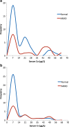
Graphs comparing the frequency distribution of serum Co (a) and Cr (b) ions in patients with ASR THRs with and without ARMD. The patterns of distribution demonstrate no clinically useful correlation.
Fig. 10
Fig. 10
Scatterplot of grades of severity of ARMD measured with serial MRI in patients in which the first MR was normal. The plot demonstrates that most subsequent MRI is also normal but in the 10% that develop ARMD do so between 7 and 11 years after surgery.

References

    1. 9th Annual Report. Surgical data to 31 December 2011: National Joint Registry for England and Wales; 2012.
    1. Amstutz HC, Campbell P, McKellop H, et al. Metal on metal total hip replacement workshop consensus document. Clin Orthop Relat Res. Aug 1996(329 Suppl):S297–303. - PubMed
    1. Amstutz HC, Grigoris P. Metal on metal bearings in hip arthroplasty. Clin Orthop Relat Res. Aug 1996(329 Suppl):S11–34. - PubMed
    1. Anderson H, Toms AP, Cahir JG, Goodwin RW, Wimhurst J, Nolan JF. Grading the severity of soft tissue changes associated with metal-on-metal hip replacements: reliability of an MR grading system. Skeletal Radiol. 2011;40(3):303–7. doi: 10.1007/s00256-010-1000-7. - DOI - PubMed
    1. August AC, Aldam CH, Pynsent PB. The McKee–Farrar hip arthroplasty. A long-term study. J Bone Joint Surg Br. 1986;68(4):520–7. - PubMed