. 2013 Sep 6;8(9):e71041.
doi: 10.1371/journal.pone.0071041. eCollection 2013.

Myocardial injection of apelin-overexpressing bone marrow cells improves cardiac repair via upregulation of Sirt3 after myocardial infarction

Affiliations

Myocardial injection of apelin-overexpressing bone marrow cells improves cardiac repair via upregulation of Sirt3 after myocardial infarction

Lanfang Li et al. PLoS One. .

Abstract

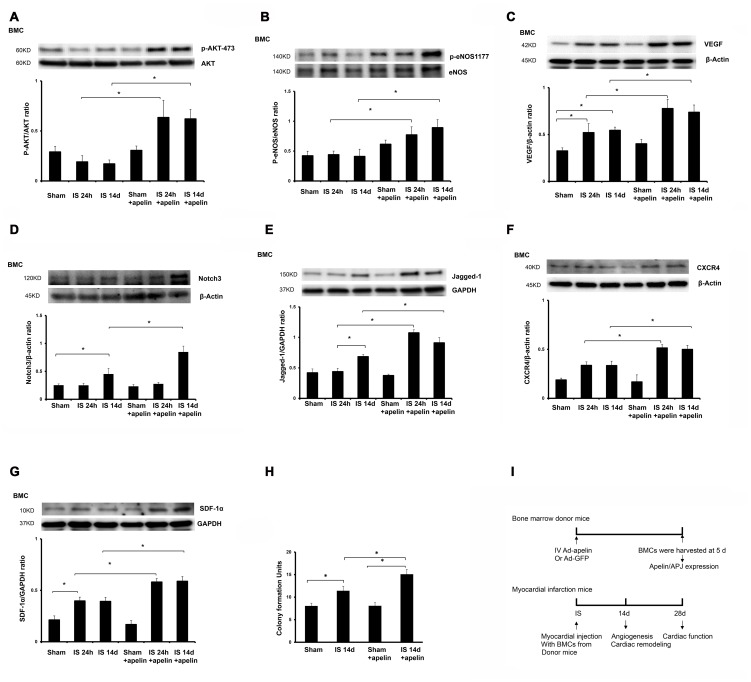
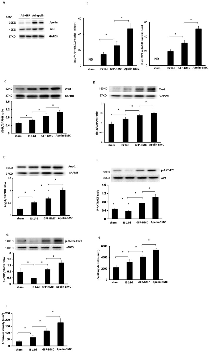
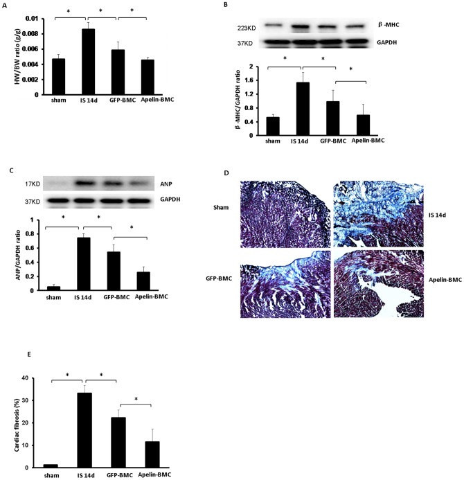
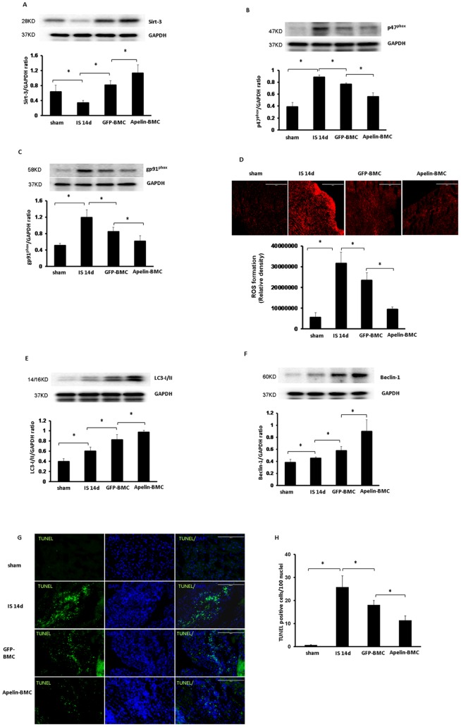
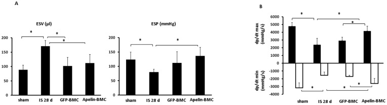
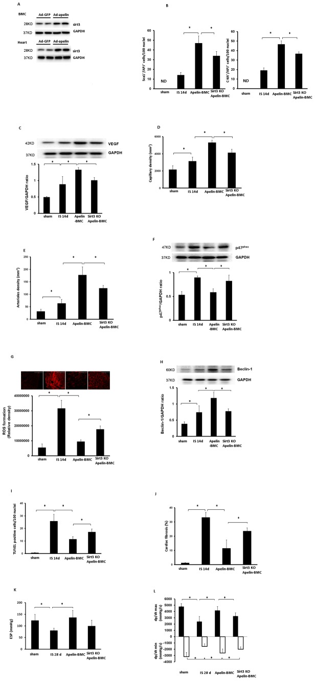
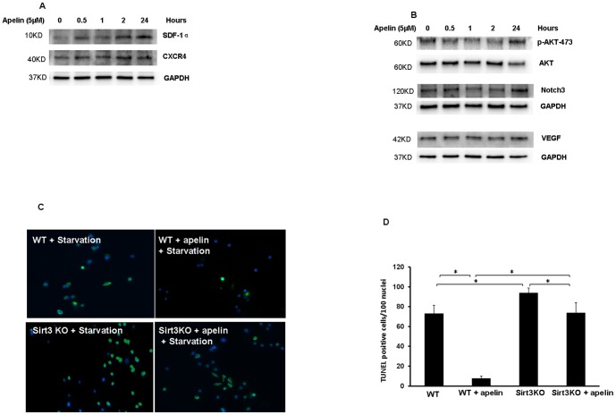
Our previous study shows that treatment with apelin increases bone marrow cells (BMCs) recruitment and promotes cardiac repair after myocardial infarction (MI). The objective of this study was to investigate whether overexpression of apelin in BMCs improved cell therapy and accelerated cardiac repair and functional recovery in post-MI mice. Mouse myocardial infarction was achieved by coronary artery ligation and BMCs overexpressing apelin (apelin-BMCs) or GFP (GFP-BMCs) were injected into ischemic area immediately after surgery. In vitro, exposure of cultured BMCs to apelin led to a gradual increase in SDF-1á and CXCR4 expression. Intramyocardial delivery of apelin-BMCs in post-MI mice resulted in a significant increase number of APJ⁺/c-kit⁺/Sca1⁺ cells in the injected area compared to GFP-BMCs treated post-MI mice. Treatment with apelin-BMCs increased expression of VEGF, Ang-1 and Tie-2 in post-MI mice. Apelin-BMCs treatment also significantly increased angiogenesis and attenuated cardiac fibrosis formation in post-MI mice. Most importantly, treatment with apelin-BMCs significantly improved left ventricular (LV) systolic function in post-MI mice. Mechanistically, Apelin-BMCs treatment led to a significant increase in Sirtuin3 (Sirt3) expression and reduction of reactive oxygen species (ROS) formation. Treatment of cultured BMCs with apelin also increased Notch3 expression and Akt phosphorylation. Apelin treatment further attenuated stress-induced apoptosis whereas knockout of Sirt3 abolished anti-apoptotic effect of apelin in cultured BMCs. Moreover, knockout of Sirt3 significantly attenuated apelin-BMCs-induced VEGF expression and angiogenesis in post-MI mice. Knockout of Sirt3 further blunted apelin-BMCs-mediated improvement of cardiac repair and systolic functional recovery in post-MI mice. These data suggest that apelin improves BMCs therapy on cardiac repair and systolic function in post-MI mice. Upregulation of Sirt3 may contribute to the protective effect of apelin-BMCs therapy.


Conflict of interest statement

Competing Interests: The authors have declared that no competing interests exist.

Figures

Figure 1
Figure 1. Apelin treatment increases expression of pro-survival and pro-angiogenic growth factors in BMCs.
A and B. Representative phosphorylation levels of Akt-(473) and eNOS-(1177) in cultured BMCs of post-MI mice. The phosphorylation of Akt-(473) and eNOS-(1177) was significantly enhanced in cultured BMCs from apelin-treated post-MI mice (n = 4 mice, *p<0.05). C. Expression of VEGF in cultured BMCs of post-MI mice. VEGF expression in cultured BMCs was significantly upregulated in apelin treated post-MI mice compared to saline-treated mice at 24 hour and 14 days of post-MI (n = 4 mice, *p<0.05). D and E. Expression of Notch3 and Jagged1 in cultured BMCs of post-MI mice. Notch3 expression in cultured BMCs was significantly increased in control post-MI mice compared with sham control mice at 14 days. Treatment with apelin led to a significant increase in Notch3 expression in cultured BMCs of post-MI mice compared to control post-MI mice at 14 days (n = 4 mice, *p<0.05). Jagged1 expression in cultured BMCs was significantly increased in control post-MI mice compared with sham control mice at 14 days. Treatment with apelin resulted in a significant increase in Jagged1 expression compared to control post-MI mice (n = 4 mice, *p<0.05). F and G. CXCR-4 and SDF-1α expression in cultured BMCs of post-MI mice. The expression of CXCR-4 and SDF-1α was significantly upregulated in cultured BMCs of the apelin treated post-MI mice compared to saline-treated post-MI mice at 24 hour and 14 days (n = 4 mice, *p<0.05). H. BMC colony formation units. Apelin treatment led to a significantly increase in colony formation in cultured BMCs compared to saline-treated mice at 14 days of post-MI (n = 6 mice, *p<0.05). I. Schematic diagram of apelin-BMCs treatment and experimental endpoints in vivo.
Figure 2
Figure 2. Apelin-BMC treatment augments expression of angiogenic growth factors and promotes myocardial angiogenesis in post-MI mice.
A. Western blot analysis revealing that systemic delivery of Ad-apelin in WT mice for 5 days upregulated apelin and APJ expression in the BMCs. n = 2 mice. B. Quantitative analysis of APJ+/Sca1+/c-kit+ cells demonstrating that Apelin-BMCs treated post-MI mice had a significant higher number of APJ+/Sca1+/c-kit+ cells in the injected area compared to GFP-BMCs treated mice. All data represent mean ± SD; n = 5, *p<0.05. ND = not detected. C to G. Treatment with GFP-BMCs significantly increased VEGF, Tie2 and Ang-1 expression in post-MI mice compared to that of control post-MI mice. The expression of VEGF, Tie2 and Ang-1 was significantly upregulated in the apelin-BMCs treated mice compared to GFP-BMCs treatment (n = 5 mice, *p<0.05). GFP-BMCs treatment significantly increased phosphorylation of Akt-473 and eNOS-1177 in post-MI mice compared to that of control post-MI mice. The phosphorylation of Akt-473 and eNOS-1177 was significantly increased in the apelin-BMCs treated mice compared to GFP-BMCs treatment. n = 5 mice; *p<0.05. H. GFP-BMCs treatment significantly increased capillary formation compared to control post-MI mice. Treatment with apelin-BMCs led to a further increase in myocardial capillary density compared to GFP-BMCs treatment. n = 5 mice; *p<0.05. I. GFP-BMCs treatment significantly increased myocardial arteriole density compared to control post-MI mice at 14 days. Treatment with apelin-BMCs significantly increased myocardial arteriole density compared to GFP-BMCs treatment. n = 5 mice; *p<0.05.
Figure 3
Figure 3. Apelin-BMCs treatment inhibits cardiac hypertrophy and fibrosis formation.
A. Treatment with GFP-BMCs significantly reduced HW/BW ratio compared to saline treatment. Treatment with apelin-BMCs led to a further significant reduction of HW/BW ratio compared to GFP-BMCs treated group. n = 4–6 mice, *p<0.05. B and C. GFP-BMCs treatment significantly decreased heart β-MHC and ANP expression compared to saline treatment. Treatment with apelin-BMCs led to a further decrease in β-MHC and ANP expression compared to GFP-BMCs group. n = 4 mice, *p<0.05. D and E. Representative images of cardiac fibrosis in the infarction zone and quantitative analysis of fibrotic area in mice (Masson's trichrome). The area of cardiac fibrosis (blue) in post-MI mice was increased significantly at 14 days of MI. GFP-BMCs treatment significantly reduced the area of cardiac fibrosis compared to those treated with saline. Apelin-BMCs treatment significantly decreased cardiac fibrosis area compared to GFP-BMCs treated group. n = 5 mice; *p<0.05.
Figure 4
Figure 4. Apelin-BMCs treatment upregulates Sirt3 expression and attenuates ROS formation.
A. Treatment with GFP-BMCs significantly increased Sirt-3 expression compared to mice treated with saline. Treatment with apelin-BMCs led to a further significant increase in Sirt-3 expression compared to GFP-BMCs treatment. n = 4 mice, *p<0.05. B and C. The expression p47phox and gp91phox was significantly increased at 14 days of post-MI mice compared to sham control mice. GFP-BMCs treatment significantly reduced p47phox and gp91phox expression compared to post-MI mice treated with saline. Apelin-BMCs treatment led to a significant reduction in p47phox and gp91phox expression compared to GFP-BMCs treatment. n = 4 mice, *p<0.05. D. DHE staining and quantitative analysis showing that ROS formation was significantly increased in post-MI mouse hearts at 14 days compared to sham control. Treatment with GFP-BMCs significantly decreased ROS formation compared to that of control post-MI mice. ROS formation was significantly reduced in the apelin-BMCs treated mice compared to GFP-BMCs treated mice. n = 5 mice; *p<0.05. E and F. LC3-I/II and Beclin-1 expression was significantly increased in the mouse hearts of post-MI compared to sham control. GFP-BMCs treatment significantly increased LC3-I/II and Beclin-1 expression compared to that of control post-MI mice. The expression of LC3-I/II and Beclin-1 was significantly increased in the apelin-BMCs treated mice compared to GFP-BMCs treated mice. n = 4 mice; *p<0.05. G and H. Apoptotic cells in the infarcted area of the left ventricle were identified by TUNEL staining (green, 10×) and total nuclei by DAPI counterstaining (blue, 10×). GFP-BMCs significantly decreased TUNEL+ nuclei in post-MI mice compared to that of control post-MI mice. TUNEL+ nuclei were significantly decreased in the apelin-BMCs treated mice compared to GFP-BMCs treated mice. n = 6 mice; *p<0.05.
Figure 5
Figure 5. Treatment with apelin-BMCs improves heart systolic function at 28 days of post-MI mice.
A. The end-systolic volume (ESV) was significantly increased whereas the end-systolic pressure (ESP) was decreased in post-MI mice. GFP-BMCs and apelin-BMCs treatment significantly decreased ESV compared to saline-treated mice. Treatment with apelin-BMCs, but not GFP-BMCs, led to a significant increase in ESP compared to saline-treated post-MI mice. n = 5–8 mice, *p<0.05. B. Maximum +dP/dt pressure was increased whereas minimum −dP/dt pressure was decreased in the apelin-BMCs treated mice compared to saline-treated and GFP-BMCs-treated mice. n = 5–8 mice, *p<0.05.
Figure 6
Figure 6. Knockout of Sirt3 blunts apelin-BMCs-mediated cardiac repair and functional recovery after MI.
A. Systemic delivery of Ad-apelin for 5 days increased levels of Sirt3 in the BMCs and hearts. n = 2 mice. B. The number of Sca1+/APJ+ cells and c-kit+/APJ+ cells in the injected area was significantly reduced in post-MI mice treated with Sirt3KO-apelin-BMCs compared to apelin-BMCs treatment. n = 5, *p<0.05. ND = not detected. C. VEGF expression was significantly reduced in the mice treated with Sirt3KO-apelin-BMCs compared to apelin-BMCs treatment. n = 6 mice, *p<0.05. D. Myocardial capillary density in the border zone of ischemia area was significantly decreased in the mice treated with Sirt3KO-apelin-BMCs compared to apelin-BMCs treatment. n = 6 mice, *p<0.05. E. Arteriole density in the border zone of ischemia area was significantly decreased in the mice treated with Sirt3KO-apelin-BMCs compared to apelin-BMCs treatment. n = 6 mice, *p<0.05. F. The expression of p47phox was significantly elevated in the hearts of post-MI mice treated with Sirt3KO-apelin-BMCs compared to apelin-BMCs treatment. n = 6 mice, *p<0.05. G. DHE staining and quantitative analysis showing that myocardial ROS formation was significantly increased in the ischemia area of post-MI mice treated with Sirt3KO-apelin-BMCs compared to apelin-BMCs treatment. n = 5 mice, *p<0.05. H. The expression of beclin-1 was significantly reduced in the mice treated with Sirt3KO-apelin-BMCs compared to apelin-BMCs treatment. n = 6 mice, *p<0.05. I. Quantitative analysis of apoptotic cells by TUNEL staining showing that TUNEL+ nuclei in the ischemia area was significantly increased in post-MI mice treated with Sirt3KO-apelin-BMCs compared to apelin-BMCs treatment. n = 6 mice, *p<0.05. J. The cardiac fibrosis was significantly increased in post-MI mice treated with Sirt3KO-apelin-BMCs compared to apelin-BMCs treatment. n = 3–5 mice, *p<0.05. K. Treatment with apelin-BMCs resulted in a significant increase in ESP compare to the saline-treated ischemic mice. But there were no obvious difference between Sirt3KO-apelin-BMCs treatment with saline-treated ischemic mice. n = 4–6 mice, *p<0.05. L. Sirt3KO-apelin-BMCs treatment significantly decreased +dP/dtmax pressure and increased –dp/dtmin pressure compared to apelin-BMCs treatment. n = 4–6 mice,*p<0.05.
Figure 7
Figure 7. Apelin increases pro-survival and pro-angiogenic growth factors and attenuates cell apoptosis in cultured BMCs.
A. Representative Western blot analysis showing that treatment of cultured BMCs with apelin (5 µM) upregulates SDF-1α and CXCR-4 expression at 0.5 hours up to 24 hours. Experiments were repeated in two cell lines. B. Representative Western blot analysis showing that treatment of cultured BMCs with apelin (5 µM) for 24 hours increases Akt phosphorylation and upregulates VEGF and Notch3 expression. Experiments were repeated in two cell lines. C and D. Representative TUNEL staining images and quantitative analysis showing that knockout of Sirt3 significantly increased starvation-induced BMCs apoptosis. Treatment with apelin resulted in a significant reduction of BMCs apoptosis whereas knockout of Sirt3 diminished apelin-mediated protective effect against cell apoptosis in cultured BMCs. n = 3, *p<0.05.

Similar articles

Cited by

References

    1. Schachinger V, Assmus B, Britten MB, Honold J, Lehmann R, et al. (2004) Transplantation of progenitor cells and regeneration enhancement in acute myocardial infarction: final one-year results of the TOPCARE-AMI Trial. J Am Coll Cardiol 44: 1690–1699. - PubMed
    1. Britten MB, Abolmaali ND, Assmus B, Lehmann R, Honold J, et al. (2003) Infarct remodeling after intracoronary progenitor cell treatment in patients with acute myocardial infarction (TOPCARE-AMI): mechanistic insights from serial contrast-enhanced magnetic resonance imaging. Circulation 108: 2212–2218. - PubMed
    1. Jujo K, Ii M, Losordo DW (2008) Endothelial progenitor cells in neovascularization of infarcted myocardium. J Mol Cell Cardiol 45: 530–544. - PMC - PubMed
    1. Hu CH, Li ZM, Du ZM, Zhang AX, Rana JS, et al. (2010) Expanded human cord blood-derived endothelial progenitor cells salvage infarcted myocardium in rats with acute myocardial infarction. Clin Exp Pharmacol Physiol 37: 551–556. - PubMed
    1. Cho J, Zhai P, Maejima Y, Sadoshima J (2011) Myocardial injection with GSK-3beta-overexpressing bone marrow-derived mesenchymal stem cells attenuates cardiac dysfunction after myocardial infarction. Circ Res 108: 478–489 CIRCRESAHA.110.229658 [pii]; doi:10.1161/CIRCRESAHA.110.229658 - DOI - PMC - PubMed

Publication types

MeSH terms