. 2013 Sep 5;8(9):e71935.
doi: 10.1371/journal.pone.0071935. eCollection 2013.

Sulphamoylated 2-methoxyestradiol analogues induce apoptosis in adenocarcinoma cell lines

Affiliations

Sulphamoylated 2-methoxyestradiol analogues induce apoptosis in adenocarcinoma cell lines

Michelle Visagie et al. PLoS One. .

Abstract

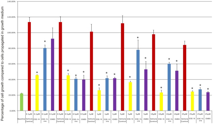
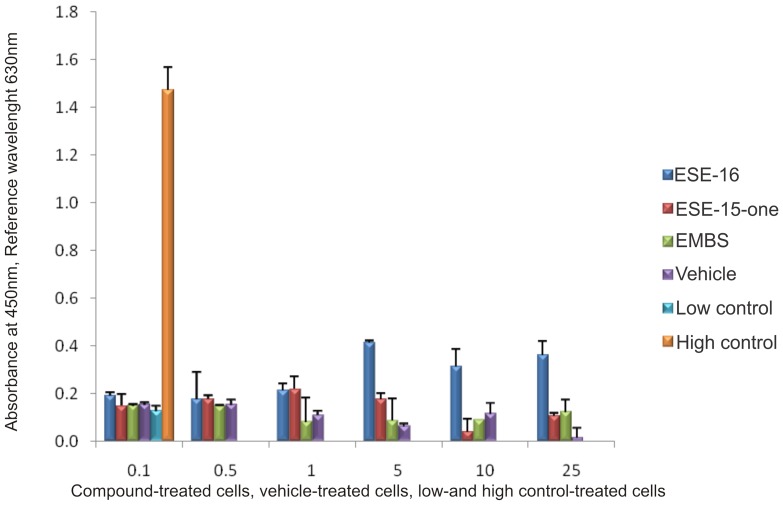
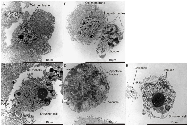
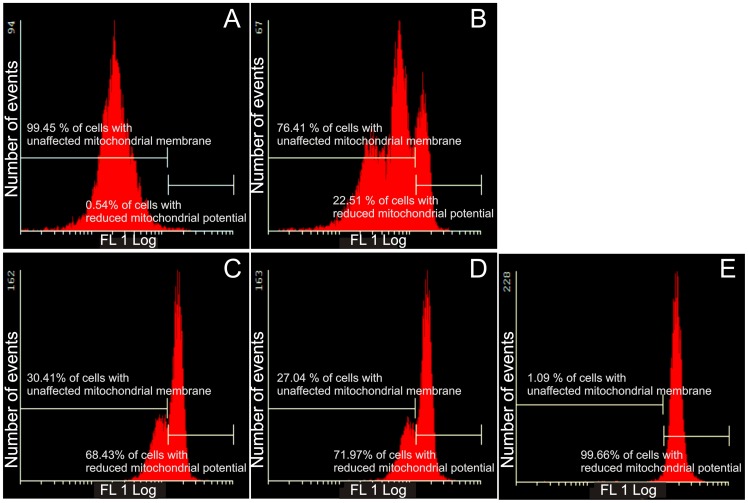
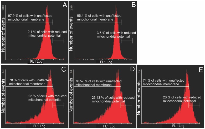
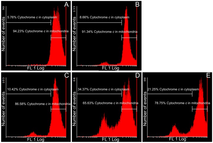
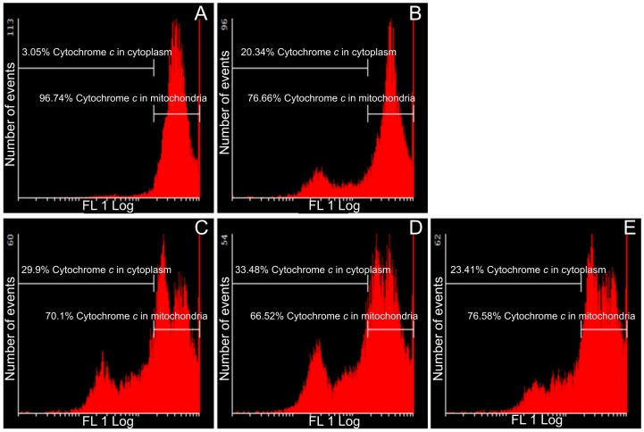
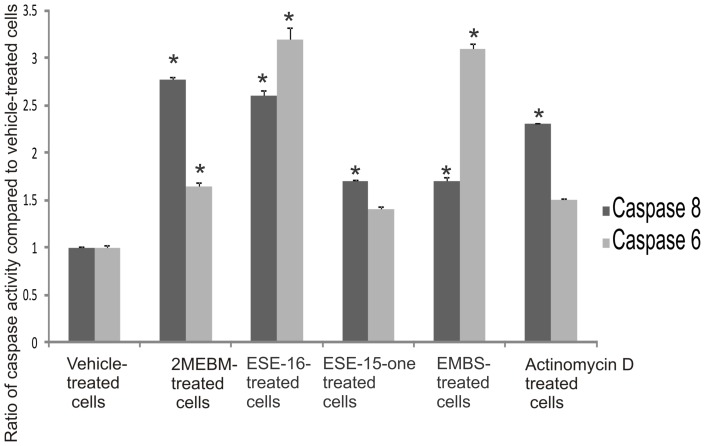
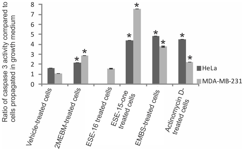
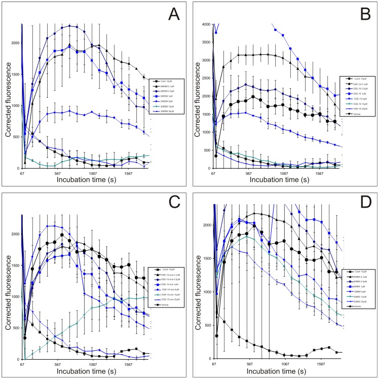
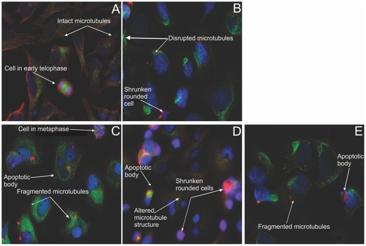
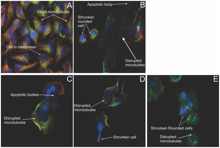
2-Methoxyestradiol (2ME2) is a naturally occurring estradiol metabolite which possesses antiproliferative, antiangiogenic and antitumor properties. However, due to its limited biological accessibility, synthetic analogues have been synthesized and tested in attempt to develop drugs with improved oral bioavailability and efficacy. The aim of this study was to evaluate the antiproliferative effects of three novel in silico-designed sulphamoylated 2ME2 analogues on the HeLa cervical adenocarcinoma cell line and estrogen receptor-negative breast adenocarcinoma MDA-MB-231 cells. A dose-dependent study (0.1-25 μM) was conducted with an exposure time of 24 hours. Results obtained from crystal violet staining indicated that 0.5 μM of all 3 compounds reduced the number of cells to 50%. Lactate dehydrogenase assay was used to assess cytotoxicity, while the mitotracker mitochondrial assay and caspase-6 and -8 activity assays were used to investigate the possible occurrence of apoptosis. Tubulin polymerization assays were conducted to evaluate the influence of these sulphamoylated 2ME2 analogues on tubulin dynamics. Double immunofluorescence microscopy using labeled antibodies specific to tyrosinate and detyrosinated tubulin was conducted to assess the effect of the 2ME2 analogues on tubulin dynamics. An insignificant increase in the level of lactate dehydrogenase release was observed in the compounds-treated cells. These sulphamoylated compounds caused a reduction in mitochondrial membrane potential, cytochrome c release and caspase 3 activation indicating apoptosis induction by means of the intrinsic pathway in HeLa and MDA-MB-231 cells. Microtubule depolymerization was observed after exposure to these three sulphamoylated analogues.


Conflict of interest statement

Competing Interests: The authors have declared that no competing interests exist.

Figures

Figure 1
Figure 1. Cell number determination.
HeLa cell numbers expressed as a % of cells relative to the control (cells propagated in growth medium) after exposure to ESE-15-one, EMBS and ESE-16 for 24 h. An asterisk (*) indicates a statistically significant P-value of <0.05 when compared to cells propagated in growth medium.
Figure 2
Figure 2. Cytotoxicity by means of lactate dehydrogenase assay.
LDH levels of ESE-15-one, EMBS and ESE-16 -treated HeLa cells and vehicle-treated control cells after 24 h exposure. The background control consists of growth medium only. The low control refers to cells resuspended in growth medium and the high control to cells resuspended in growth medium with cell lysis solution added to the cells shortly before the experiment was terminated (according to the manufacturer's instructions). No statistically significant increase in LDH levels was observed in treated cells. These results showed that the sulphamoylated compounds were not toxic to the cells.
Figure 3
Figure 3. Morphology investigation using transmission electron microscopy.
TEM revealed vehicle-treated cells displaying no signs of distress (A). Cells treated with 2-methoxyestradiol-bis-sulphamate (B), ESE-16 (C) and ESE-15-one (D) demonstrated the occurrence of apoptotic bodies and vacuole formation. Cells treated with EMBS (E) displayed an increase in the number of vacuoles.
Figure 4
Figure 4. Mitochondrial membrane potential assay of HeLa cells.
Mitotracker-stained vehicle-treated control cells (A), 2-methoxyestradiol-bis-sulphamate-treated cells (B), ESE-16-treated cells (C), ESE-15-one-treated cells (D) and EMBS-treated cells (E) after 24 h exposure. An increase in the number of cells with reduced mitochondrial potential treated with 2ME2 analogues compared to the vehicle-treated control cells was observed.
Figure 5
Figure 5. Mitochondrial membrane potential assay of MDA-MB-231 cells.
Mitocapture-stained vehicle-treated control cells (A), 2-methoxyestradiol-bis-sulphamate-treated cells (B), ESE-16-treated cells (C), ESE-15-one-treated cells (D) and EMBS-treated cells (E) after 24 h exposure. An increase in the number of cells presenting with compromised mitochondrial potential was demonstrated in all three compound-treated cells when compared to the vehicle-treated control cells.
Figure 6
Figure 6. Assay demonstrating cytochrome c release in HeLa cells.
Flow cytometry was used to show the effects of these compounds on cytochrome c release from the mitochondria into the cytoplasm in HeLa cells. Vehicle-treated control cells (A), 2-methoxyestradiol-bis-sulphamate-treated cells (B), ESE-16-treated cells (C), ESE-15-one-treated cells (D) and EMBS-treated cells (E) following 24 h of exposure. Cytochrome c release was evident in all three compound-treated cells when compared to the vehicle-treated cells.
Figure 7
Figure 7. Assay demonstrating cytochrome c release in MDA-MB-231 cells.
Flow cytometry was conducted to present the effects of these sulphamoylated compounds on cytochrome c release from the mitochondria into the cytoplasm. Vehicle-treated control cells (A), 2-methoxyestradiol-bis-sulphamate-treated cells (B), ESE-16-treated cells (C), ESE-15-one-treated cells (D) and EMBS-treated cells (E) following 24 h of exposure. All three compound-treated cells demonstrated increased levels of cytochrome c release when compared to the vehicle-treated cells.
Figure 8
Figure 8. Determination of caspase activation.
Caspase 8 and caspase 6 activity ratios of compound- and actinomycin D-treated cells compared to vehicle-treated cells. Caspase 6 and caspase 8 activities in all compound-treated cells increased when compared to vehicle-treated cells. EMBS-treated cells demonstrated the most prominent increase in caspase 8 activity and ESE-16-treated cells the most prominent increase in caspase 6 activity when compared to vehicle-treated cells. 2-Methoxyestradiol-bis-sulphamate-treated cells are illustrated in the figure as 2MEBM due to limited space. An asterisk (*) indicates a statistically significant P-value of <0.05 when compared to vehicle-treated cells.
Figure 9
Figure 9. Caspase 3 activity in HeLa and MDA-MB-231 cells.
Spectrophotometry results of caspase 3 activity indicated that after exposure to these sulphamoylated compounds caspase 3 activity increased significantly. An asterisk (*) indicates a statistically significant P-value <0.05 when compared to cells propagated in growth medium.
Figure 10
Figure 10. Tubulin polymerization assay.
Tubulin was incubated with the compounds ((A), ESE-16 (B), ESE-15-one (C) and EMBS (D)), as described in the methods section. This assay showed that ESE-16 had a pronounced effect comparable with that of colchicine.
Figure 11
Figure 11. Immunofluorescent determination of microtubule dynamics in HeLa cells.
Double immunoflourecence was conducted to determine the compounds' effect after 24 h on microtubule dynamics within HeLa cells. Tyrosinated (dynamic) microtubules are visualized as red, whereas the detyrosinated (stable or stabilized) microtubules are stained in green. The vehicle-treated cells (A) demonstrated an intact dynamic microtubule structure. Both the colchicine positive control (B), as well as the ESE-16-treated cells (C) showed complete microtubule depolymerisation with few detyrosinated microtubule fragments remaining. ESE-15-one-treated cells (D) demonstrated altered microtubule morphology. EMBS-exposed cells (E) revealed fragmented microtubules. All treated cell samples demonstrated a decrease in cell density and rounded cells (X63 oil objective).
Figure 12
Figure 12. Immunofluorescent determination of microtubule dynamics in MDA-MB-231 cells.
Double immunoflourecence was done to determine the compounds' effect after 24 h on microtubule dynamics within MDA-MB-231 cells. Tyrosinated (dynamic) microtubules are visualized as red, whereas the detyrosinated microtubules are stained green. The vehicle-treated cells (A) demonstrated an intact dynamic microtubule structure. The colchicine-exposed positive control cells (B) showed complete microtubule depolymerisation with few detyrosinated microtubule fragments remaining. ESE-16 (C), ESE-15-one (D) and EMBS-treated cells (E) demonstrated detyrosinated microtubule fragments. All treated cell samples demonstrated a decrease in cell density with rounded, shrunken cells. Apoptotic bodies are evident in the ESE-16 treated cells (X63 oil objective).

References

    1. Banerjeei SK, Zoubine MN, Sarkar DK, Weston AP, Shah JH, et al. (2000) 2-Methoxyestradiol blocks estrogen-induced rat pituitary tumor growth and tumor angiogenesis: possible role of vascular endothelial growth factor. Anticancer Res 20: 2641–2645. - PubMed
    1. Pribluda VS, Gubish ER, La Vallee TM, Treston A, Swartz GM, et al. (2000) 2-Methylestradiol: An endogenous antiangiogenic and antiproliferative drug candidate. Cancer Metastasis Rev 19: 173–179. - PubMed
    1. Lavallee TM, Zhan XH, Herbstritt CJ, Kough EC, Green SJ, et al. (2002) 2-Methoxyestradiol inhibits proliferation and induces apoptosis independently of estrogen receptors alpha and beta. Cancer Res 62: 3691–3697. - PubMed
    1. Lippert TH, Adlercreutz H, Berger MR, Seeger H, Elger W, et al. (2003) Effect of 2-methoxyestradiol on the growth of methyl-nitroso-urea (MNU)-induced rat mammary carcinoma. J Steroid Biochem Mol Biol 84: 51–56. - PubMed
    1. Bhati R, Gokmen-Polar Y, Sledge GW, Fan C, Nakshatri H, et al. (2007) 2-Methoxyestradiol inhibits the anaphase-promoting complex and protein translation in human breast cancer cells. Cancer Res 67: 702–708. - PubMed

Publication types

MeSH terms