. 2013 Sep 9;8(9):e71982.
doi: 10.1371/journal.pone.0071982. eCollection 2013.

CTR1 silencing inhibits angiogenesis by limiting copper entry into endothelial cells

Affiliations

CTR1 silencing inhibits angiogenesis by limiting copper entry into endothelial cells

Gomathy Narayanan et al. PLoS One. .

Abstract

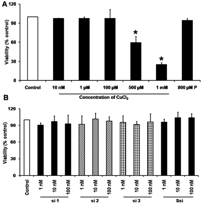
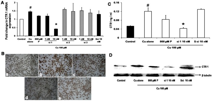
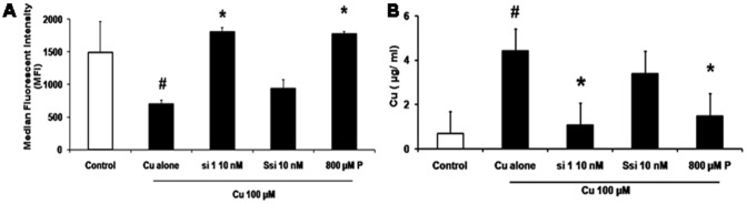
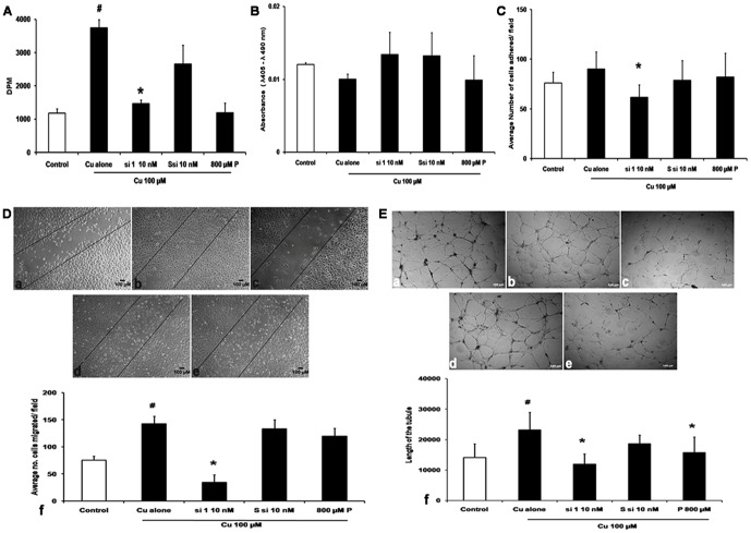
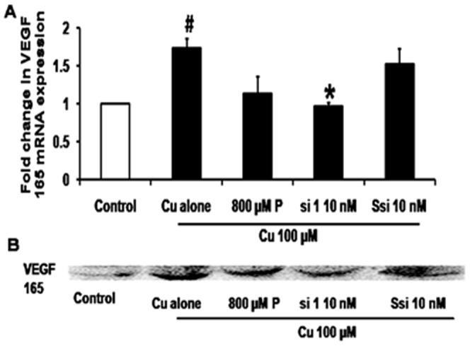
Increased levels of intracellular copper stimulate angiogenesis in human umbilical vein endothelial cells (HUVECs). Copper transporter 1 (CTR1) is a copper importer present in the cell membrane and plays a major role in copper transport. In this study, three siRNAs targeting CTR1 mRNA were designed and screened for gene silencing. HUVECs when exposed to 100 µM copper showed 3 fold increased proliferation, migration by 1.8-fold and tube formation by 1.8-fold. One of the designed CTR1 siRNA (si 1) at 10 nM concentration decreased proliferation by 2.5-fold, migration by 4-fold and tube formation by 2.8-fold. Rabbit corneal packet assay also showed considerable decrease in matrigel induced blood vessel formation by si 1 when compared to untreated control. The designed si 1 when topically applied inhibited angiogenesis. This can be further developed for therapeutic application.


Conflict of interest statement

Competing Interests: The bioactive oligonucleotide si 1 has been filed for patency as an inhibitor of angiogenesis in India (File no: 3221/che'2012). This does not alter the authors' adherence to the PLOS guidelines on sharing data and materials.

Figures

Figure 1
Figure 1. Cytotoxicity assay.
A. MTT assay for varying Cu concentrations: Cu concentration of 10 nM–1 mM in HUVECs cells showed that the IC50 for Cu to be 500 µM. B. MTT assay for different siRNAs: Cytotoxic effect of different siRNAs concentration from 1 nM–100 nM were tested and found to have no cytotoxic effect.
Figure 2
Figure 2. CTR1 silencing inhibits mRNA and protein expression.
A. Real time PCR for CTR1: qRT-PCR shows that Cu 100 µM treatment showed an increase in 45% CTR1 mRNA expression (# p = 0.01) compared to cells without Cu. CTR1 si 1 at 10 nM concentration significantly (* p = 0.01) decreased the CTR1mRNA expression by 71% when compared to 100 µM Cu whereas si 2, si 3 at 10 nM showed no inhibition. P 800 µM showed 14% decreased in expression of CTR1 when compared to Cu treated. B. Immuno cytochemistry for CTR1: a - Control, b - Cu 100 µM, c- Cu 100 µM+P 800 µM, d - Cu 100 µM+si 110 nM. CTR1 protein staining showed an increase in staining intensity with Cu treatment, (shown as brown colour) and decrease in staining intensity with si 110 nM treatment. C. CTR1 ELISA: HUVECs were treated with Cu 100 µM showed a significant (# p = 0.05) increase in CTR1 protein level compared to control and si 1 10 nM showed a significant decrease (* p = 0.04) in protein level when compared to Cu. D. Western blot for CTR1: Immunoblot showed an inhibition of CTR1 protein with si 110 nM treated condition compared to Cu which was not observed in 800 µM P treatment.
Figure 3
Figure 3. CTR1 silencing inhibits Cu entry.
A. Cu estimation by FACS: Histogram for Phen Green analysis was plotted with the mean fluorescent intensity obtained. Cu estimation by FACS, showed that treatment with Cu (# p = 0.04) increased the intracellular Cu levels (indicated by a decrease fluorescent intensity) and si 1 - 10 nM (*p = 0.02) decreased intracellular Cu levels in the presence of Cu, (indicated by an increase in fluorescent intensity). Ssi showed no significant effect and P 800 µM also showed significant inhibition (*p = 0.03) of Cu inside the cell. B. Cu estimation by AAS: Intracellular Cu levels increased significantly with Cu100 µM treatment (# p = 0.0005) and si 1 - 10 nM treatment decreased Cu levels (* p = 0.0008), even though P 800 µM also showed (* p = 0.01), a decrease in the Cu level statistically significant.
Figure 4
Figure 4. CTR1 silencing inhibits angiogenesis.
A. Proliferation assay: Triated thymidine was used for proliferation assay. Cu treatment showed an increase in proliferation (# p = 0.0003) and si 1 - 10 nM showed inhibition of proliferation (* p = 0.003), Ssi at 10 nM concentration did not show any effect, whereas Cu+P also significantly inhibited proliferation (* p = 0.02). B. Apoptosis assay: Apoptosis assay by Cell death ELISA kit showed no apoptosis in all conditions tested. C. Tube formation assay: a - Control, b- Cu 100 µM, c- Cu 100 µM+si 1 - 10 nM, d- Cu 100 µM+Ssi 10 nM, e- Cu 100 µM+P 800 µM. Cu 100 µM increased tube formation seen in the length of the tubule in Cu treated when compared with cells without Cu. Cu+si 1 - 10 nM reduced tube formation by decreasing the length of the tubule (*p = 0.01), when compared to Cu treated, which was not observed in Cu 100 µM+Ssi 10 nM. P 800 µM treatment decrease in length of the tubule (*p = 0.01) when compared to Cu. g- Histogram representing the length of the tubule in various conditions. D. Migration assay: Scratch assay a -Control, b- Cu 100 µM, c- Cu 100 µM+si 1 - 10 nM, d- Cu 100 µM+Ssi 10 nM, e- Cu 100 µM+P 800 µM (Magnification - 10×). HUVECs treated with Cu 100 µM increased the cell migration whereas si 1 - 10 nM inhibited migration, and no inhibition was in the presence of Ssi 10 nM and P 800 µM treatment. f- Transwell migration assay: Histogram showed that Cu 100 µM increased the cell migration significantly (# p = 0.00001), and Cu+si 1 - 10 nM reduced migration significantly (* p = 0.00001), Ssi 10 nM showed no effect, whereas P 800 µM treated did not inhibit migration. E. Attachment assay: a -Control, b- Cu 100 µM, c- Cu 100 µM+si 1 - 10 nM, d- Cu 100 µM+Ssi 10 nM, e- Cu 100 µM+P 800 µM HUVECs, f- histogram of cells counted in average of five fields. Cells showed that Cu 100 µM increased the cell attachment to the gelatin matrix significantly (# p = 0.02) and Cu+si 1 - 10 nM treatment reduced the number of cells attaching (* p = 0.00009), Ssi 10 nM showed no effect; whereas P 800 µM treated showed reduction in the number of cells attached which not significant.
Figure 5
Figure 5. CTR1 siRNA inhibited angiogenesis.
A. Untreated eye. B. Vehicle control- showed matrigel induced microvascular blood vessel formation (indicated in arrow). C. si RNA (si 1 100 ng) treated – showed decreased blood vessel formation after treatment with siRNA (indicated in arrow).
Figure 6
Figure 6. CTR 1 silencing inhibits VEGF 165.
A.VEGF real time PCR: qRT PCR shows that Cu 100 µM treatment showed a 1.73 fold increase in VEGF165 mRNA expression (#p = 0.013) compared to control. CTR1 si 1 at 10 nM concentration decreased the VEGF expression by 50% when compared to Cu (*p = 0.015), P 800 µM showed a mild decrease when compared with Cu, whereas Ssi 10 nM did not show any significant change B. Western blot: Cu increased VEGF protein levels, si 1 - 10 nM decreased of VEGF 165 protein when compared with Cu 100 µM treatment. P 800 µM showed slight reduction in protein level whereas Ssi 10 nM did not show any significant change when compared to Cu treatment.

References

    1. Folkman J (1972) Anti-angiogenesis: new concept for therapy of solid tumors. Ann Surg 175: 409–416. - PMC - PubMed
    1. van Hinsbergh VW, Koolwijk P (2008) Endothelial sprouting and angiogenesis: matrix metalloproteinases in the lead. Cardiovasc Res 78: 203–212. - PubMed
    1. Papetti M, Herman IM (2002) Mechanisms of normal and tumor-derived angiogenesis. Am J Physiol Cell Physiol 282: C947–970. - PubMed
    1. Carmeliet P (2005) Angiogenesis in life, disease and medicine. Nature 438: 932–936. - PubMed
    1. Riely GJ, Miller VA (2007) Vascular endothelial growth factor trap in non small cell lung cancer. Clin Cancer Res 13: s4623–4627. - PubMed

Publication types