. 2013 Sep 11;8(9):e72456.
doi: 10.1371/journal.pone.0072456. eCollection 2013.

Breaking of plant stomatal one-cell-spacing rule by sugar solution immersion

Affiliations

Breaking of plant stomatal one-cell-spacing rule by sugar solution immersion

Kae Akita et al. PLoS One. .

Abstract

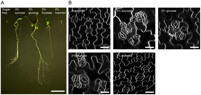
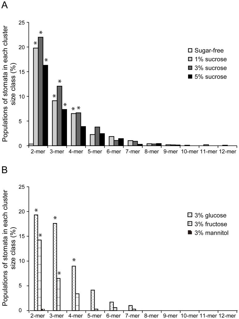
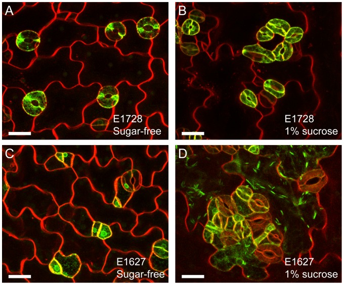
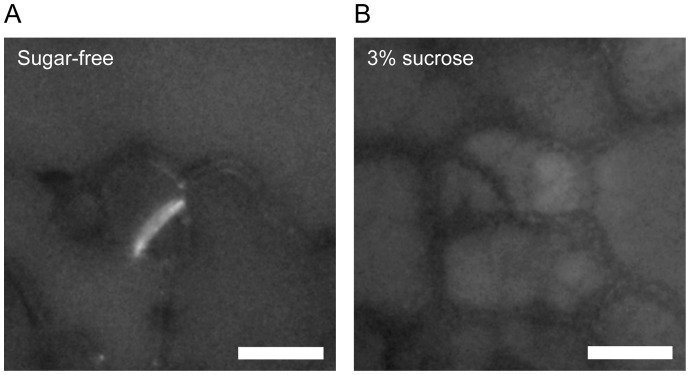
The spatial distribution of plant stomata is a model system to study epidermal cell pattern formation. Molecular genetic approaches have identified several key genes required for stomatal distribution patterning, but environmental conditions that perturb the stomatal spacing distribution have not yet been identified. We found that immersing hydroponic cultures in 1-5% sucrose solution induced abnormally clustered stomata in the cotyledons of Arabidopsis seedlings. Clustered stomata were also induced by treatment with glucose or fructose solution but not by mannitol solution, suggesting that osmotic stress was not a cause of the disturbed stomatal patterns. Stomatal lineage cell-specific enhancer trap lines revealed that the sugar solution treatment led to ectopic expression of stomatal lineage cell-specific genes in non-stomatal lineage cells. Aniline blue staining also showed that there was reduced deposition of callose, a plant cell wall component, in new cell walls during formation of stomatal precursor cells (meristemoids). These results suggested that the immersion treatment with sugar solution permitted ectopic guard cell differentiation through dysfunction of the cell wall dividing stomatal- and non-stomatal lineage cells. Our simple induction system for clustered stomata provides a suitable tool for further studies to investigate the one-cell-spacing rule during plant stomatal development.


Conflict of interest statement

Competing Interests: The authors have declared that no competing interests exist.

Figures

Figure 1
Figure 1. Effects of immersion in sugar-free, 3% sucrose, glucose, fructose and mannitol solutions on Arabidopsis seedlings and cotyledon guard cell distributions.
(A) Representative 14 day-old seedlings. Scale bar  = 1 cm. (B) Fluorescence microscopy images of abaxial cotyledon epidermis stained with FM4-64 dye. Representative images from 10–24 independent seedlings were shown. The maximum intensity projections were constructed with a 0.5 µm step size. Scale bars  = 20 μm.
Figure 2
Figure 2. Percentage of stomata in each cluster size class.
Abaxial cotyledons from 12- to 15 day-old seedlings grown in sugar-free, 1, 3 or 5% sucrose (A), and 3% glucose, fructose or mannitol (B) solutions were subjected to quantitative analysis. Data are mean values of 20–68 independent observations. Significance with sugar-free conditions was determined using Mann–Whitney's U-test. p-value *<0.0001. Total number of stomata counted: n = 281–1843.
Figure 3
Figure 3. Effects of sucrose exposure on stomatal lineage cell markers.
(A and B) Mature guard cell marker E1728-labeled and FM4-64-stained cotyledon epidermis from sugar-free control (A) and 1% sucrose (B) treatments. (C and D) Stomatal cell lineage marker E1627-labeled and FM4-64-stained cotyledon epidermis from sugar-free control (C) and 3% sucrose (D) treatments. Representative images from 10–15 independent seedlings were shown. Note that the jigsaw puzzle-shaped epidermal cells were labeled with E1627 in the sucrose treatment but not in the sugar-free control. Scale bars  = 20 μm.
Figure 4
Figure 4. Aniline blue staining of cotyledon epidermal cells.
Four day-old cotyledons immersed in sugar-free (A) or 3% sucrose (B) solutions were stained with 0.02% aniline blue for 1 week. Representative images from 24 (sugar-free) and 38 (3% sucrose) independent seedlings were shown. Note that aniline blue fluorescence was clearly detected in new cell walls forming in meristemoids immersed in sucrose-free solutions but not in 3% sucrose solutions. Scale bars  = 10 μm.

References

    1. Sachs T (1991) Pattern formation in plant tissues. Cambridge University Press, Cambridge, UK.
    1. Hara K, Kajita R, Torii KU, Bergmann DC, Kakimoto T (2007) The secretory peptide gene EPF1 enforces the stomatal one-cell-spacing rule. Genes Dev 21: 1720–1725. - PMC - PubMed
    1. Hara K, Yokoo T, Kajita R, Onishi T, Yahata S, et al. (2009) Epidermal cell density is autoregulated via a secretory peptide, EPIDERMAL PATTERNING FACTOR 2 in Arabidopsis leaves. Plant Cell Physiol 50: 1019–1031. - PubMed
    1. Yang M, Sack FD (1995) The too many mouths and four lips mutations affect stomatal production in Arabidopsis . Plant Cell 7: 2227–2239. - PMC - PubMed
    1. Geisler M, Nadeau J, Sack FD (2000) Oriented asymmetric divisions that generate the stomatal spacing pattern in Arabidopsis are disrupted by the too many mouths mutation. Plant Cell 12: 2075–2086. - PMC - PubMed

Publication types

MeSH terms

LinkOut - more resources