. 2013 Sep 5;8(9):e72907.
doi: 10.1371/journal.pone.0072907. eCollection 2013.

Age of onset of RNA toxicity influences phenotypic severity: evidence from an inducible mouse model of myotonic dystrophy (DM1)

Affiliations

Age of onset of RNA toxicity influences phenotypic severity: evidence from an inducible mouse model of myotonic dystrophy (DM1)

Jordan T Gladman et al. PLoS One. .

Abstract

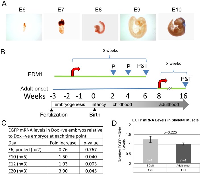
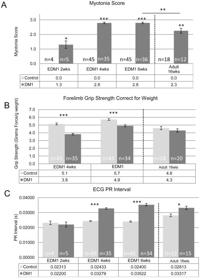
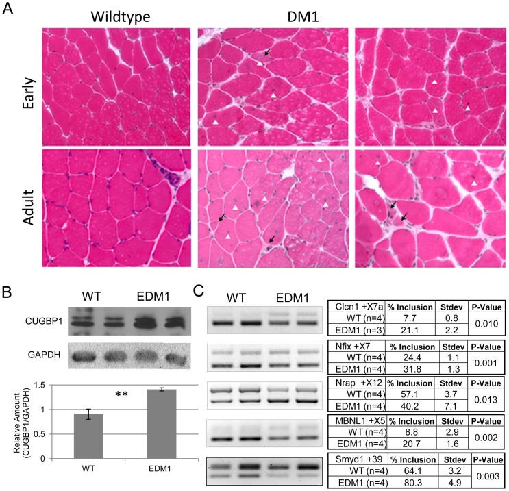
Myotonic dystrophy type 1 (DM1) is the most common muscular dystrophy in adults. It is caused by an expanded (CTG)n tract in the 3' UTR of the Dystrophia Myotonica Protein Kinase (DMPK) gene. This causes nuclear retention of the mutant mRNA into ribonuclear foci and sequestration of interacting RNA-binding proteins (such as muscleblind-like 1 (MBNL1)). More severe congenital and childhood-onset forms of the disease exist but are less understood than the adult disease, due in part to the lack of adequate animal models. To address this, we utilized transgenic mice over-expressing the DMPK 3' UTR as part of an inducible RNA transcript to model early-onset myotonic dystrophy. In mice in which transgene expression was induced during embryogenesis, we found that by two weeks after birth, mice reproduced cardinal features of myotonic dystrophy, including myotonia, cardiac conduction abnormalities, muscle weakness, histopathology and mRNA splicing defects. Notably, these defects were more severe than in adult mice induced for an equivalent period of exposure to RNA toxicity. Additionally, the utility of the model was tested by over-expressing MBNL1, a key therapeutic strategy being actively pursued for treating the disease phenotypes associated with DM1. Significantly, increased MBNL1 in skeletal muscle partially corrected myotonia and splicing defects present in these mice, demonstrating the responsiveness of the model to relevant therapeutic interventions. Furthermore, these results also represent the first murine model for early-onset DM1 and provide a tool to investigate the effects of RNA toxicity at various stages of development.


Conflict of interest statement

Competing Interests: The authors have declared that no competing interests exist.

Figures

Figure 1
Figure 1. Generation of an early-induction DM1 mouse model.
A. Whole mount in-situ hybridization of embryos at different time points showing onset of Dmpk expression (represented by the blue color) is visible only by the 10th day of development (E10). B. Schematic diagram representing the protocol used to generate and study the EDM1 mice and adult-onset mice. The green line represents administration of doxycycline. The red arrow represents induction of the transgene resulting in an increase in toxic RNA. Arrowheads with a P represent phenotypic analysis while the final arrowhead P&T represents phenotypic analysis and tissues collection. C. Induction of the transgene was measured in whole embryos by qRT-PCR at varying time points as indicated. Significant doxycycline mediated transgene induction starts at embryonic day 10, similar to results for endogenous Dmpk (see Figure 1A.). D. Graph of toxic RNA levels in skeletal muscle shows no significant difference between the EDM1 and adult-onset DM1 mice. Number of samples (n) provided on the graph, averages listed in the tables below the graphs, error bars are SEM. Two Sample T-Test was used to determine statistical significance.
Figure 2
Figure 2. Characterization of EDM1 mice.
A. Myotonia was measured and scored on a 3 point scale with 0 being no myotonia and 3 being strong, persistent, and prevalent myotonia. EDM1 mice show myotonia as early as 2 weeks of age and reached maximal severity by 4 weeks. B. EDM1 mice have a reduction in grip strength as early as 4 weeks of age while the adult-onset mice did not show a significant reduction. C. PR interval was measured in the EDM1 mice. No statistical difference was measured at 2 weeks of age; however the subsequent time points showed a significant lengthening of the interval. Similar but less severe results were observed in the adult-onset DM1 model. Number of samples (n) provided on the graph, averages listed in the tables below the graphs, error bars are SEM. Two Sample T-Test was used to determine statistical significance with p-values of 0.05 (*), 0.01 (**) or <0.001 (***) displayed.
Figure 3
Figure 3. Histopathology, RNA splicing and CUGBP1 are all altered in the EDM1 mice.
A. H&E staining shows uniform fibers and peripheral nuclei in the wildtype sections. With RNA toxicity, there is an increase in central nuclei (white arrowhead), increase in fiber size variability, and of atrophic fibers (black arrow). B. CUGBP1 is increased in the skeletal muscles of EDM1 mice. C. Clcn1, Nfix, Nrap, Mbnl1, and SmyD1 splicing alterations found in human DM1 are present in the EDM1 mice. Number of samples (n) provided in the tables, averages listed in the tables along with standard deviation. Two Sample T-Test was used to determine statistical significance with p-values displayed.
Figure 4
Figure 4. Age of onset of toxic RNA expression affects DM1 disease phenotypes.
RNA toxicity animals were compared to control groups for both A) PR interval and B) grip strength. Effects on grip strength and PR interval were more severe in the early induction mice (EDM1) compared to the adult onset mice (Adult-onset DM1). Averages are listed in the tables below the graphs. Error bars are SEM. Groups that do not share a letter are significantly different (p<0.05) using a One-Way ANOVA with Tukey’s Multiple Comparisons. N = 10 or more for each control group used to generate the control average and N = 17 or more for each DM1 group.
Figure 5
Figure 5. Partial Rescue of EDM1 phenotype in skeletal muscle by over-expression of MBNL1.
A. RNA-IP using a monoclonal antibody against MBNL1 pulls down the GFP-DMPK 3′UTR mRNA in both the EDM1 and adult-onset DM1 mice but not mice expressing EGFP only. Beads, IgG and RT -ve controls were used to show MBNL1 specificity. EGFP was amplified to detect the transgene. B. MBNL1 was confirmed to be up-regulated by qRT-PCR and C. by western blot analysis in the MBNL1 treated leg but not the EGFP treated leg. D. Median myotonia score in EDM1 mice expressing MBNL1 and EGFP in their TA muscles shows a slight reduction in myotonia due the presence of MBNL1 but not EGFP. E. mRNA splicing was also partially improved for SmyD1. Clcn1-exon7a showed no change. Number of samples (n) provided along with average or median in the tables along with standard deviation. Two Sample T-Test was used to determine statistical significance with p-values displayed.

References

    1. Dansithong W, Jog SP, Paul S, Mohammadzadeh R, Tring S, et al. (2011) RNA steady-state defects in myotonic dystrophy are linked to nuclear exclusion of SHARP. EMBO Rep. 12: 735–742. - PMC - PubMed
    1. Yadava RS, Frenzel-McCardell CD, Yu Q, Srinivasan V, Tucker AL, et al. (2008) RNA toxicity in myotonic muscular dystrophy induces NKX2–5 expression. Nat Genet. 40: 61–68. - PMC - PubMed
    1. Ravel-Chapuis A, Belanger G, Yadava RS, Mahadevan MS, DesGroseillers L, et al. (2012) The RNA-binding protein Staufen1 is increased in DM1 skeletal muscle and promotes alternative pre-mRNA splicing. J Cell Biol. 196: 699–712. - PMC - PubMed
    1. Philips AV, Timchenko LT, Cooper TA (1998) Disruption of splicing regulated by a CUG-binding protein in myotonic dystrophy. Science. 280: 737–741. - PubMed
    1. Day JW, Ranum LP (2005) Genetics and molecular pathogenesis of the myotonic dystrophies. Curr Neurol Neurosci Rep. 5: 55–59. - PubMed

Publication types

MeSH terms

LinkOut - more resources