. 2013 Sep 6;8(9):e73146.
doi: 10.1371/journal.pone.0073146. eCollection 2013.

c-Myc is essential to prevent endothelial pro-inflammatory senescent phenotype

Affiliations

c-Myc is essential to prevent endothelial pro-inflammatory senescent phenotype

Victoria Florea et al. PLoS One. .

Abstract

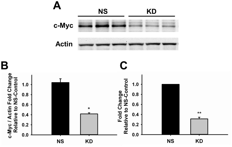
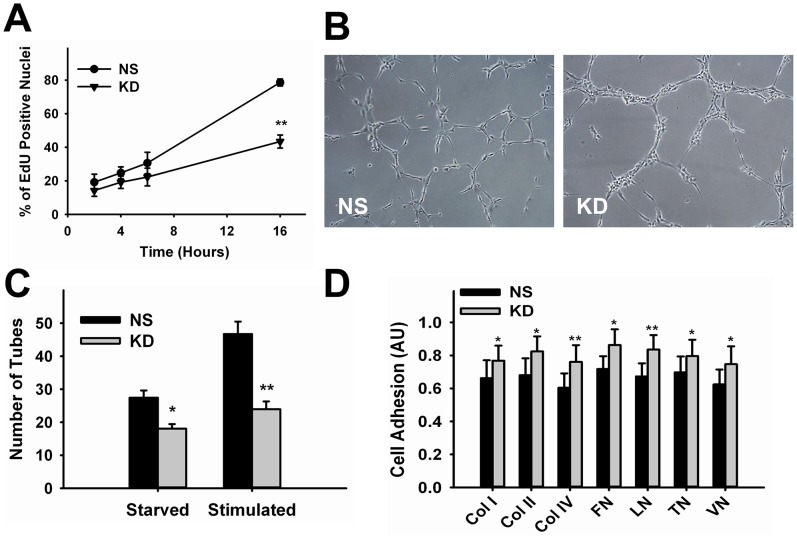
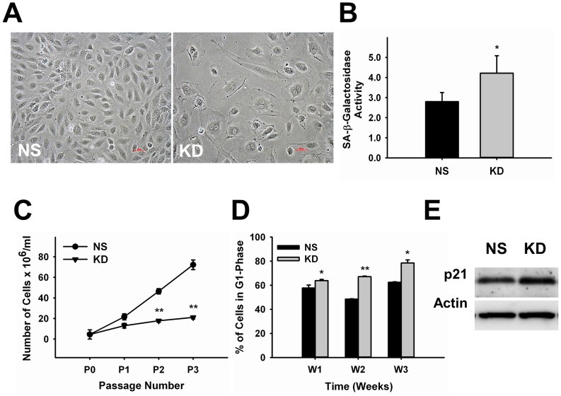
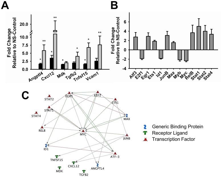
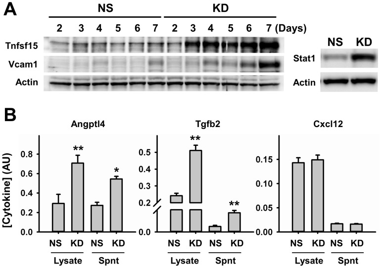
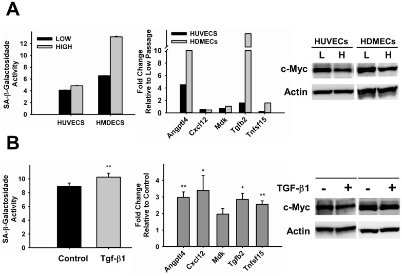
The proto-oncogene c-Myc is vital for vascular development and promotes tumor angiogenesis, but the mechanisms by which it controls blood vessel growth remain unclear. In the present work we investigated the effects of c-Myc knockdown in endothelial cell functions essential for angiogenesis to define its role in the vasculature. We provide the first evidence that reduction in c-Myc expression in endothelial cells leads to a pro-inflammatory senescent phenotype, features typically observed during vascular aging and pathologies associated with endothelial dysfunction. c-Myc knockdown in human umbilical vein endothelial cells using lentivirus expressing specific anti-c-Myc shRNA reduced proliferation and tube formation. These functional defects were associated with morphological changes, increase in senescence-associated-β-galactosidase activity, upregulation of cell cycle inhibitors and accumulation of c-Myc-deficient cells in G1-phase, indicating that c-Myc knockdown in endothelial cells induces senescence. Gene expression analysis of c-Myc-deficient endothelial cells showed that senescent phenotype was accompanied by significant upregulation of growth factors, adhesion molecules, extracellular-matrix components and remodeling proteins, and a cluster of pro-inflammatory mediators, which include Angptl4, Cxcl12, Mdk, Tgfb2 and Tnfsf15. At the peak of expression of these cytokines, transcription factors known to be involved in growth control (E2f1, Id1 and Myb) were downregulated, while those involved in inflammatory responses (RelB, Stat1, Stat2 and Stat4) were upregulated. Our results demonstrate a novel role for c-Myc in the prevention of vascular pro-inflammatory phenotype, supporting an important physiological function as a central regulator of inflammation and endothelial dysfunction.


Conflict of interest statement

Competing Interests: The authors have declared that no competing interests exist.

Figures

Figure 1
Figure 1. Knockdown of c-Myc in Endothelial Cells.
A. Representative western blot image of control and knockdown samples of three independent experiments. Actin was used as loading control (n = 3) B. Quantification of c-Myc knockdown at protein level by densitometry analysis of western blot shown in A (n = 3). C. Quantitative RT-PCR analysis of control and knockdown samples (n = 6). Results are expressed as fold-change relative to NS-Control. Data were normalized to actin endogenous control gene (n = 4). *p<0.01, **p<0.001. NS, control; KD, knockdown.
Figure 2
Figure 2. Effect of c-Myc Knockdown in Endothelial Cell Proliferation and Morphogenesis.
A. Time-course analysis of DNA synthesis in control and knockdown cells from 2–16 hours (n = 3–7). B. Representative images of stimulated control and knockdown cells collected 5 hours after incubation in Matrigel. C. Quantitative analysis of tube formation (n = 3–6). D. Quantification of control and knockdown cell adhesion to extracellular-matrix proteins (n = 5). *p<0.05, **p<0.005. NS, control; KD, knockdown; Col, collagen; FN, fibronectin; LN, laminin; TN, tenascin; VN, vitronectin. Magnification = 10X.
Figure 3
Figure 3. c-Myc Knockdown in Endothelial Cells Leads to Senescence.
A. Representative images showing morphological changes associated with senescence in c-Myc deficient endothelial cells compared to NS-control. B. Quantitative analysis of senescence-associated-β-galactosidase activity in control and knockdown endothelial cells (n = 8). C. Determination of population doubling along passage (n = 8). D. Time-course cell cycle analysis of control and knockdown endothelial cells showing accumulation in G1-phase (n = 3–7). E. Representative western blot image of control and knockdown cells showing upregulation of the cell cycle inhibitor p21, 3–4 days after knockdown. Actin was used as loading control (n = 3). *p<0.05, **p<0.005. NS, control; KD, knockdown. Magnification = 20X.
Figure 4
Figure 4. Gene Expression Analysis of Inflammatory Mediators and Transcription Factors after c-Myc knockdown.
A. Time-dependent changes in the expression of pro-inflammatory genes Angptl4, Cxcl12, Mdk, Tgfb2, Tnfsf15 and Vcam1 in endothelial cells three (black bars) and six (gray bars) days after c-Myc knockdown. B. Transcription factor gene expression profiling 6 days after c-Myc knockdown. Results are expressed as fold-change relative to NS-Control. Gene expression data were normalized to at least two endogenous control genes. C. Network pathway analysis of inflammatory mediators and transcription factors induced by c-Myc knockdown. (n = 4–5) (*p<0.05, **p<0.005).
Figure 5
Figure 5. Protein Expression Analysis of Pro-Inflammatory Markers.
A. Representative western blot image showing time-dependent changes in the expression of Tnfsf15, Vcam1, and Stat1 after c-Myc knockdown. Expression of Stat1 was analyzed 6 days after knockdown. Actin was used as loading control (n = 4). B. Expression analysis of Angptl4, Tgfb2 and Cxcl12 in cell lysates and concentrated supernatants (Spnt) by ELISA 6 days after knockdown (n = 4). NS, control; KD, knockdown. *p<0.05, **p<0.005.
Figure 6
Figure 6. Analysis of c-Myc Expression in Endothelial Cells Undergoing Replicative- or Stress-Induced Senescence.
A. HUVECs and HMDECs at low (P6) and high (P12–11) passages. B. HUVECs treated with TGF-β1 for three days (n = 4–6). All samples were analyzed for SA-β-galactosidase activity (right panel), expression of pro-inflammatory markers genes (middle panel) and c-Myc expression by Western Blot (right panel). Actin was used as loading control. L = Low, H = High. *p<0.05, **p<0.005.
Figure 7
Figure 7. Effect of TNF-α in c-Myc deficient HUVECs Pro-inflammatory Response.
Control and Knockdown HUVECs were treated with TNF-α for three hours and analyzed for expression of pro-inflammatory genes by RT-PCR. Results are expressed as fold-change relative to NS-Control. Data were normalized to at least two endogenous control genes. *p<0.05, **p<0.005. ***p<0.0005, (n = 3).

References

    1. Pelengaris S, Khan M (2003) The many faces of c-MYC. Arch Biochem Biophys 416: 129–136. - PubMed
    1. Coller HA, Grandori C, Tamayo P, Colbert T, Lander ES, et al. (2000) Expression analysis with oligonucleotide microarrays reveals that MYC regulates genes involved in growth, cell cycle, signaling, and adhesion. Proc Natl Acad Sci U S A 97: 3260–3265. - PMC - PubMed
    1. Nie Z, Hu G, Wei G, Cui K, Yamane A, et al. (2012) c-Myc is a universal amplifier of expressed genes in lymphocytes and embryonic stem cells. Cell 151: 68–79. - PMC - PubMed
    1. Napoli C, Lerman LO, de Nigris F, Sica V (2002) c-Myc oncoprotein: a dual pathogenic role in neoplasia and cardiovascular diseases? Neoplasia 4: 185–190. - PMC - PubMed
    1. de Nigris F, Balestrieri ML, Napoli C (2006) Targeting c-Myc, Ras and IGF cascade to treat cancer and vascular disorders. Cell Cycle 5: 1621–1628. - PubMed

Publication types

MeSH terms