. 2013 Sep 9;8(9):e73562.
doi: 10.1371/journal.pone.0073562. eCollection 2013.

Paraquat poisoning induces TNF-α-dependent iNOS/NO mediated hyporesponsiveness of the aorta to vasoconstrictors in rats

Affiliations

Paraquat poisoning induces TNF-α-dependent iNOS/NO mediated hyporesponsiveness of the aorta to vasoconstrictors in rats

Rosária D Aires et al. PLoS One. .

Abstract

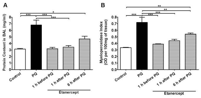
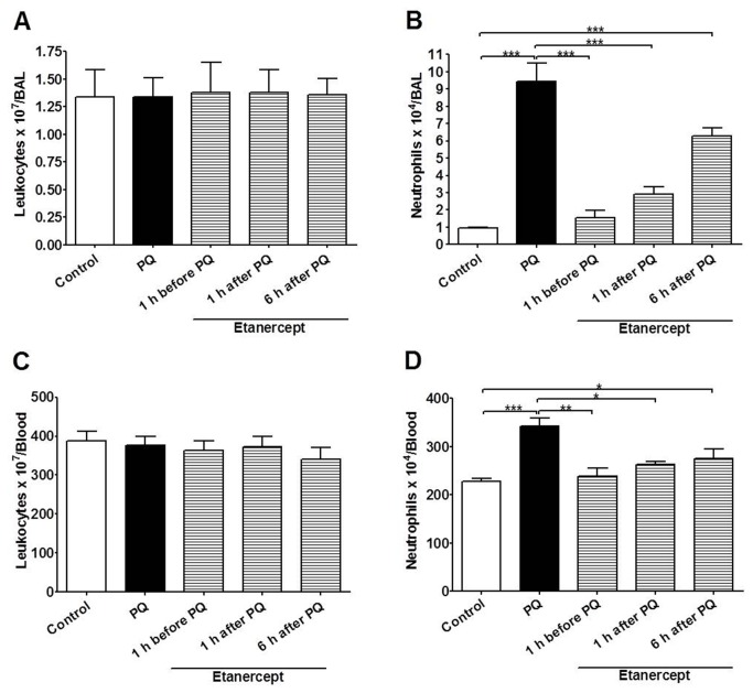
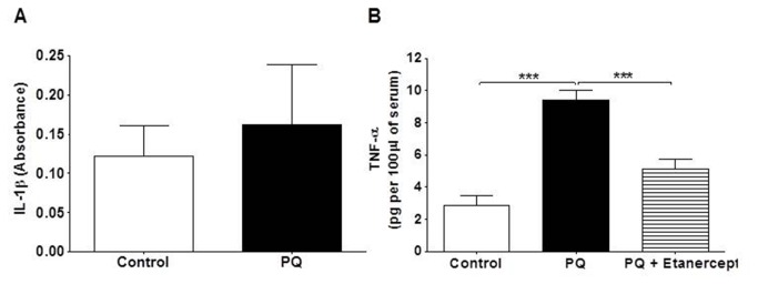
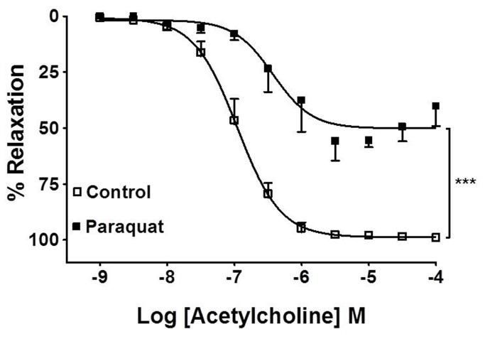
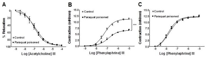
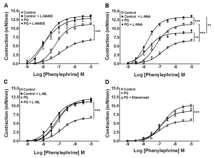
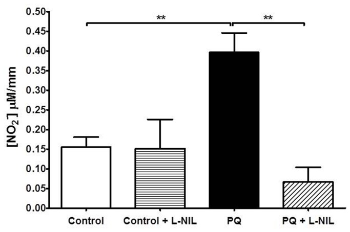
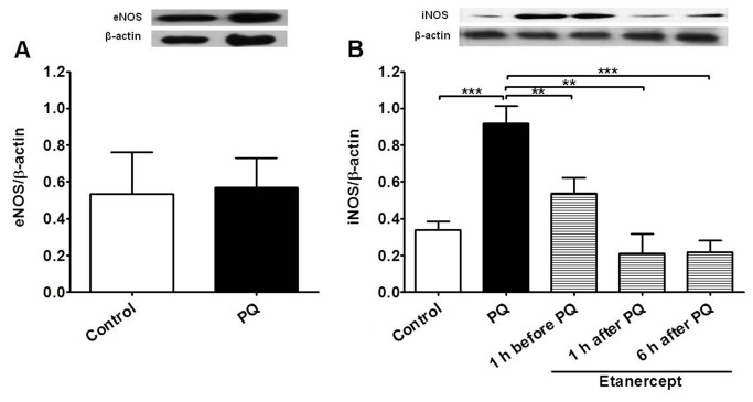
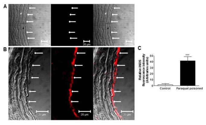
Paraquat is a toxic herbicide that may induce acute lung injury, circulatory failure and death. The present work aimed at investigating whether there is systemic inflammation and vascular dysfunction after paraquat exposure and whether these parameters were related. There was neutrophilia and accumulation of neutrophils in lung and bronchoalveolar lavage of animals given paraquat. This was associated with an increase in serum levels of TNF-α. In rats given paraquat, the relaxant response of aortic rings to acetylcholine was not modified but the contractile response to phenylephrine was greatly reduced. Endothelium removal or treatment with non-selective (L-NAME) or selective (L-NIL) inhibitors of inducible nitric oxide synthase (iNOS) restored contraction of aortas. There was greater production of nitric oxide (NO), which was restored to basal level by L-NIL, and greater expression of iNOS in endothelial cells, as seen by Western blot analyses and confocal microscopy. Blockade of TNF-α reduced pulmonary and systemic inflammation and vascular dysfunction. Together, our results clearly show that paraquat causes pulmonary and systemic inflammation, and vascular dysfunction in rats. Vascular dysfunction is TNF-α dependent, associated with enhanced expression of iNOS in aortic endothelial cells and greater NO production, which accounts for the decreased responsiveness of aortas to vasoconstrictors. Blockers of TNF-α may be useful in patients with paraquat poisoning.


Conflict of interest statement

Competing Interests: The authors have declared that no competing interests exist.

Figures

Figure 1
Figure 1. Paraquat-poisoning produces TNF-α-mediated edema and neutrophil accumulation in the lungs.
Pulmonary permeability (A) and neutrophil accumulation in the lungs (B) of control, paraquat-poisoned (PQ) and paraquat-poisoned rats after treatment with etanercept. The total protein content was used as an index of protein leakage due to alveolar-microvascular membrane injury. Myeloperoxidase (MPO) activity was used as an index of neutrophil influx into the lungs. Results are shown as protein content (mg/ml) or MPO index and represent the mean ± SE of five animals in each group. *p<0.05; **p<0.01 and ***p<0.001.
Figure 2
Figure 2. Paraquat produces accumulation of neutrophils in bronchoalveolar lavage (BAL) and neutrophilia.
Effect of paraquat-poisoning (PQ) on total number of leukocytes and neutrophil in BAL (A, B) or in blood (C, D). TNF-α blockade by etanercept restore neutrophil number to the control level in BAL (B) and blood (D). Results are shown as the mean ± SE of five animals in each group. *p<0.05; **p<0.01 and ***p<0.001.
Figure 3
Figure 3. TNF-α concentration is increased in the serum of paraquat-poisoned animals.
Effect of paraquat-poisoning (PQ) on (A) IL-1β (n=11-12) and (B) TNF-α (n=5) concentration in serum. Results are shown as the mean ± SE ***p<0.001.
Figure 4
Figure 4. In vitro effect of paraquat in aortic rings.
Effects of in vitro treatment of endothelium-intact aortic rings with paraquat (5 µM) for 20 min on vasodilation induced by acetylcholine. The values are mean ± SE from five experiments. ***p<0.001.
Figure 5
Figure 5. Ex vivo vascular effects of paraquat-poisoning.
Vasodilator effect of acetylcholine in endothelium-intact aortic rings from control and paraquat-poisoned animals (A). Contractile response to phenylephrine in endothelium-intact (B) and endothelium-denuded (C) aortic rings from control and paraquat-poisoned animals. The values are mean ± SE from five experiments. ***p<0.001.
Figure 6
Figure 6. Nitric oxide and TNF-α mediate vascular dysfunction in paraquat-poisoned animals.
Effect of (A) L-NAME (300 µM), (B) L-NNA (1 µM), (C) L-NIL (10 µM) and (D) in vivo treatment with etanercept on phenylephrine-induced contraction in endothelium-intact aortic rings from control and paraquat-poisoned (PQ) animals. The values are mean ± SE from five experiments. **p<0.01; ***p<0.001.
Figure 7
Figure 7. Paraquat-poisoning increases basal production of nitric oxide (NO) in the aorta.
Basal production of NO in endothelium-intact aortic rings removed from control and paraquat-poisoned (PQ) animals in the presence or in the absence of L-NIL (10 µM). Results are shown as the mean ± SE from at least five experiments. **p<0.01.
Figure 8
Figure 8. TNF-α-dependent increases of iNOS expression in aortas from paraquat-poisoned animals.
Western blot analysis of (A) eNOS in endothelium-intact aortic rings from control and paraquat-poisoned (PQ) rats and (B) iNOS in endothelium-intact aortic rings from control, paraquat-poisoned and paraquat-poisoned animals after in vivo treatment with etanercept. Bar graphs represent mean ± SE of five experiments. Images are representative blots from four separate experiments. **p<0.01 and ***p<0.001.
Figure 9
Figure 9. Paraquat-poisoning increases expression of iNOS in the vascular endothelial cell layer.
Imunofluorescence detection of iNOS (A) in endothelium-intact aortic rings from control and (B) paraquat-poisoned animals. Immunostaining for iNOS is shown only in endothelial cells (arrows) of vessels from paraquat-poisoned animals. (C) Graphical representation of the relative iNOS fluorescence intensity in endothelial cells. Images are representative of five animals for each group. ***p<0.001.

References

    1. Dinis-Oliveira RJ, Duarte JA, Sánchez-Navarro A, Remião F, Bastos ML et al. (2008) Paraquat poisonings: mechanisms of lung toxicity, clinical features, and treatment. Crit Rev Toxicol 38: 13-71. doi:10.1080/10408440701669959. PubMed: 18161502. - DOI - PubMed
    1. Mainwaring G, Lim FL, Antrobus K, Swain C, Clapp M et al. (2006) Identification of early molecular pathways affected by paraquat in rat lung. Toxicology 225: 157-172. doi:10.1016/j.tox.2006.05.017. PubMed: 16854511. - DOI - PubMed
    1. Rose MS, Lock EA, Smith LL, Wyatt I (1976) Paraquat accumulation: tissue and species specificity. Biochem Pharmacol 25: 419-423. doi:10.1016/0006-2952(76)90344-0. PubMed: 820354. - DOI - PubMed
    1. Rose MS, Smith LL, Wyatt I (1974) Evidence for Energy-Dependent Accumulation of Paraquat into Rat Lung. Nature 252: 314-315. doi:10.1038/252314a0. PubMed: 4431454. - DOI - PubMed
    1. Day BJ, Patel M, Calavetta L, Chang LY, Stamler JS (1999) A mechanism of paraquat toxicity involving nitric oxide synthase. Proc Natl Acad Sci U S A 96: 12760-12765. doi:10.1073/pnas.96.22.12760. PubMed: 10535996. - DOI - PMC - PubMed

Publication types

LinkOut - more resources