

The long coiled-coil protein NECC2 is associated to caveolae and modulates NGF/TrkA signaling in PC12 cells [corrected]
- PMID: 24040018
- PMCID: PMC3765260
- DOI: 10.1371/journal.pone.0073668
The long coiled-coil protein NECC2 is associated to caveolae and modulates NGF/TrkA signaling in PC12 cells [corrected]
Erratum in
- PLoS One. 2013;8(9). doi:10.1371/annotation/55c4c5bd-99e8-435a-94f4-7e151043bb51
Abstract
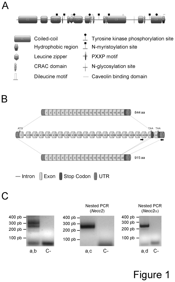
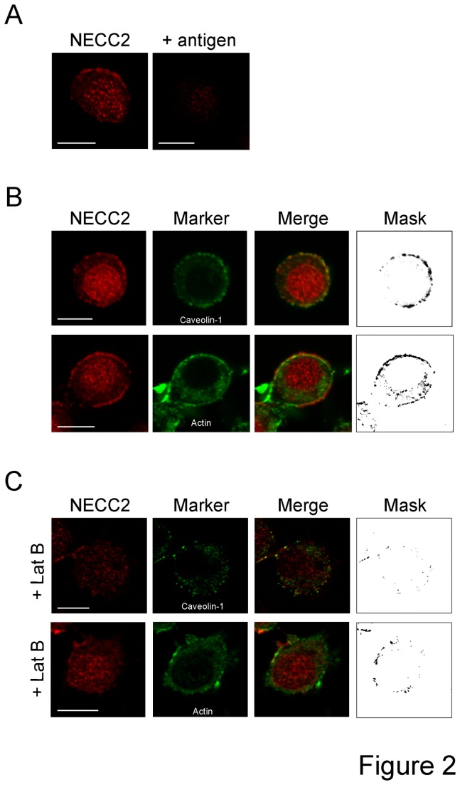
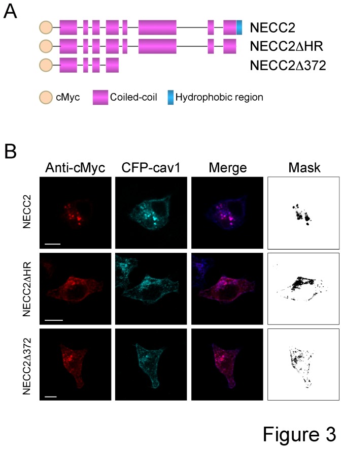
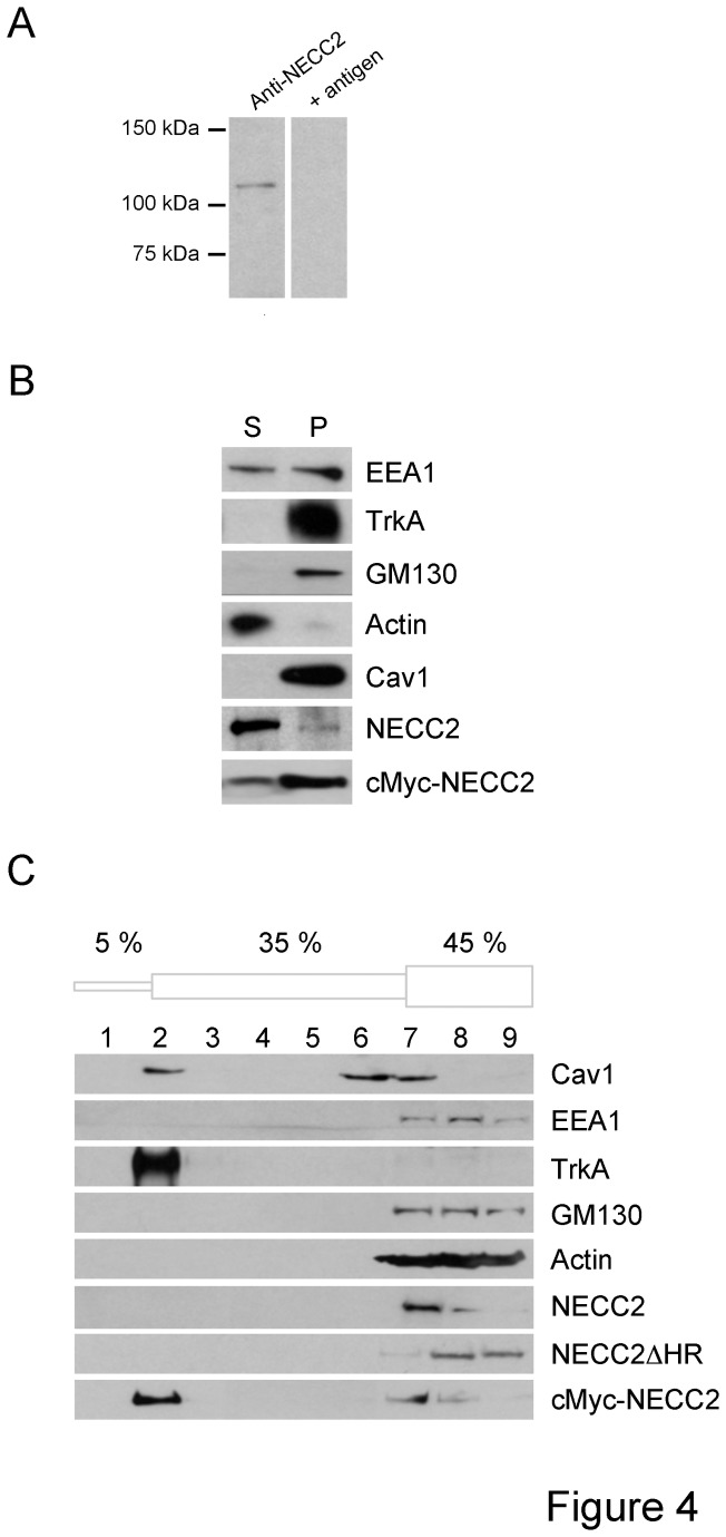
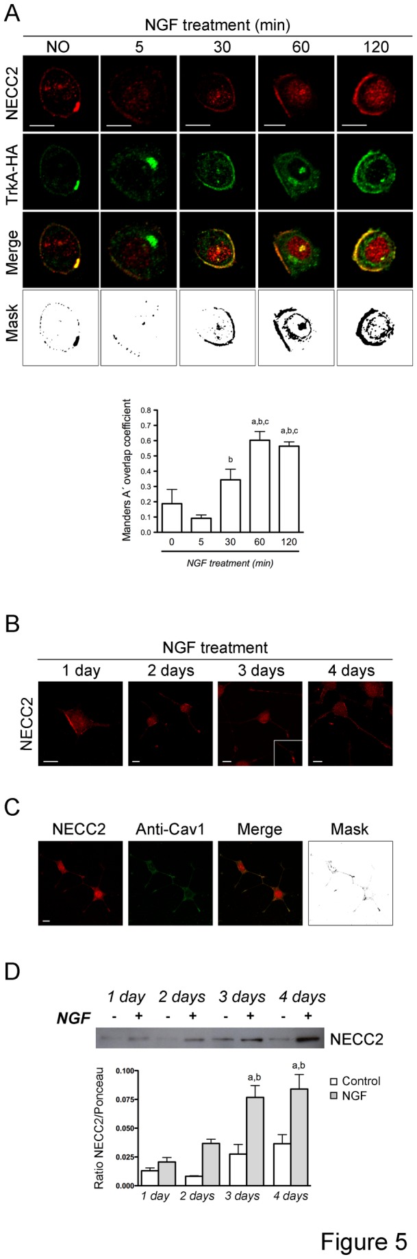
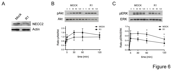
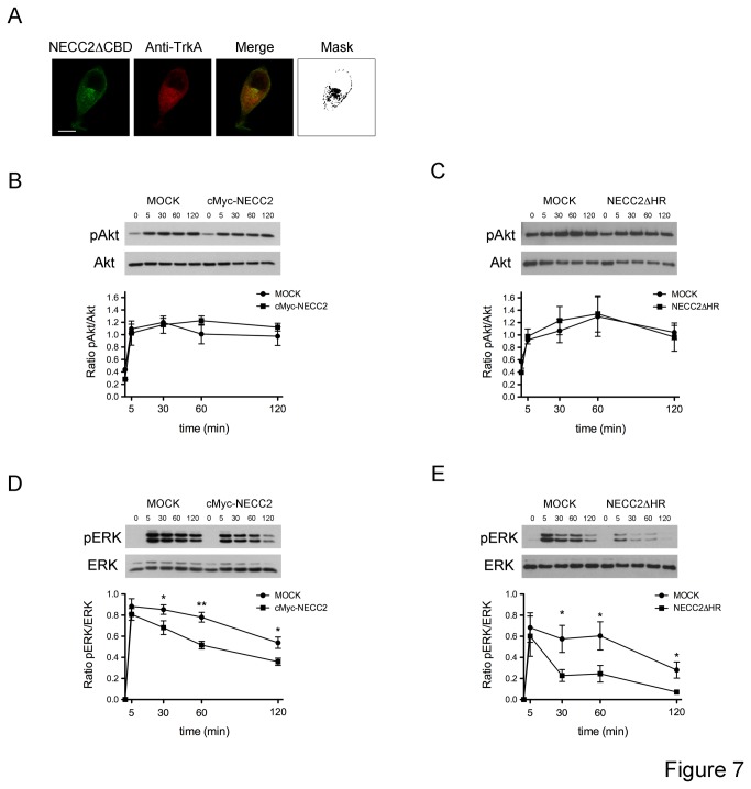
TrkA-mediated NGF signaling in PC12 cells has been shown to be compartimentalized in specialized microdomains of the plasma membrane, the caveolae, which are organized by scaffold proteins including the member of the caveolin family of proteins, caveolin-1. Here, we characterize the intracellular distribution as well as the biochemical and functional properties of the neuroendocrine long coiled-coil protein 2 (NECC2), a novel long coiled-coil protein selectively expressed in neuroendocrine tissues that contains a predicted caveolin-binding domain and displays structural characteristics of a scaffolding factor. NECC2 distributes in caveolae, wherein it colocalizes with the TrkA receptor, and behaves as a caveolae-associated protein in neuroendocrine PC12 cells. In addition, stimulation of PC12 cells with nerve growth factor (NGF) increased the expression and regulated the distribution of NECC2. Interestingly, knockdown as well as overexpression of NECC2 resulted in a reduction of NGF-induced phosphorylation of the TrkA downstream effector extracellular signal-regulated kinases 1 and 2 (ERK1/ERK2) but not of Akt. Altogether, our results identify NECC2 as a novel component of caveolae in PC12 cells and support the contribution of this protein in the maintenance of TrkA-mediated NGF signaling.
Conflict of interest statement
Figures
Similar articles
-
The caveolae-associated coiled-coil protein, NECC2, regulates insulin signalling in Adipocytes.J Cell Mol Med. 2018 Nov;22(11):5648-5661. doi: 10.1111/jcmm.13840. Epub 2018 Aug 30. J Cell Mol Med. 2018. PMID: 30160359 Free PMC article.
-
PC12 cells have caveolae that contain TrkA. Caveolae-disrupting drugs inhibit nerve growth factor-induced, but not epidermal growth factor-induced, MAPK phosphorylation.J Biol Chem. 2000 Dec 1;275(48):37846-52. doi: 10.1074/jbc.M000487200. J Biol Chem. 2000. PMID: 10982788
-
Nerve Growth Factor Signaling from Membrane Microdomains to the Nucleus: Differential Regulation by Caveolins.Int J Mol Sci. 2017 Mar 24;18(4):693. doi: 10.3390/ijms18040693. Int J Mol Sci. 2017. PMID: 28338624 Free PMC article.
-
Methylmercury inhibits TrkA signaling through the ERK1/2 cascade after NGF stimulation of PC12 cells.Brain Res Dev Brain Res. 2004 Mar 22;149(1):53-61. doi: 10.1016/j.devbrainres.2003.10.017. Brain Res Dev Brain Res. 2004. PMID: 15013629
-
Nerve growth factor stimulates the concentration of TrkA within lipid rafts and extracellular signal-regulated kinase activation through c-Cbl-associated protein.Mol Cell Biol. 2007 Aug;27(16):5686-98. doi: 10.1128/MCB.01109-06. Epub 2007 Jun 4. Mol Cell Biol. 2007. PMID: 17548467 Free PMC article.
Cited by
-
Integrated Quantitative Transcriptome Maps of Human Trisomy 21 Tissues and Cells.Front Genet. 2018 Apr 24;9:125. doi: 10.3389/fgene.2018.00125. eCollection 2018. Front Genet. 2018. PMID: 29740474 Free PMC article.
-
The cytoskeletal protein septin 11 is associated with human obesity and is involved in adipocyte lipid storage and metabolism.Diabetologia. 2017 Feb;60(2):324-335. doi: 10.1007/s00125-016-4155-5. Epub 2016 Nov 19. Diabetologia. 2017. PMID: 27866222
-
Microarray Analysis Reveals Increased Transcriptional Repression and Reduced Metabolic Activity but Not Major Changes in the Core Apoptotic Machinery during Maturation of Sympathetic Neurons.Front Cell Neurosci. 2016 Mar 16;10:66. doi: 10.3389/fncel.2016.00066. eCollection 2016. Front Cell Neurosci. 2016. PMID: 27013977 Free PMC article.
-
Divergent Contribution of the Golgi Apparatus to Microtubule Organization in Related Cell Lines.Int J Mol Sci. 2022 Dec 19;23(24):16178. doi: 10.3390/ijms232416178. Int J Mol Sci. 2022. PMID: 36555819 Free PMC article.
-
The caveolae-associated coiled-coil protein, NECC2, regulates insulin signalling in Adipocytes.J Cell Mol Med. 2018 Nov;22(11):5648-5661. doi: 10.1111/jcmm.13840. Epub 2018 Aug 30. J Cell Mol Med. 2018. PMID: 30160359 Free PMC article.
References
-
- Steindler C, Li Z, Algarté M, Alcover A, Libri V et al. (2004) Jamip1 (marlin-1) defines a family of proteins interacting with janus kinases and microtubules. J Biol Chem 279: 43168-43177. doi:10.1074/jbc.M401915200. PubMed: 15277531. - DOI - PubMed
-
- Vidal RL, Ramírez OA, Sandoval L, Koenig-Robert R, Härtel S et al. (2007) Marlin-1 and conventional kinesin link GABAB receptors to the cytoskeleton and regulate receptor transport. Mol Cell Neurosci 35: 501-512. doi:10.1016/j.mcn.2007.04.008. PubMed: 17532644. - DOI - PubMed
-
- Vidal RL, Valenzuela JI, Luján R, Couve A (2009) Cellular and subcellular localization of Marlin-1 in the brain. BMC Neurosci 10: 37. doi:10.1186/1471-2202-10-S1-P37. PubMed: 19386132. - DOI - PMC - PubMed
-
- Vidal RL, Fuentes P, Valenzuela JI, Alvarado-Diaz CP, Ramírez OA et al. (2012) RNA interference of Marlin-1/Jakmip1 results in abnormal morphogenesis and migration of cortical pyramidal neurons. Mol Cell Neurosci 51: 1-11. doi:10.1016/j.mcn.2012.07.007. PubMed: 22828129. - DOI - PubMed
-
- Cruz-Garcia D, Vazquez-Martinez R, Peinado JR, Anouar Y, Tonon MC et al. (2007) Identification and characterization of two novel (neuro)endocrine long coiled-coil proteins. FEBS Lett 581: 3149-3156. doi:10.1016/j.febslet.2007.06.002. PubMed: 17572408. - DOI - PubMed
Publication types
MeSH terms
Substances
LinkOut - more resources
Full Text Sources
Other Literature Sources
Molecular Biology Databases
Miscellaneous
