. 2013 Sep 9;8(9):e73706.
doi: 10.1371/journal.pone.0073706. eCollection 2013.

Statins decrease lung inflammation in mice by upregulating tetraspanin CD9 in macrophages

Affiliations

Statins decrease lung inflammation in mice by upregulating tetraspanin CD9 in macrophages

Yingji Jin et al. PLoS One. .

Abstract

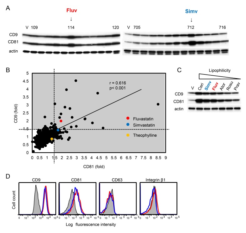
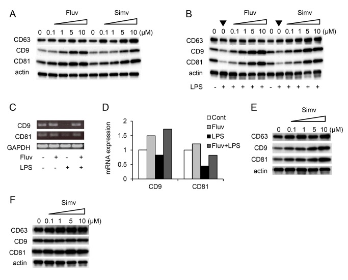
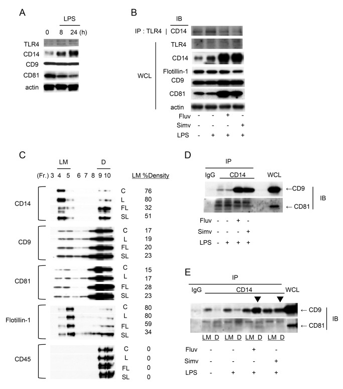
Tetraspanins organize protein complexes in tetraspanin-enriched membrane microdomains that are distinct from lipid rafts. Our previous studies suggested that reduction in the levels of tetraspanins CD9 and CD81 may be involved in the progression of inflammatory lung diseases, especially COPD. To search for agents that increase the levels of these tetraspanins, we screened 1,165 drugs in clinical use and found that statins upregulate CD9 and CD81 in RAW264.7 macrophages. The lipophilic statins, fluvastatin and simvastatin, reversed LPS-induced downregulation of CD9 and CD81, simultaneously preventing TNF-α and matrix metalloproteinase-9 production and spreading of RAW264.7 cells. These statins exerted anti-inflammatory effects in vitro in wild-type macrophages but not in CD9 knockout macrophages, and decreased lung inflammation in vivo in wild-type mice but not in CD9 knockout mice, suggesting that their effects are dependent on CD9. Mechanistically, the statins promoted reverse transfer of the LPS-signaling mediator CD14 from lipid rafts into CD9-enriched microdomains, thereby preventing LPS receptor formation. Finally, upregulation of CD9/CD81 by statins was related to blockade of GTPase geranylgeranylation in the mevalonate pathway. Our data underscore the importance of the negative regulator CD9 in lung inflammation, and suggest that statins exert anti-inflammatory effects by upregulating tetraspanin CD9 in macrophages.


Conflict of interest statement

Competing Interests: The study was performed on the basis of collaboration between Osaka University and Dainippon Sumitomo Co., Ltd. One of the authors (MI) is affiliated with the latter pharmaceutical company. The authors received funding to do the collaborative research from the company, but this does not alter the authors' adherence to all the PLOS ONE policies on sharing data and materials. There are no patents, products in development, or marketed products to declare.

Figures

Figure 1
Figure 1. Screening of a drug library for agents that upregulate CD9 or CD81 in RAW264.7 macrophages.
(A) RAW264.7 cells were cultured for 24 h in the absence (V, vehicle alone) and presence of each drug (10 µM). The cells were lysed, and levels of CD9 and CD81 were examined by immunoblotting. Blots of results with fluvastatin (Fluv) and simvastatin (Simv) are shown. Anti-actin blots show that comparable amounts of protein were loaded in each lane. (B) After testing 1,165 drugs, levels of CD9 and CD81 relative to actin were quantified by densitometry. Fold changes of the expression levels compared with vehicle alone were calculated and plotted. Drugs that increased the level of either CD9 or CD81 more than 1.5-fold compared with vehicle alone were regarded as positive. Correlation between fold changes in CD9 and CD81 levels was analyzed using Pearson’s correlation coefficient. (C) RAW264.7 cells were cultured in the absence (V) or presence of multiple statins (10 µM) and levels of CD9 and CD81 were examined by immunoblotting. The statins are arranged in order of decreasing lipophilicity. Ceri, cerivastatin; Simv, simvastatin; Fluv, fluvastatin; Ator, atorvastatin; Rosu, rosuvastatin; Prav, pravastatin. (D) RAW264.7 cells were cultured in the absence (shaded histograms) or presence (10 µM) of fluvastatin (open red histograms) and simvastatin (open blue histograms). Surface levels of CD9, CD63, CD81, and the integrin β1 subunit were analyzed by flow cytometry.
Figure 2
Figure 2. Fluvastatin and simvastatin increase CD9 and CD81 levels in RAW264.7 cells.
(A) RAW264.7 cells were cultured for 24 h in the absence or presence of increasing concentrations of fluvastatin (Fluv) or simvastatin (Simv). The cells were lysed, and levels of CD9, CD63, and CD81 were examined by immunoblotting. Anti-actin blots show that comparable amounts of protein were loaded in each lane. (B) RAW264.7 cells were untreated (-) or cultured in the absence or presence of increasing concentrations of fluvastatin or simvastatin and stimulated for 24 h with 0.1 µg/ml LPS (+). Levels of CD9, CD63, and CD81 were examined by immunoblotting. Note that LPS downregulates CD9 and CD81 in the absence of statins (arrowheads). (C) RAW264.7 cells were cultured in the absence (-) or presence of 3 µM fluvastatin (+), and unstimulated (-) or stimulated for 24 h with 1 µg/ml LPS (+). mRNA levels of CD9 and CD81 were examined by reverse transcription PCR. GAPDH is an internal loading control. (D) RAW264.7 cells were cultured in the absence or presence of fluvastatin, and unstimulated or stimulated with LPS. Control (Cont) was an untreated culture. mRNA levels of CD9 and CD81 were examined by real-time PCR. Data shown are from one representative of three similar experiments. (E) Human monocytic THP-1 cells were treated for 4 h with 1 µg/ml phorbol 12-myristate 13-acetate, allowed to attach to a plate, and then cultured in the absence or presence of increasing concentrations of simvastatin. Levels of CD9, CD63, and CD81 were examined by immunoblotting. (F) Mouse 3T3 fibroblasts were cultured in the absence or presence of increasing concentrations of simvastatin. Levels of CD9, CD63, and CD81 were examined by immunoblotting.
Figure 3
Figure 3. Fluvastatin and simvastatin prevent TNF-α and MMP-9 production and cell spreading in LPS-stimulated RAW264.7.
(A) RAW264.7 cells were untreated (-) or cultured for 24 h in the absence or presence of increasing concentrations of fluvastatin (Fluv) or simvastatin (Simv) and stimulated for 15 min with 1 µg/ml LPS (+). The cells were lysed and levels of IκBα were examined by immunoblotting. Anti-actin blots show that comparable amounts of protein were loaded in each lane. (B) RAW264.7 cells were cultured in the absence or presence of increasing concentrations of fluvastatin (top) or simvastatin (bottom), and stimulated for 5 h with 0.1 µg/ml LPS (+). Activities of MMP-9 in culture supernatants were analyzed by gelatin zymography. (C) RAW264.7 cells were untreated (-) or cultured in the absence or presence of increasing concentrations of fluvastatin (left) or simvastatin (right) and stimulated for 5 h with 0.1 µg/ml LPS (+). Concentrations of TNF-α in culture supernatants were measured by ELISA. (D) RAW264.7 cells were untreated (Cont, control) or cultured in the absence or presence of 5 µM fluvastatin or simvastatin and stimulated for 4 h with 0.1 µg/ml LPS, and then stained and photographed (upper panel). Scale bar, 100 µm. Percentages of spread cells were determined according to their longest diameters (lower panel). Each bar represents the mean ± SEM. P < 0.05; ⋆ ⋆ P < 0.01.
Figure 4
Figure 4. Statins transfer CD14 from lipid rafts into CD9-enriched microdomains.
(A) RAW264.7 cells were stimulated with 0.1 µg/ml LPS and, after the indicated times, the cells were lysed and protein levels were examined by immunoblotting. Anti-actin blots show that comparable amounts of protein were loaded in each lane. (B) RAW264.7 cells were untreated (-) or cultured for 24 h in the absence (-) or presence of 5 µM fluvastatin (Fluv) or simvastatin (Simv) (+) and stimulated for 2 h with 1 µg/ml LPS (+). Proteins in whole-cell lysate (WCL) and CD14 protein in immunoprecipitates (IP) with anti-TLR4 Ab were immunoblotted (IB). (C) RAW264.7 cells were treated as in B. Lysates of untreated (C, control) cultures or LPS-stimulated cultures in the absence (L) or presence of fluvastatin (FL) or simvastatin (SL) were fractionated by sucrose density gradients, and protein distributions were visualized by immunoblotting. The intensities of blots were quantified by densitometry, and percentages of density units of light membrane (LM) fractions are displayed to the right of the blots. Data shown are from one representative of three similar experiments. (D) Immunoblots of CD9 and CD81 proteins in whole-cell lysates and in immunoprecipitates with control IgG or anti-CD14 mAb. (E) Immunoblots of CD9 and CD81 proteins in whole-cell lysates and in immunoprecipitates with control IgG or anti-CD14 mAb from pooled LM fractions (4 and 5) and dense (D) fractions (9 and 10). In the presence of statins, more CD14/CD9 complexes were formed in dense fractions (arrowheads).
Figure 5
Figure 5. The anti-inflammatory effects of statins are CD9-dependent.
(A) BMDMs from WT mice were cultured for 24 h in the absence (-) or presence of 3 µM fluvastatin (Fluv) (+), and unstimulated (-) or stimulated for 24 h with 1 µg/ml LPS (+). The cells were lysed, and levels of CD9 and CD81 were examined by immunoblotting. Anti-actin blots show that comparable amounts of protein were loaded in each lane. (B) BMDMs from WT and CD9 KO mice were cultured in the absence or presence of the indicated concentrations of fluvastatin, and stimulated for 18 h with 10 µg/ml LPS (+). Activities of MMP-9 in culture supernatants were analyzed by gelatin zymography. (C) BMDMs from WT and CD9 KO mice were cultured in the absence (vehicle) or presence of 10 µM fluvastatin or simvastatin (Simv), and unstimulated (-) or stimulated for 18 h with 1 µg/ml LPS (+). Concentrations of TNF-α in culture supernatants were measured by ELISA. Each bar represents the mean ± SEM. P < 0.05; ⋆ ⋆ P < 0.01.
Figure 6
Figure 6. Statins protect mice from LPS-induced injury in a CD9-dependent manner.
(A) WT mice were repeatedly intraperitoneally injected with vehicle (-) or 30 mg/kg fluvastatin (Fluv), and unchallenged (-) or intraperitoneally challenged with 30 mg/kg LPS (+). After 48 h, BMDMs were isolated and the level of CD9 was examined by immunoblotting. Anti-actin blots show that comparable amounts of protein were loaded in each lane. (B) WT mice were treated as in A. CD9 mRNA levels in the BMDMs were examined by reverse transcription PCR. GAPDH is an internal loading control. (C) WT and CD9 KO mice were repeatedly intraperitoneally injected with vehicle (-) or 30 mg/kg fluvastatin (+), and intranasally challenged with 0.5 mg/kg LPS (+). After 24 h, activities of MMP-2 and MMP-9 in BALF were analyzed by gelatin zymography. (D) WT and CD9 KO mice were untreated (Cont, control) or intraperitoneally injected with vehicle or 20 mg/kg simvastatin (Simv) and intranasally challenged with 0.5 mg/kg LPS. After 4 days, total cells in BALF from the mice from each group (n = 9) were counted using a hemocytometer. Each bar represents the mean ± SEM. (E) WT and CD9 KO mice were treated as in D. Histological lung sections collected at 4 days were stained with hematoxylin and eosin. Scale bar, 100 µm. (F) WT and CD9 KO mice were intraperitoneally injected with vehicle or 20 mg/kg simvastatin, and intraperitoneally challenged with 40 mg/kg LPS. Survival of the mice from each group (n = 12) was monitored and analyzed by the Kaplan-Meier method. P < 0.05; ⋆ ⋆ P < 0.01.
Figure 7
Figure 7. Blockade of the mevalonate pathway increases CD9 and CD81.
(A) RAW264.7 cells were untreated (-) or treated for 48 h with 50 ng/ml TSA (+) in the absence (-) or presence of 50 µM theophylline or 0.5 µM fluvastatin (Fluv) (+). The cells were lysed, and levels of CD9 and CD81 were examined by immunoblotting. Anti-actin blots show that comparable amounts of protein were loaded in each lane. (B) The mevalonate pathway and inhibitors. n-BP, nitrogenous bisphosphonate. (C) RAW264.7 cells were cultured for 24 h in the presence of indicated concentrations of fluvastatin, simvastatin (Simv), zoledronate (Zol), or risedronate (Ris). Levels of CD9 and CD81 were examined by immunoblotting. (D) RAW264.7 cells were cultured for 24 h in the absence (V, vehicle alone) or presence of mevalonate (Mev), farnesyl pyrophosphate (FPP), squalene (Squ), or geranylgeranyl pyrophosphate (GGPP). Although the actin level in the GGPP lane appears to be lower, an equal amount of protein was loaded. (E) RAW264.7 cells were cultured for 24 h in the absence (V) or presence of fluvastatin, zoledronate, farnesyl transferase inhibitor (FTI), or geranylgeranyl transferase inhibitor (GGTI). (F) RAW264.7 cells were untreated (-) or treated with fluvastatin (+) in the absence (V) or presence of mevalonate, FPP, squalene, or GGPP. (G) RAW264.7 cells were untreated (-) or treated with zoledronate (+) in the absence (V) or presence of mevalonate, FPP, squalene, or GGPP. (H) RAW264.7 cells were untreated (-) or treated with fluvastatin (+) in the absence (V) or presence of mevalonate, FPP, squalene, or GGPP and stimulated for 15 min with 0.1 µg/ml LPS (+). The cells were lysed, and levels of IκBα were examined by immunoblotting. (I) RAW264.7 cells were cultured for 24 h in the indicated concentrations of HA1077. Levels of CD9 and CD81 were examined by immunoblotting.
Figure 8
Figure 8. A schematic model illustrating CD9-dependent regulation of LPS-induced inflammatory signaling.
CD9 expression is downregulated by either inactivation of HDAC (e.g., by LPS or cigarette smoke exposure) or activation of the mevalonate pathway. The loss of CD9 causes the transfer of the LPS-signaling mediator CD14 from tetraspanin-enriched microdomains to lipid rafts and thereby leads to augmentation of LPS-induced inflammatory signaling in macrophages. Theophylline and dexamethasone (which activate HDACs) or statins (which inhibit the mevalonate pathway) upregulate CD9 and may reverse this cascade in inflammatory lung diseases, including COPD.

Similar articles

Cited by

References

    1. Barnes PJ (2009) Role of HDAC2 in the pathophysiology of COPD. Annu Rev Physiol 71: 451-464. doi:10.1146/annurev.physiol.010908.163257. PubMed: 18817512. - DOI - PubMed
    1. Fabbri LM, Rabe KF (2007) From COPD to chronic systemic inflammatory syndrome? Lancet 370: 797-799. doi:10.1016/S0140-6736(07)61383-X. PubMed: 17765529. - DOI - PubMed
    1. Barnes PJ, Celli BR (2009) Systemic manifestations and comorbidities of COPD. Eur Respir J 33: 1165-1185. doi:10.1183/09031936.00128008. PubMed: 19407051. - DOI - PubMed
    1. Fabbri LM, Luppi F, Beghé B, Rabe KF (2008) Complex chronic comorbidities of COPD. Eur Respir J 31: 204-212. doi:10.1183/09031936.00114307. PubMed: 18166598. - DOI - PubMed
    1. Clowes JA, Riggs BL, Khosla S (2005) The role of the immune system in the pathophysiology of osteoporosis. Immunol Rev 208: 207-227. doi:10.1111/j.0105-2896.2005.00334.x. PubMed: 16313351. - DOI - PubMed

Publication types

MeSH terms