. 2013 Sep 11;8(9):e73741.
doi: 10.1371/journal.pone.0073741. eCollection 2013.

Cold atmospheric plasma for selectively ablating metastatic breast cancer cells

Affiliations

Cold atmospheric plasma for selectively ablating metastatic breast cancer cells

Mian Wang et al. PLoS One. .

Abstract

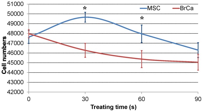
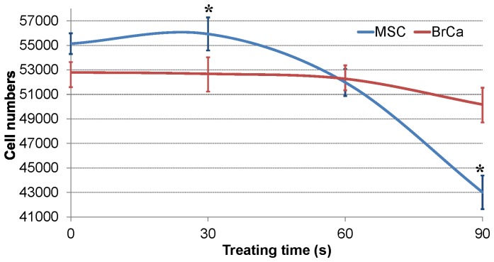
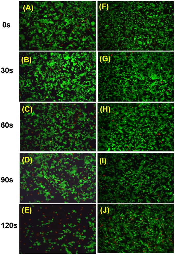
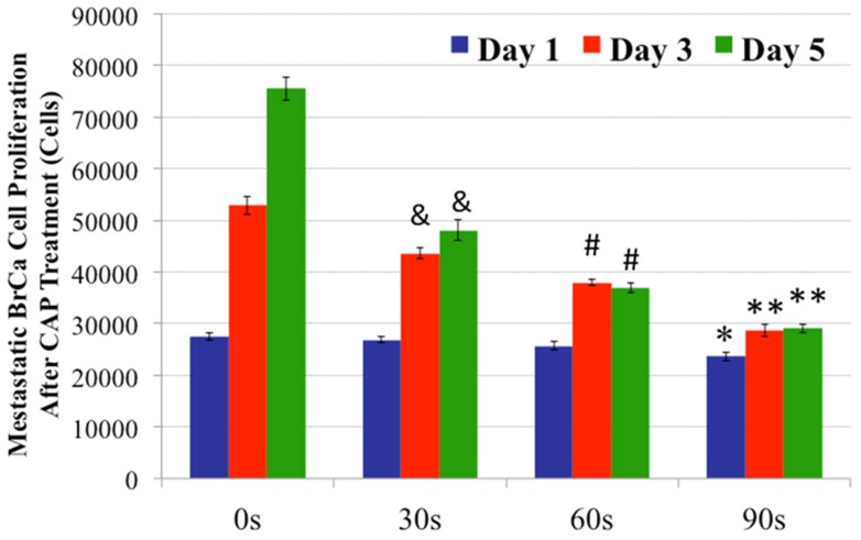
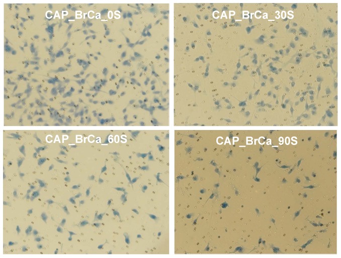
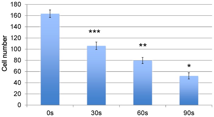
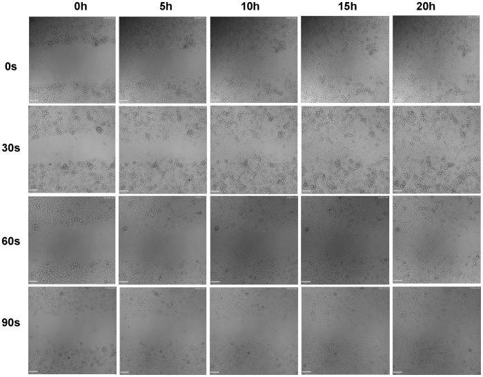
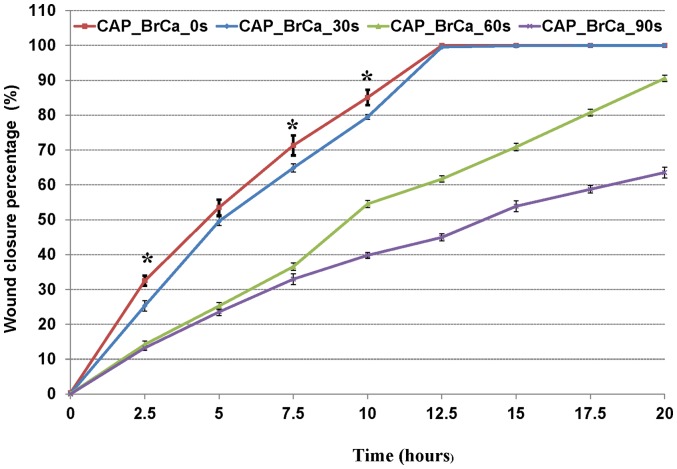
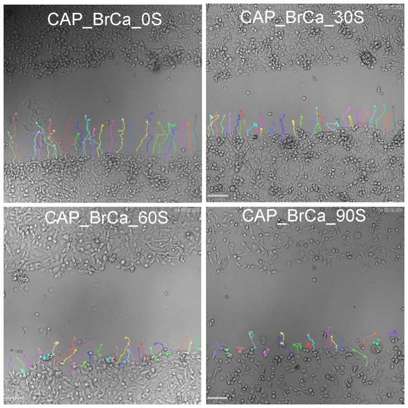
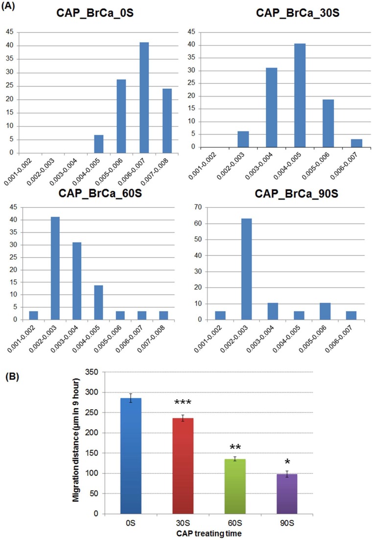
Traditional breast cancer treatments such as surgery and radiotherapy contain many inherent limitations with regards to incomplete and nonselective tumor ablation. Cold atmospheric plasma (CAP) is an ionized gas where the ion temperature is close to room temperature. It contains electrons, charged particles, radicals, various excited molecules, UV photons and transient electric fields. These various compositional elements have the potential to either enhance and promote cellular activity, or disrupt and destroy them. In particular, based on this unique composition, CAP could offer a minimally-invasive surgical approach allowing for specific cancer cell or tumor tissue removal without influencing healthy cells. Thus, the objective of this research is to investigate a novel CAP-based therapy for selectively bone metastatic breast cancer treatment. For this purpose, human metastatic breast cancer (BrCa) cells and bone marrow derived human mesenchymal stem cells (MSCs) were separately treated with CAP, and behavioral changes were evaluated after 1, 3, and 5 days of culture. With different treatment times, different BrCa and MSC cell responses were observed. Our results showed that BrCa cells were more sensitive to these CAP treatments than MSCs under plasma dose conditions tested. It demonstrated that CAP can selectively ablate metastatic BrCa cells in vitro without damaging healthy MSCs at the metastatic bone site. In addition, our study showed that CAP treatment can significantly inhibit the migration and invasion of BrCa cells. The results suggest the great potential of CAP for breast cancer therapy.


Conflict of interest statement

Competing Interests: The authors have declared that no competing interests exist.

Figures

Figure 1
Figure 1. CAP set-up: (a) Configuration of CAP generation device. (b) electrical circuit diagram.
Figure 2
Figure 2. Radial measurement of helium plasma jet: (a) plasma tube aligned with optical probe (b) schematic diagram of holder and probe; (c) intensity of major species drop along radius.
Figure 3
Figure 3. Emission spectrum of helium cold plasma at an output voltage of 3.68 kV.
Figure 4
Figure 4. CAP effects on cell growth: MSC and BrCa cell growth under condition 5 (RA = 3000 Ω, RB = 3000 Ω, output voltage is 3.68 KV) in 0s, 30 s, 60 s, 90 s of CAP treatment.
Data are mean ± SEM, n = 9; *p<0.05 when compared to BrCa cells under 30 and 60 s CAP treatments.
Figure 5
Figure 5. CAP effects on cell growth: MSC and BrCa cell growth under condition 4 (RA = 3700 Ω, RB = 3700 Ω, output voltage is 3.28 KV) in 0 s, 30 s, 60 s, 90 s of CAP treatment.
Data are mean ± SEM, n = 9; *p<0.05 when compared to BrCa cells under 30 and 90 s CAP treatments.
Figure 6
Figure 6. Fluorescence microscopy images. Live (green) and dead (red) BrCa cells (A–E) and MSCs (F–J) under 0, 30, 60, 90 and 120 s of CAP treatment.
Figure 7
Figure 7. CAP treated MSC proliferation.
MSC 1, 3 and 5 day proliferation under different daily CAP treatments. Data are mean ± SEM, n = 9; *p<0.05 and #p<0.01 when compared to 60 and 90 s daily treatment after day 1 and day 3; **p<0.05 and ##p<0.01 when compared to 90s treatment after day 1 and day 3; &p<0.01 when compared to 90 s daily treatment after day 5 and &&p<0.05 when compared to 60s daily treatment after day 5.
Figure 8
Figure 8. CAP treated BrCa cell proliferation.
Significantly inhibited BrCa cell 1, 3 and 5 day proliferation under different daily CAP treatments. Data are mean ± SEM, n = 9; *p<0.01 when compared to 0 s and 30 s daily treatment after day 1; **p<0.01 when compared to all other samples after day 3 and day 5. #p<0.01 when compared to 0 s and 30 s daily treatment after day 3 and day 5; and &p<0.01 when compared to untreated samples (0 s) after day 3 and day 5.
Figure 9
Figure 9. BrCa cell migration via Transwell Migration assay.
CAP treatment decreased invasion of BrCa in matrigel transwell. Cells were treated with CAP in 0 s, 30 s, 60 s and 90 s. CAP treatment influenced invasion of BrCa cell in matrigel transwell.
Figure 10
Figure 10. Quantified BrCa cell invasion in matrigel transwell.
Data are mean ± SEM, n = 9; *p<0.001 when compared to all other treatments. **p<0.01 when compared to 0 s, and 30 s daily treatment. ***p<0.001 when compared to 0 s controls.
Figure 11
Figure 11. Wound healing assay: BrCa cells treated with different CAP time were captured every 5 hours.
Figure 12
Figure 12. Representation of the rate of closure for each condition: untreated BrCa, CAP treated 30 s, 60 s, 90 s.
Data are mean ± SEM, n = 9; *p<0.01 when comparing the untreated group with CAP treatment of 30 s, 60 s and 90 s.
Figure 13
Figure 13. Representative microscopy images of BrCa migration pathway.
Cell tracks of the first line of bottom of scratch for each condition in first 9 hours: untreated BrCa cells, CAP treated 30 s, 60 s, and 90 s.
Figure 14
Figure 14. BrCa cell migration velocity after CAP treatment.
(A) Cell velocity distribution of the first line of bottom of scratch for each condition in first 9 hours. X axis represents velocity distribution (µm/second) and Y axis represents cell percentage (%). (B) Quantification of average migration distance of BrCa cells under different CAP treatments in 9 hours. Data are mean ± SEM, n = 9; *p<0.001, **p<0.001 and ***p<0.001 when compared to all other treatments.

References

    1. Lipton A, Uzzo R, Amato RJ, Ellis GK, Hakimian B, et al... (2009) The science and practice of bone health in oncology: managing bone loss and metastasis in patients with solid tumors. J Natl Compr Canc Netw 7 Suppl 7: S1-29; quiz S30. - PMC - PubMed
    1. Fridman A (2008) Plasma Chemistry. Cambridge: Cambridge University Press. 1017 p.
    1. Cooper M, Fridman G, Staack D, Gutsol A, Vasilets V, et al. (2009) Decontamination of Surfaces From Extremophile Organisms Using Nonthermal Atmospheric-Pressure Plasmas. IEEE Trans Plasma Sci 37: 866–871.
    1. Feng H, Wang R, Sun P, Wu H, Liu Q, et al... (2010) Atmospheric pressure cold plasma as an antifungal therapy. Appl Phys Lett 97.
    1. Shimizu T, Steffes B, Pompl R, Jamitzky F, Bunk W (2008) Characterization of microwave plasma torch for decontamination. Plasma Processes and Polymers 5: 577–582.

Publication types