. 2013 Sep 10;8(9):e74010.
doi: 10.1371/journal.pone.0074010. eCollection 2013.

Staphylococcus aureus activates the NLRP3 inflammasome in human and rat conjunctival goblet cells

Affiliations

Staphylococcus aureus activates the NLRP3 inflammasome in human and rat conjunctival goblet cells

Victoria E McGilligan et al. PLoS One. .

Abstract

The conjunctiva is a moist mucosal membrane that is constantly exposed to an array of potential pathogens and triggers of inflammation. The NACHT, leucine rich repeat (LRR), and pyrin domain-containing protein 3 (NLRP3) is a Nod-like receptor that can sense pathogens or other triggers, and is highly expressed in wet mucosal membranes. NLRP3 is a member of the multi-protein complex termed the NLRP3 inflammasome that activates the caspase 1 pathway, inducing the secretion of biologically active IL-1β, a major initiator and promoter of inflammation. The purpose of this study was to: (1) determine whether NLRP3 is expressed in the conjunctiva and (2) determine whether goblet cells specifically contribute to innate mediated inflammation via secretion of IL-1β. We report that the receptors known to be involved in the priming and activation of the NLRP3 inflammasome, the purinergic receptors P2X4 and P2X7 and the bacterial Toll-like receptor 2 are present and functional in conjunctival goblet cells. Toxin-containing Staphylococcus aureus (S. aureus), which activates the NLRP3 inflammasome, increased the expression of the inflammasome proteins NLRP3, ASC and pro- and mature caspase 1 in conjunctival goblet cells. The biologically active form of IL-1β was detected in goblet cell culture supernatants in response to S. aureus, which was reduced when the cells were treated with the caspase 1 inhibitor Z-YVAD. We conclude that the NLRP3 inflammasome components are present in conjunctival goblet cells. The NRLP3 inflammasome appears to be activated in conjunctival goblet cells by toxin-containing S. aureus via the caspase 1 pathway to secrete mature IL1-β. Thus goblet cells contribute to the innate immune response in the conjunctiva by activation of the NLRP3 inflammasome.


Conflict of interest statement

Competing Interests: The authors have declared that no competeing interests exist.

Figures

Figure 1
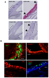
Figure 1. Human conjunctiva and rat conjunctiva constitutively express the NLRP3 inflammasome components.
Human conjunctiva was analyzed by immunohistochemistry and NLRP3 was shown to be highly expressed in the epithelium as indicated by pink staining (A). Arrows indicate epithelial layer of conjunctiva; arrowheads indicate goblet cells. Isotype controls were negative. Rat conjunctiva was analyzed by immunofluorescence microscopy (B) and all three of the inflammasome components NLRP3, caspase 1, and ASC were identified as demonstrated by the red peri-nuclear staining. The green UEA staining indicates goblet cell secretory product, denoting the location of goblet cells in the conjunctiva. Arrows indicate goblet cells. The mouse isotype controls (B) were negative as were the rabbit isotype controls (not shown). Epi, epithelium.
Figure 2
Figure 2. Constitutive expression of the NLRP3 inflammasome components in human and rat conjunctival goblet cells.
Primary cultures of human or rat goblet cells were analyzed by immunofluorescence microscopy after staining with antibodies against inflammasome components NLRP3, caspase 1, and ASC (A). All three components were identified as indicated by the red peri-nuclear staining pattern. Rabbit isotype controls (shown) and mouse isotype controls (not shown) were negative. The presence of NLRP3 (B), caspase 1 (C), ASC (D) and IL-1β (E) were confirmed by western blot analysis in lysates from human goblet cells and rat conjunctiva and goblet cells. Please note that the positive controls for NLRP3 and ASC are transfected cells lines overexpressing the protein and thus the amount of protein loaded for the western blots was below the detection level of the β-actin antibody. In addition, the NLRP3 is tagged with His tag while the ASC is tagged with a FLAG tag causing these proteins to run at a higher molecular weight than the native molecules.
Figure 3
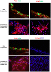
Figure 3. Purinergic receptors P2X4, P2X7, and TLR2 are expressed in the rat conjunctiva and rat goblet cell cultures.
All three receptors were identified by red immunofluorescent staining. UEA stains goblet cell secretory products green, allowing the identification of goblet cells within the conjunctiva. Rabbit isotype controls were negative. Arrows indicate location of goblet cells.
Figure 4
Figure 4. Purinergic receptors P2X4, P2X7 and TLR2 are expressed in rat goblet cell cultures.
All three receptors were identified by western blot. Lanes 1–3 represent separate animals. B is the positive control of rat brain. The β-actin blot is for both P2X4 and P2X7 receptors blots.
Figure 5
Figure 5. The purinergic receptors P2X4, P2X7, and TLR2 are functional in cultured rat goblet cells.
Rat goblet cells were loaded with fura-2 and then stimulated with ATP and intracellular calcium response measured. A typical trace from one experiment with ATP (5 mM, A) or lipoteichoic acid (LTA, 10 µg/ml, B) is representative of 3 animals. Representative photographs of a single field of cells after treatment with ATP or LTA, with warmer colors indicating intracellular Ca2+ increase, are shown as insets. Peak [Ca2+]i calculated for ATP (0.1 µM–5 mM) from 3 experiments, is shown in C. Peak [Ca2+]i for goblet cells, also preincubated with LTA for 5 h and then loaded with fura-2 for 1 additional h before addition of ATP (5 mM), calculated from 3 experiments, is shown in D. Results are expressed as mean ± SEM. * indicates significance of p<0.05 from no addition (0).
Figure 6
Figure 6. Effect of S. aureus on Goblet Cell Viability.
Cultured rat goblet cells were incubated with S. aureus at MOIs of 20, 40, and 60 for 0–24 h, and cell viability was determined by trypan pan blue exclusion. Data is mean ± SEM from 3 independent experiments.
Figure 7
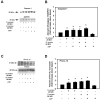
Figure 7. Effect of S. aureus on NLRP3 and ASC expression by cultured rat goblet cells.
Cultured rat goblet cells were incubated with S. aureus (MOI 20 or 60) for 6 h. Cultures were treated for an additional 2 h with ATP (5 mM) or buffer alone. Cell lysates collected and analysed by western blot. Representative blots are shown in A and C. Lower, major band in ASC blot was scanned. Blots were scanned and quantified. Data from a single experiment, representative of two experiments, is shown in B and means ± SEM from 3 independent experiments are shown in D. * indicates significance of p<0.05 compared to no addition which was set to 1.
Figure 8
Figure 8. Effect of S. aureus on Caspase 1 and proIL-1β protein expression.
Cultured rat goblet cells were incubated with S. aureus (MOI 20 or 60) for 6 h. Cultures were treated for an additional 2 h with ATP (5 mM) or buffer alone. Cell lysates were collected and analyzed by western blot. Representative blots are shown in A and C. Blots were scanned and mean ± SEM from 3 independent experiments are shown in B and D. * indicates significance of p<0.05 compared to no addition which was set to 1.
Figure 9
Figure 9. Active caspase 1 expression in rat goblet cells treated with S. aureus and ATP.
Primary cultures of rat goblet cells were incubated with S. aureus (MOI 20 or 60) for 6 h. Cultures were treated for an additional 2 h with ATP (5 mM) or buffer alone. The FLICA reagent, which detects only active caspase 1, was added followed by the nuclear Hoescht stain and viewed by immunofluorescence microscopy. Representative micrographs are shown in A. The total number of nuclei in four fields of view and the number of cells with staining green (indicative of active caspase 1) were counted. Data is expressed as mean ± SEM from 3 independent experiments, and are shown in B. * indicates significance of p<0.05 compared to no addition, which was set to 1. Magnification 40×.
Figure 10
Figure 10. Effect of Inhibition of Caspase 1 on IL-1β secretion in response to S. aureus and ATP.
Primary cultures of rat goblet cells were treated with or without the caspase 1 inhibitor Z-YVAD for 1 h and then incubated with S. aureus (MOI 20 or 60) for 6 h. Cultures were treated for an additional 2 h with ATP (5 mM) or buffer alone. Culture supernatant was removed and analyzed for IL-1β by ELISA. Data is expressed as mean ± SEM from 3 independent experiments. * indicates significance of p<0.05 compared to no addition. # indicates significance of p<0.05 compared to no inhibitor.

References

    1. Martinon F, Burns K, Tschopp J (2002) The inflammasome: a molecular platform triggering activation of inflammatory caspases and processing of proIL-beta. Mol Cell 10: 417–426. - PubMed
    1. Anders HJ, Muruve DA (2011) The inflammasomes in kidney disease. J Am Soc Nephrol 22: 1007–1018. - PubMed
    1. Franchi L, Eigenbrod T, Munoz-Planillo R, Nunez G (2009) The inflammasome: a caspase-1-activation platform that regulates immune responses and disease pathogenesis. Nat Immunol 10: 241–247. - PMC - PubMed
    1. Mariathasan S (2007) ASC, Ipaf and Cryopyrin/Nalp3: bona fide intracellular adapters of the caspase-1 inflammasome. Microbes Infect 9: 664–671. - PubMed
    1. Hornung V, Bauernfeind F, Halle A, Samstad EO, Kono H, et al. (2008) Silica crystals and aluminum salts activate the NALP3 inflammasome through phagosomal destabilization. Nat Immunol 9: 847–856. - PMC - PubMed

Publication types

MeSH terms