. 2013 Sep 11;8(9):e74377.
doi: 10.1371/journal.pone.0074377. eCollection 2013.

Rapid antemortem detection of CWD prions in deer saliva

Affiliations

Rapid antemortem detection of CWD prions in deer saliva

Davin M Henderson et al. PLoS One. .

Abstract

Chronic wasting disease (CWD) is an efficiently transmitted prion disease of cervids, now identified in 22 United States, 2 Canadian provinces and Korea. One hallmark of CWD is the shedding of infectious prions in saliva, as demonstrated by bioassay in deer. It is also clear that the concentration of prions in saliva, blood, urine and feces is much lower than in the nervous system or lymphoid tissues. Rapid in vitro detection of CWD (and other) prions in body fluids and excreta has been problematic due to the sensitivity limits of direct assays (western blotting, ELISA) and the presence of inhibitors in these complex biological materials that hamper detection. Here we use real-time quaking induced conversion (RT-QuIC) to demonstrate CWD prions in both diluted and prion-enriched saliva samples from asymptomatic and symptomatic white-tailed deer. CWD prions were detected in 14 of 24 (58.3%) diluted saliva samples from CWD-exposed white-tailed deer, including 9 of 14 asymptomatic animals (64.2%). In addition, a phosphotungstic acid enrichment enhanced the RT-QuIC assay sensitivity, enabling detection in 19 of 24 (79.1%) of the above saliva samples. Bioassay in Tg[CerPrP] mice confirmed the presence of infectious prions in 2 of 2 RT-QuIC-positive saliva samples so examined. The modified RT-QuIC analysis described represents a non-invasive, rapid ante-mortem detection of prions in complex biologic fluids, excreta, or environmental samples as well as a tool for exploring prion trafficking, peripheralization, and dissemination.


Conflict of interest statement

Competing Interests: The authors have declared that no competing interests exist.

Figures

Figure 1
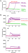
Figure 1. RT-QuIC analysis of CWD-positive clinically ill white-tailed deer.
Both 1:10 diluted and PTA precipitated saliva show positive results by RT-QuIC. Each line represents an individual experiment with the average of four replicates. Blue and light blue lines represent 1:10 diluted saliva experiments and red and dark red lines represent PTA precipitated counterpart of the same saliva samples. The left axis (blue) denotes ThT fluorescence for 1:10 diluted saliva samples and the right axis (red) denotes ThT fluorescence for PTA saliva samples. Dashed lines represent threshold for a positive sample. Blue for 1:10 diluted saliva experiments and red for PTA saliva experiments. Positive RT-QuIC results were observed for saliva from clinically ill white-tailed deer in 5 of 9 1:10 diluted saliva samples and 8 of 9 PTA precipitated saliva samples. Each saliva sample was from an individual deer.
Figure 2
Figure 2. SHrPrP substrate can detect CWD-positive brain over a range of dilutions from 10-4 to 10-7.
CWD positive and negative brain was serially diluted and analyzed by RT-QuIC assay. Each line for positive CWD samples represents the average of 4 individual wells from 4 independent experiments.
Figure 3
Figure 3. PTA precipitation increases sensitivity of the RT-QuIC assay.
A. Each data point shows the ThT fluorescence average of 4 replicate wells loaded with either 2 µl of brain homogenate dilution or PTA pellets from the same brain homogenate equivalents. Lines with triangles represent PTA precipitated samples while smooth lines are non-precipitated counterparts or controls (in the latter case, invisible under the green triangles). The data for spiked PTA precipitated deer saliva (purple triangles) comes from a separate experiment performed under the same experimental conditions. B. End-point dilution analysis of a CWD (+) brain homogenate including 10-7 to 10-9 dilutions with or without PTA precipitation. PTA precipitated samples were done in triplicate (purple) and represent the ThT fluorescence average of 4 replicate wells each. Non-precipitated samples (red) represent the ThT fluorescence average of all 12 replicate wells. Normal deer brain homogenates (green) are shown for PTA precipitated (3 overlapping curves of data points representing the average of 4 replicate wells) and non-precipitated (average of 12 replicate wells) samples.
Figure 4
Figure 4. Kaplan-Meier survival curve for mice inoculated with CWD+ saliva.
The same saliva samples from deer assayed in Fig. 1 (#133 and 144) were inoculated into cervidized mice (Tg[CerPrP] 5037) as well as saliva from a sham inoculated deer. 50% of mice were euthanized due to clinical disease after ~500 days.
Figure 5
Figure 5. RT-QuIC comparison of diluted saliva vs. PTA precipitated saliva harvested from pre-clinical* white-tailed deer.
PTA precipitation of saliva samples increased sensitivity and consistency of RT-QuIC prion detection of pre-clinical white-tailed deer. All colors and axis are the same as in Figure 1. (*) refers to either no signs of CWD or subtle behavioral symptoms detectable only by observers very familiar with individual deer and the very early signs of CWD.
Figure 6
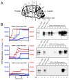
Figure 6. Correlation of RT-QuIC analysis of terminal saliva samples with western blot PrPRes signal in brains.
A. Cartoon representation of sections analyzed for PrPRes by western blotting. Seven rostral to caudal sections were analyzed plus the obex/brainstem region. B. (left) RT-QuIC analysis of saliva samples from white-tailed deer. Colors and axis are the same as in Figure 1 (right). Western blot analysis of 8 brain sections including CWD (+) and (-) control samples. PK was added to all samples except one CWD (-) brain sample. Antibody BAR-224-HRP used for detection.

References

    1. Williams ES, Young S (1980) Chronic wasting disease of captive mule deer: a spongiform encephalopathy. J Wildl Dis 16: 89-98. PubMed: 7373730. - PubMed
    1. Williams ES, Young S (1982) Spongiform encephalopathy of Rocky Mountain elk. J Wildl Dis 18: 465-471. PubMed: 7154220. - PubMed
    1. Mathiason CK, Nalls AV, Seelig DM, Kraft SL, Carnes K et al. (2013) Susceptibility of domestic cats to chronic wasting disease. J Virol, 87: 1947–56. PubMed: 23236066. - PMC - PubMed
    1. Heisey DM, Mickelsen NA, Schneider JR, Johnson CJ, Johnson CJ et al. (2010) Chronic wasting disease (CWD) susceptibility of several North American rodents that are sympatric with cervid CWD epidemics. J Virol 84: 210-215. doi:10.1128/JVI.00560-09. PubMed: 19828611. - DOI - PMC - PubMed
    1. Sigurdson CJ, Mathiason CK, Perrott MR, Eliason GA, Spraker TR et al. (2008) Experimental chronic wasting disease (CWD) in the ferret. J Comp Pathol 138: 189-196. doi:10.1016/j.jcpa.2008.01.004. PubMed: 18387626. - DOI - PubMed

Publication types