. 2013 Sep 9;8(9):e74569.
doi: 10.1371/journal.pone.0074569. eCollection 2013.

Myocarditis, disseminated infection, and early viral persistence following experimental coxsackievirus B infection of cynomolgus monkeys

Affiliations

Myocarditis, disseminated infection, and early viral persistence following experimental coxsackievirus B infection of cynomolgus monkeys

Cheryl E Cammock et al. PLoS One. .

Abstract

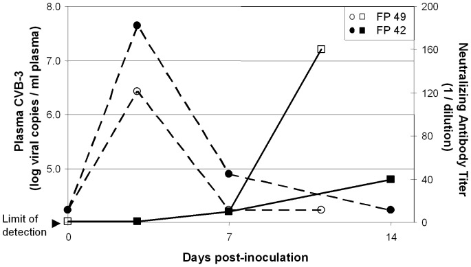
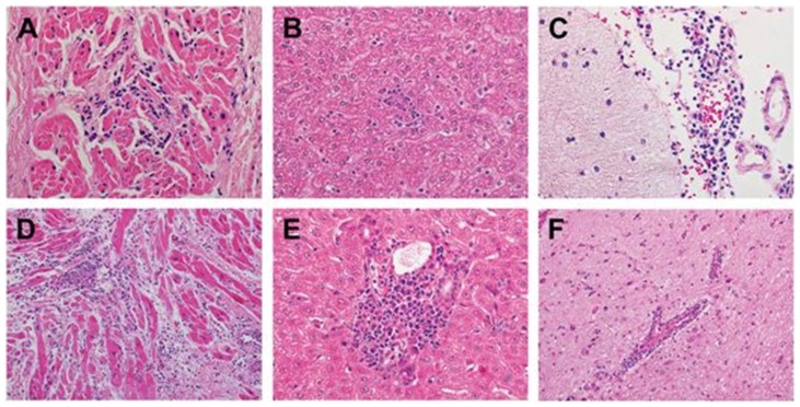
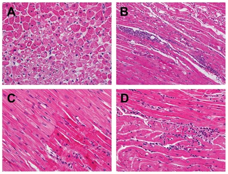
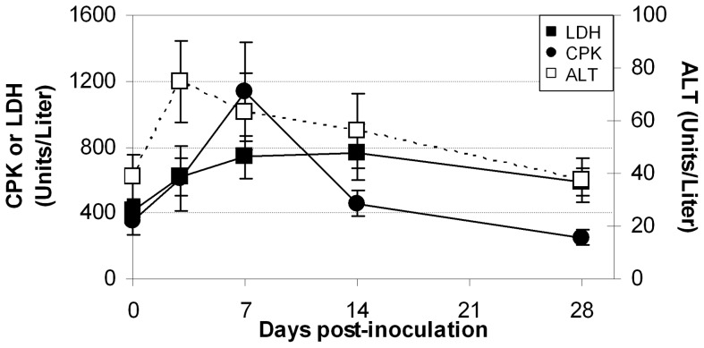
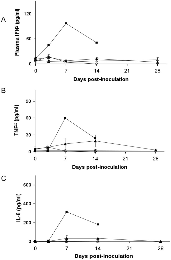
Coxsackievirus B (CVB) infection is a common cause of acute viral myocarditis. The clinical presentation of myocarditis caused by this enterovirus is highly variable, ranging from mildly symptoms to complete hemodynamic collapse. These variations in initial symptoms and in the immediate and long term outcomes of this disease have impeded development of effective treatment strategies. Nine cynomolgus monkeys were inoculated with myocarditic strains of CVB. Virological studies performed up to 28 days post-inoculation demonstrated the development of neutralizing antibody in all animals, and the presence of CVB in plasma. High dose intravenous inoculation (n = 2) resulted in severe disseminated disease, while low dose intravenous (n = 6) or oral infection (1 animal) resulted in clinically unapparent infection. Transient, minor, echocardiographic abnormalities were noted in several animals, but no animals displayed signs of significant acute cardiac failure. Although viremia rapidly resolved, signs of myocardial inflammation and injury were observed in all animals at the time of necropsy, and CVB was detected in postmortem myocardial specimens up to 28 days PI. This non-human primate system replicates many features of illness in acute coxsackievirus myocarditis and demonstrates that myocardial involvement may be common in enteroviral infection; it may provide a model system for testing of treatment strategies for enteroviral infections and acute coxsackievirus myocarditis.


Conflict of interest statement

Competing Interests: The authors have declared that no competing interests exist.

Figures

Figure 1
Figure 1. Coxsackievirus viral load (plasma viral RNA copies/ml) (dashed lines) and neutralizing antibody titers (reciprocal of dilution) (solid lines) after intravenous infection of two cynomolgus monkeys: FP42 (closed figures) and FP49 (open figures).
Figure 2
Figure 2. Postmortem histopathologic findings (hematoxylin-eosin (H&E) stain) of two cynomolgus monkeys injected with 107 pfu of CVB3-H3.
Myocardium from both FP42 (A (200X)) and FP49 (D (200X)) showed evidence of viral myocarditis, consisting of focal predominantly lymphocytic mononuclear inflammatory infiltrates with myocyte injury. In addition, representative sections of the liver from both animals exhibited changes of mild viral hepatitis including focal mononuclear inflammatory infiltrates within the hepatic lobules (B (FP42)) and portal tracts with associated interface hepatitis (E (FP49)). The central nervous system exhibited perivascular lymphocytic infiltrates within the leptomeninges (C (FP42)) and intracerebral blood vessels (F (FP49)) with microglial proliferation and nodules (not shown). These changes are indicative of viral meningitis and encephalitis (meningoencephalitis).
Figure 3
Figure 3. Histopathological changes in the myocardium of animals infected with CVB-H3 (FP41, FP44, and FP46) and CVB3-MCH (FP47).
FP41 (A) exhibiting prominent cytoplasmic vacuolization indicative of myocytolysis (400X; H&E stain). FP44 (B) revealing focal, predominantly lymphocytic, mononuclear inflammatory infiltrate with associated myocyte injury consistent with myocarditis (400X; H&E stain). FP46 (C) showing hypereosinophilic, hypercontracted myofibrils indicative of contraction band necrosis of cardiac myocytes (right lower corner) (400X; H&E stain). FP47 (D) demonstrating focal mononuclear inflammatory infiltrate without associated myocardial injury (400X; H&E stain).
Figure 4
Figure 4. Biomarkers measured in animals inoculated with low dose (106 pfu) CVB-H3 or CVB-MCH.
Serum, unfractionated LDH (▪), total CPK (•), and ALT (□) are represented as mean +/− SEM for all 6 animals at each timepoint.
Figure 5
Figure 5. Immunohistochemistry of myocardial tissue.
Upper left panel: H&E stain. Antibody specificity used for immunoperoxidase staining is listed below remaining panels. (All sections at 400X)
Figure 6
Figure 6. Plasma cytokine concentrations following intravenous inoculation with CVB3.
Concentrations of single cytokines IFN-g (panel A), TNF-a (Panel B), and IL-6 (panel C) are demonstrated. Mean cytokine concentrations for the two animals receiving high dose (107 pfu) CVB-H3 by i.v. (▪) or the single animal infected by enteral infection () are shown. Mean cytokine concentration +/− SEM for animals receiving low dose (106 pfu) CVB-H3 (▴) or CVB-MCH (Δ) by i.v. are shown

References

    1. Dennert R, Crijns HJ, Heymans S (2008) Acute viral myocarditis. Eur Heart J 29: 2073–2082. - PMC - PubMed
    1. Cherry JD, Krogstad P (2009) Enteroviruses and Parechoviruses. In: Feigin RD, Cherry JD, Demmler-Harrison GJ, Kaplan SL, editors. Textbook of Pediatric Infectious Diseases. 6th ed. Philadelphia, PA: WB Saunders Co. pp. 2110−2169.
    1. Verboon-Maciolek MA, Krediet TG, van Loon AM, Kaan J, Galama JM, et al. (2002) Epidemiological survey of neonatal non-polio enterovirus infection in the Netherlands. J Med Virol 66: 241–245. - PubMed
    1. Freund MW, Kleinveld G, Krediet TG, van Loon AM, Verboon-Maciolek MA (2010) Prognosis for neonates with enterovirus myocarditis. Arch Dis Child Fetal Neonatal Ed 95: F206–212. - PubMed
    1. Centers for Disease Control (2008) Increased detections and severe neonatal disease associated with coxsackievirus B1 infection—United States, 2007. MMWR Morb Mortal Wkly Rep 57: 553–556. - PubMed

Publication types

MeSH terms