. 2013 Sep 10;8(9):e74580.
doi: 10.1371/journal.pone.0074580. eCollection 2013.

Stepwise assembly of fibrinogen is assisted by the endoplasmic reticulum lectin-chaperone system in HepG2 cells

Affiliations

Stepwise assembly of fibrinogen is assisted by the endoplasmic reticulum lectin-chaperone system in HepG2 cells

Taku Tamura et al. PLoS One. .

Abstract

The endoplasmic reticulum (ER) plays essential roles in protein folding and assembly of secretory proteins. ER-resident molecular chaperones and related enzymes assist in protein maturation by co-operated interactions and modifications. However, the folding/assembly of multimeric proteins is not well understood. Here, we show that the maturation of fibrinogen, a hexameric secretory protein (two trimers from α, β and γ subunits), occurs in a stepwise manner. The αγ complex, a precursor for the trimer, is retained in the ER by lectin-like chaperones, and the β subunit is incorporated into the αγ complex immediately after translation. ERp57, a protein disulfide isomerase homologue, is involved in the hexamer formation from two trimers. Our results indicate that the fibrinogen hexamer is formed sequentially, rather than simultaneously, using kinetic pause by lectin chaperones. This study provides a novel insight into the assembly of most abundant multi-subunit secretory proteins.


Conflict of interest statement

Competing Interests: The authors have declared that no competing interests exist.

Figures

Figure 1
Figure 1. Stepwise assembly of fibrinogen in HepG2 cells.
(A) Cys and Met-starved HepG2 cells were pulse-labeled for 20 min and chased for 0 or 5 hr. Cells were lysed and media were supplemented with TX-100 (final 0.5%) then immunoprecipitated using anti-fibrinogen antibody after prewash with Pansorbin cells. Immunoprecipitated samples were resolved with 4–15% SDS-PAGE in non-reducing conditions. The molecular mass of marker proteins (lane M) is shown on the left in kDa. Band positions of fibrinogen molecules are indicated on the right: (αβγ)2 and αβγ represent fibrinogen hexamer and trimer, respectively. Lane numbers are shown at the bottom. (B) Separated fibrinogen molecules of 0 or 5 hr chase with non-reduced SDS-PAGE in 4–15% gradient gel (horizontal axis), as described in A, were resolved by 9% SDS-PAGE after denaturing with β-mercaptoethanol (vertical axis). Of note, a 97 kDa endogenous protein, which associates with fibrinogen, is represented by an arrow in A and B. (C) HepG2 cells were exposed to the indicated concentration of DTT (mM, at the top) in culture medium for 10 min at 37°C. After alkylation of the free thiol group with IAA, the fibrinogen proteins were separated by non-reducing 9% SDS-PAGE and detected by immunoblotting using anti-fibrinogen antibody.
Figure 2
Figure 2. Fibrinogen molecules mainly localize at the early secretory pathway in HepG2 cells.
(A) HepG2 cells split onto coverslips in 35 mm dishes were immunostained with anti-fibrinogen antibody (green) and then with anti-GM130 or HSP47 antibody (red), as described in the Materials and Methods. Scale bars represent 10 µm. (B) HepG2 cells were lysed in the lysis buffer (50 mM Hepes pH 7.4 containing 100 mM NaCl, 1 mM EDTA, 5% glycerol and 1% TX-100) and protein concentration was measured by standard Bradford assay. After denaturing with 0.5% SDS, samples were treated with PNGase F (lane 3) or Endoglycosidase H (Endo H, lane 2) according to the manufacturer’s protocol. Samples were separated with 9% SDS-PAGE (loaded 10 µg in each lane) under reducing conditions, and fibrinogen subunits were visualized by immunoblotting using anti-fibrinogen antibody. Migration of the undigested fibrinogen subunits is marked on the left. Positions of deglycosylated or Endo H sensitive (Endo Hs) or resistant (Endo Hr) forms of β or γ subunits are indicated on the right.
Figure 3
Figure 3. Cellular retention of fibrinogen αγ complex by ER lectin chaperone through monoglucosylated N-glycans.
HepG2 cells were starved for Met and Cys and then pulse-labeled for 20 min. In the chase experiment, the medium was replaced with non-radiolabeled media every 20 min. These experiments were conducted in the presence or absence of 1 mM CAS. Samples were immunoprecipitated using anti-fibrinogen antibody and then resolved by a 4–15% gradient gel under non-reducing conditions (A), or by a 9% gel under reducing conditions (B). The lane “−160 (Cell)” denotes the immunoprecipitation samples of cell lysate after chase for 160 min. (C) Quantification of each fibrinogen molecule in A. The percentage of each fibrinogen band relative to the sum of all species (100%) is noted inside the bars. Lane numbers are shown at the bottom. White, light gray, dark gray and black bars from the top to the bottom are represented as (αβγ)2, αβγ, αγ and γ chains, respectively. Of note, the bands not reaching 5% intensity are not stated in the bars.
Figure 4
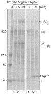
Figure 4. Transient binding of ERp57 to fibrinogen trimer and hexamer during the assembly.
Cys and Met starved HepG2 cells were pulse-labeled for 3 min and then chased for 0, 5 and 10 min. The cell lysate was divided into two aliquots and immunoprecipitated using anti-fibrinogen (lanes 1–3) or anti-ERp57 (lanes 4–6) antibodies. Samples were separated with 4–15% gradient SDS-PAGE under non-reducing conditions and visualized with BAS-2000.
Figure 5
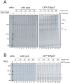
Figure 5. Acceleration of fibrinogen assembly by ERp57 in a reglucosylation-independent manner.
Wild type or CFP-ERp57 stably overexpressing HepG2 cells were starved for Met and Cys for 3 hr. After pulse labeling for 5 min, the microsomes were prepared at 4°C and then suspended in the cytosolic condition buffer . Refolding reaction was initiated by incubation at 37°C for the indicated times (min) in the presence (+) or absence (-) of 1 mM UDP-glucose. After immunoprecipitation using anti-fibrinogen antibody, the samples were separated by a non-reducing SDS-PAGE in a 4–15% gel (A) or reducing SDS-PAGE in a 9% gel (B). The molecular mass of marker proteins is shown on the left and each fibrinogen subunit is indicated on the right. The asterisk represents a non-specific band derived from Pansorbin cells.
Figure 6
Figure 6. Down regulation of ERp57 retards fibrinogen hexamer formation.
(A) HepG2 cells were transfected with indicated double strand RNA (C, control: 57, ERp57) for 96 hr. Cells were lysed in SDS-PAGE sample buffer and subjected to a reducing 9% SDS-PAGE. Proteins were visualized using indicated antibodies. Knock down effects of ERp57 are quantitated using ImageJ and shown at the bottom of panel A versus GAPDH. (B) HepG2 cells were transfected as described in A, and fibrinogen molecules were detected by western blotting using anti-fibrinogen antibody in non-reducing conditions.
Figure 7
Figure 7. A working model of ER chaperone assistance in fibrinogen assembly.
(A) Calnexin (CNX) holds the fibrinogen αγ complex through monoglucosylated N-linked glycans. (B) The newly synthesized fibrinogen β chain is integrated into the αγ complex. (C) A fibrinogen trimer is formed and handed off to ERp57 from CNX. (D) ERp57 facilitates the integration of the two trimers into the hexamer. (E) The properly assembled fibrinogen hexamer is moved forward to the later secretory pathway. Noteworthy, N-glycosylated β and γ chains are illustrated by the Y motif.

References

    1. Eriksson KK (2004) EDEM Contributes to Maintenance of Protein Folding Efficiency and Secretory Capacity. Journal of Biological Chemistry 279: 44600–44605 doi:10.1074/jbc.M407972200 - DOI - PubMed
    1. Braakman I, Bulleid NJ (2011) Protein folding and modification in the mammalian endoplasmic reticulum. Annu Rev Biochem 80: 71–99 doi:10.1146/annurev.biochem.69.1.69 - DOI - PubMed
    1. Trombetta ES (2003) The contribution of N-glycans and their processing in the endoplasmic reticulum to glycoprotein biosynthesis. Glycobiology 13: 77R–91 doi:10.1093/glycob/cwg075 - DOI - PubMed
    1. Schrag JD, Procopio DO, Cygler M, Thomas DY, Bergeron JJM (2003) Lectin control of protein folding and sorting in the secretory pathway. Trends Biochem Sci 28: 49–57. - PubMed
    1. Ellgaard L, Molinari M, Helenius A (1999) Setting the standards: quality control in the secretory pathway. Science 286: 1882–1888. - PubMed

Publication types

MeSH terms