. 2013 Sep 11;8(9):e75766.
doi: 10.1371/journal.pone.0075766. eCollection 2013.

Protein phosphatase 1 β paralogs encode the zebrafish myosin phosphatase catalytic subunit

Affiliations

Protein phosphatase 1 β paralogs encode the zebrafish myosin phosphatase catalytic subunit

Vaishali Jayashankar et al. PLoS One. .

Abstract

Background: The myosin phosphatase is a highly conserved regulator of actomyosin contractility. Zebrafish has emerged as an ideal model system to study the in vivo role of myosin phosphatase in controlling cell contractility, cell movement and epithelial biology. Most work in zebrafish has focused on the regulatory subunit of the myosin phosphatase called Mypt1. In this work, we examined the critical role of Protein Phosphatase 1, PP1, the catalytic subunit of the myosin phosphatase.

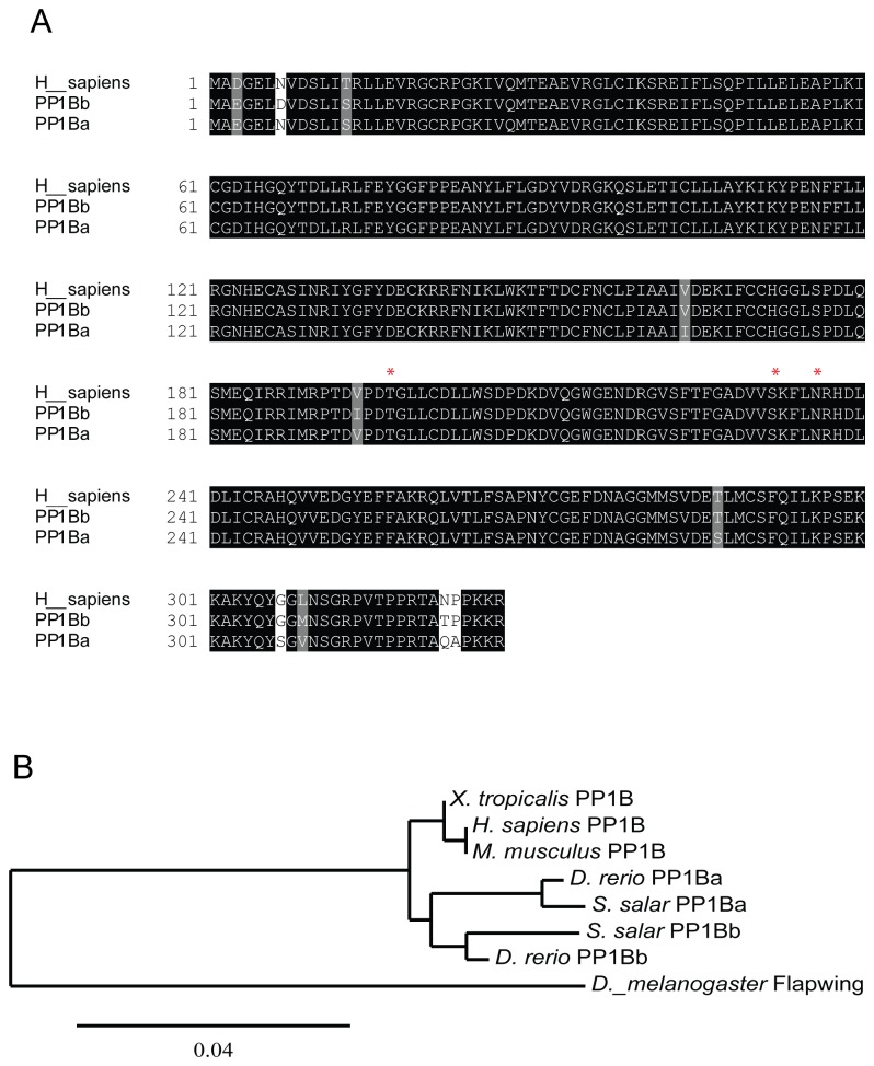
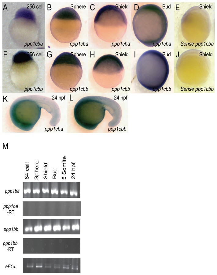
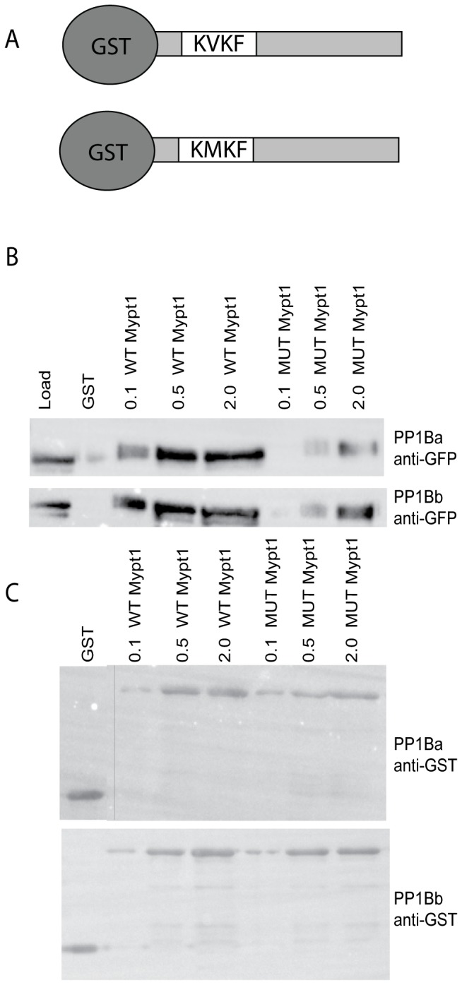
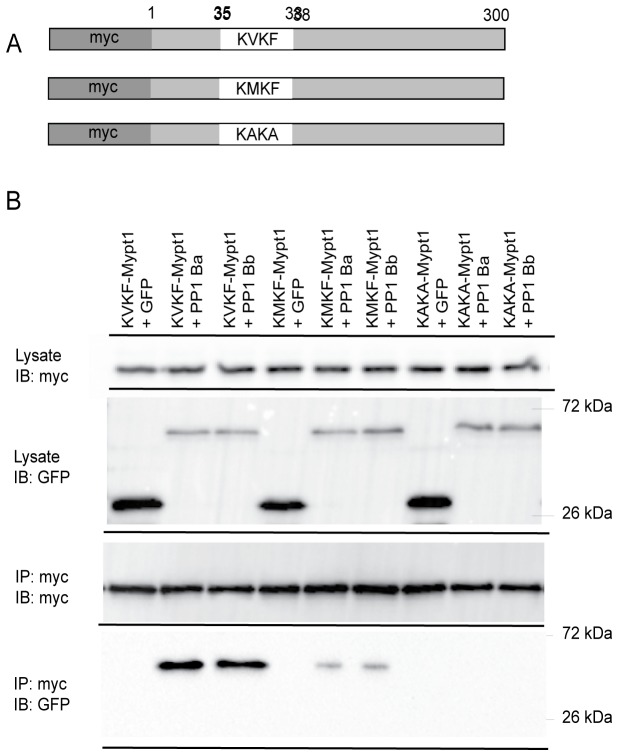
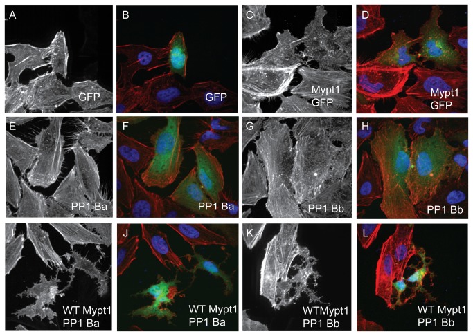
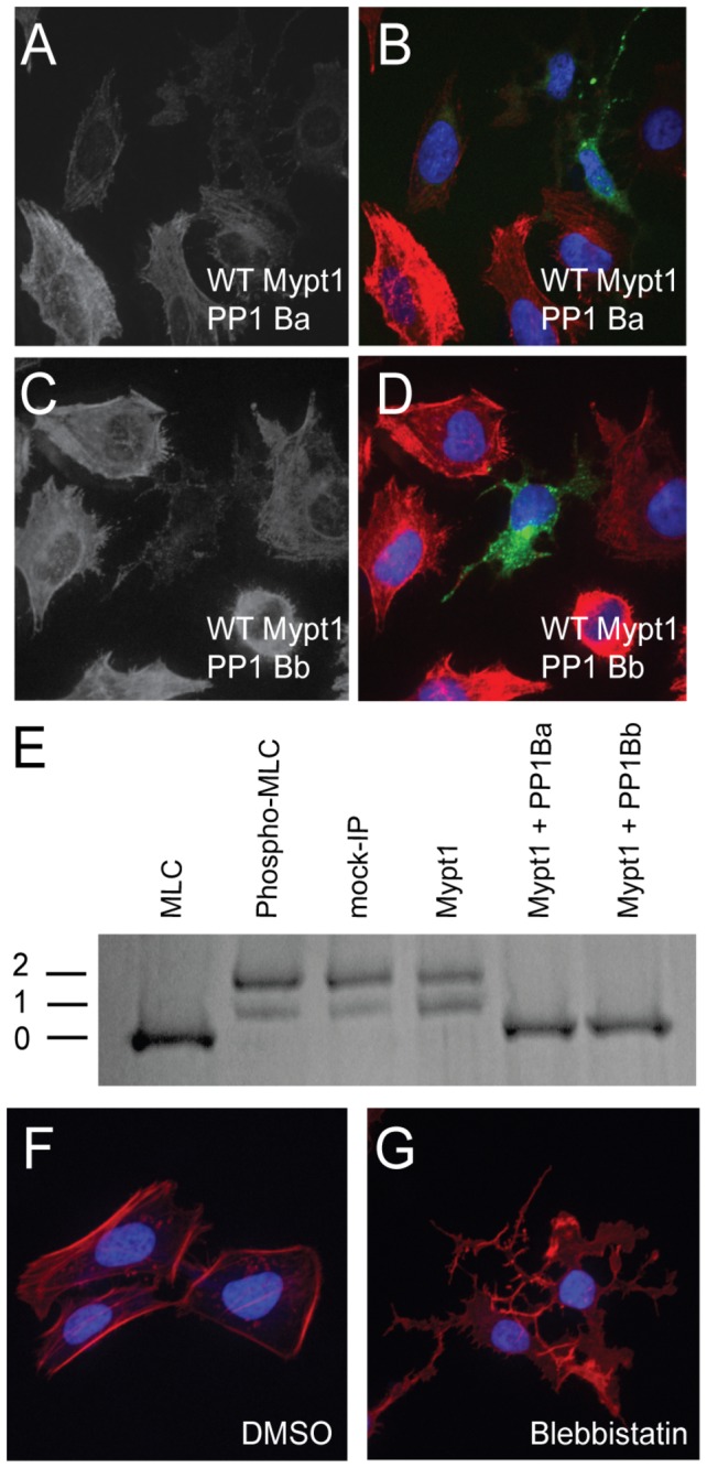
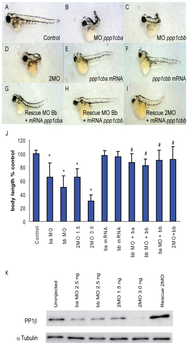
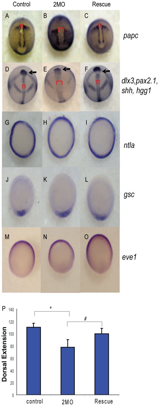
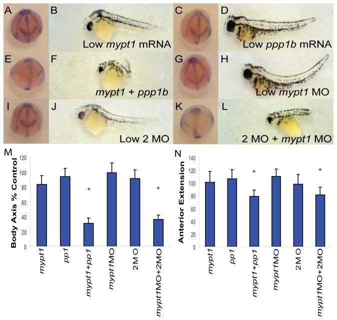
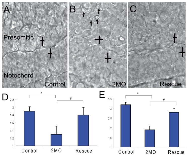
Methodology/principal findings: We observed that in zebrafish two paralogous genes encoding PP1β, called ppp1cba and ppp1cbb, are both broadly expressed during early development. Furthermore, we found that both gene products interact with Mypt1 and assemble an active myosin phosphatase complex. In addition, expression of this complex results in dephosphorylation of the myosin regulatory light chain and large scale rearrangements of the actin cytoskeleton. Morpholino knock-down of ppp1cba and ppp1cbb results in severe defects in morphogenetic cell movements during gastrulation through loss of myosin phosphatase function.

Conclusions/significance: Our work demonstrates that zebrafish have two genes encoding PP1β, both of which can interact with Mypt1 and assemble an active myosin phosphatase. In addition, both genes are required for convergence and extension during gastrulation and correct dosage of the protein products is required.


Conflict of interest statement

Competing Interests: The authors have declared that no competing interests exist.

Figures

Figure 1
Figure 1. Zebrafish have two paralogs for Protein Phosphatase 1 β.
A. A protein sequence alignment of zebrafish PP1Ba and PP1Bb with human PP1β. The amino acids marked with a * are critical for isoform specific interaction with Mypt1. B. Phylogenetic analysis of vertebrate PP1β genes using the maximum likelihood method with Drosophila Flapwing (the Drosophila PP1β) as an outgroup.
Figure 2
Figure 2. Embryonic Expression of ppp1cba and ppp1cbb during zebrafish development.
Detection of ppp1cba and ppp1cbb mRNA was carried out by whole-mount in situ hybridization using gene-specific probes on staged embryos from 256 cells to 24 hpf. Images are lateral views, animal pole at top. (A–L) ppp1cb transcripts are ubiquitously expressed at early developmental stages, 256 cells stage (A and F), sphere stage (B and G), shield stage (C and H), bud stage (D and I) and 24 hpf (K and L). A probe for ppp1cba was used in (A–D and K) while ppp1cbb was used for (F–I and L). Negative control sense probes for ppp1cba and ppp1cbb did not show staining (E and J). Gene specific primers were used to detect ppp1cba and ppp1cbb in various stages of development by RT-PCR. (M) Both genes were expressed maternally and zygotically throughout early development. Amplification of eF1α and total RNA without addition of reverse transcriptase were used as controls.
Figure 3
Figure 3. PP1Ba and PP1Bb interact with Mypt1 in vitro.
(A) Schematic of GST fusion proteins used for co-sedimentation. The N-terminal 300 amino-acids of zebrafish Mypt1 are fused to GST with either a WT KVKF or mutated KMKF PP1 binding motif. (B and C) Increasing concentrations of purified GST fusion proteins (indicated protein added in micrograms) were incubated with glutathione sepharose and used to sediment GFP-tagged PP1Ba and PP1Bb from HEK293T cell lystates. GST alone was used as a negative control. The co-sedimented proteins were analyzed by SDS-PAGE and immunoblotted using an anti-GFP (B) or anti-GST antibody (C). A vertical bar indicates the presence of a lane removed from the western blot in editing.
Figure 4
Figure 4. Interaction of Mypt1 and PP1β isoforms.
(A) A schematic of Mypt1 constructs used in immunoprecipitation experiments. The sequence of the PP1 binding domain is indicated as the WT KVKF, the partial-loss-of-function KMKF and the null KAKA. (B) Myc-tagged Mypt1 and GFP-tagged PP1β proteins were expressed in HEK293T cells and immunoprecipitated with anti-myc antibodies. The IPs were immunoblotted with anti-GFP antibodies and anti-myc antibodies.
Figure 5
Figure 5. PP1Ba and PP1Bb assemble myosin phosphatase complexes that regulate the actin cytoskeleton.
HeLa cells were transfected with either GFP alone (A, B), zebrafish Mypt1 (1-300) and GFP (C, D), zebrafish GFP-PP1Ba (E, F), zebrafish GFP-PP1Bb (G,H), Wild-type Mypt1 and PP1Ba (I, J), wild-type Mypt1 and PP1Bb (K, L). All cells were fixed and stained with DAPI and Alexa 568-phalloidin and imaged with confocal microscopy. Black and white images show phalloidin staining, while color images are a merge of DAPI (blue), GFP (green) and phalloidin (red).
Figure 6
Figure 6. PP1Ba and PP1Bb dephosphorylate the regulatory myosin light chain.
HeLa cells were transfected with zebrafish Mypt1 and GFP-PP1Ba (A, B) or zebrafish Mypt1 and GFP-PP1Bb (C, D). The HeLa cells were immunostained using an anti-phospho myosin light chain 2 antibody, co-stained with DAPI and imaged using confocal microscopy. Panels A and C show anti-phospho MLC2 staining, while B and D show a merge of GFP (green), phospho-MLC2 (red) and DAPI (blue). (E) Purified GST-MLC2 was run either as an untreated control (MLC2) or phosphorylated using ZIPK (Phosph-MLC2). The prephosphorylated MLC2 was dephosphorylated by Mypt1 (1-300)-PP1Ba or PP1Bb complexes immunoprecipitated from HEK293T cell lysates or using controls of treatment with beads from a myc-IP from untransfected cells (myc IP) or cell expressing only Mypt1 (1-300) and no additional catalytic subunit (Mypt1). Dephosphorylation (0), mono (1) and di-phosphorylation (2) was detected by band shift using a phos-tag SDS-PAGE gel as described in the materials and methods. HeLa cells were grown on fibronectin coated coverslips and treated with media containing either 0.1% DMSO (F) or 50 µM blebbistatin (G) for 4 hours. After treatment the HeLa cells were stained with DAPI (blue) and Alexa 568-phalloidin (red) and imaged with confocal microscopy and a color merged image is shown.
Figure 7
Figure 7. PP1β paralogs are required for proper zebrafish body axis elongation.
(A–I) Lateral views of representative 48 hpf zebrafish embryos injected with (A) uninjected control, (B) 2.5 ng ppp1cba MO, (C) 2.5 ng ppp1cbb MO, (D) a mixture of 0.75 ng ppp1cba MO and 0.75 ng of ppp1cbb MO (2MO), (E) 200 pg of ppp1cba mRNA, (F) 200 pg of ppp1cbb mRNA (G) a partially rescued embryo injected with 100 pg ppp1cba mRNA and 2.5 ng of ppp1cbb MO (H) a partially rescued embryo injected with 100 pg ppp1cbb mRNA and 2.5 ng of ppp1cbb MO (I) a partially rescued embryo injected with 100 pg ppp1cbb mRNA and 0.75 ng of ppp1cbb and 0.75 ppp1cba MO (J) Quantification of the truncated body axis phenotype in morphant and mRNA injected embryos. Each injection was performed multiple times with 50 embryos used to calculate body axis length and reported as % of uninjected clutch mates. Error bars are standard error and a black * indicates a statistically significant difference from control and a # indicates a statistically significant rescue compared to the corresponding morpholino injected embryos. Statistical significance was calculated using a one-factor ANOVA with Tukey post hoc analysis and is defined as p < 0.05. (K) A western blot showing PP1β levels in zebrafish embryo lysates from control and embryos injected with two doses of morpholino cocktail (1.5 ng and 3.0 ng of total MO), individual ppp1cba or ppp1cbb (2.5 ng of either) morpholinos or a rescued embryo injected with 0.75 ng of each morpholino (1.5 ng total) and 100 pg of ppp1cbb mRNA. Tubulin was used as a loading control.
Figure 8
Figure 8. PP1β knockdown blocks convergence and extension but does not alter mesodermal cell fate or dorsal-ventral patterning.
Embryos injected with either 1.5 ng of the 2 MO cocktail (0.75 ng ppp1cba MO and 0.75 ng of ppp1cbb MO) or 1.5 ng of the cocktail with 200 pg of ppp1cbb mRNA (rescue) were grown alongside uninjected clutch-mates (control) and staged and fixed for in situ hybridization. Embryos at 90% epiboly were fixed and stained with (A–C) papc (presomitic mesoderm) and imaged from an angle approximately 30 degrees from dorsal to allow visualization of the prechordal plate (marked with an arrow). Embryos at bud stage stained with hgg1 (to mark the prechordal plate), shh (midline), pax2.1 (midbrain-hindbrain boundary) and dlx3 (neural plate) (D–F) and imaged with a view from dorsal. The red bracket marks the width of the notochord and the black arrow points to the prechordal plate. Embryos at 50% epiboly were stained with ntla (G–I) to assay for mesoderm induction, gsc to assay for dorsal cell fates (J–L) and eve1 (M, N, O) for ventral cell fates, all viewed from the animal pole, with dorsal facing down. (P) Quantification of body axis elongation at bud stage for embryos injected with the indicated reagents. Each injection experiment was performed at least 3 times and the morphogenetic measurements were performed on 50 48 hpf and 50 bud stage embryos. Error bars are standard error and a black * indicates a statistically significant difference from control and a # indicates a statistically significant rescue compared to morpholino injected embryos. Statistical significance was calculated using a one-factor ANOVA with Tukey post hoc analysis and is defined as p < 0.05.
Figure 9
Figure 9. PP1β genes interact genetically with Mypt1.
(A–L) Representative embryos at bud stage (A, C, E, G, I, K) and 48 hpf (B, D, F, H, J, L) injected with 15 pg mypt1 (1-300) mRNA with 100 pg of GFP mRNA (A–B), 50 pg each of ppp1cba and ppp1cbb mRNA with 15 pg GFP mRNA (C–D), 15 pg mypt1 mRNA + 50 pg each of ppp1cba and ppp1cbb mRNA (E–F), 0.5 ng of mypt1 MO and 0.25 ng of the control MO (G–H), 0.5 ng control MO and 0.25 ng 2MO (I–J), 0.5 ng mypt1 MO and 0.25 ng 2MO (0.125 ng ppp1cba MO and 0.125 ng of ppp1cbb MO) (K–L). Quantification of the angle of body axis extension at bud stage (M) and 48 hpf (N). Error bars are standard error and a * indicates a statistically significant difference from control and a # indicates a statistically significant rescue compared to morpholino injected embryos. For statistical analysis 50 embryos were analyzed for each condition. Statistical significance was calculated using a one-factor ANOVA with Tukey post hoc analysis and is defined as p < 0.05.
Figure 10
Figure 10. PP1β paralogs are required for cell shape changes required for convergent extension.
A representative field of presomitic and notochordal mesoderm at the bud stage in control (A), 2MO (0.75 ng ppp1cba MO and 0.75 ng of ppp1cbb MO) embryos (B) or rescued embryos (C). The cell polarity of presomitic (D) and notochordal (E) mesodermal cells was determined by calculating the length width ratio (y-axis). The crossed bars indicate cells with the long axis length and the short axis width. The embryos are arranged such that dorsal is down and anterior is to the right. Arrows indicate cells that are actively producing bleb-like protrusions. Error bars are standard error and a * indicates a statistically significant difference from control. All calculations were made on 75 cells from 3–5 separate embryos. Statistical significance was calculated using a one-factor ANOVA with Tukey post hoc analysis and is defined as p < 0.05.

References

    1. Landsverk ML, Epstein HF (2005) Genetic analysis of myosin II assembly and organization in model organisms. Cell Mol Life Sci 62: 2270-2282. doi:10.1007/s00018-005-5176-2. PubMed: 16142426. - DOI - PMC - PubMed
    1. Vicente-Manzanares M, Ma X, Adelstein RS, Horwitz AR (2009) Non-muscle myosin II takes centre stage in cell adhesion and migration. Nat Rev Mol Cell Biol 10: 778-790. doi:10.1038/nrm2786. PubMed: 19851336. - DOI - PMC - PubMed
    1. Hirano K, Derkach DN, Hirano M, Nishimura J, Kanaide H (2003) Protein kinase network in the regulation of phosphorylation and dephosphorylation of smooth muscle myosin light chain. Mol Cell Biochem 248: 105-114. doi:10.1023/A:1024180101032. PubMed: 12870661. - DOI - PubMed
    1. Haystead TA (2005) ZIP kinase, a key regulator of myosin protein phosphatase 1. Cell Signal 17: 1313-1322. doi:10.1016/j.cellsig.2005.05.008. PubMed: 16005610. - DOI - PubMed
    1. Grassie ME, Moffat LD, Walsh MP, Macdonald JA (2011) The myosin phosphatase targeting protein (MYPT) family: A regulated mechanism for achieving substrate specificity of the catalytic subunit of protein phosphatase type 1delta. Arch Biochem Biophys. - PubMed

Publication types

MeSH terms