. 2013 Aug 15;6(9):1747-58.
eCollection 2013.

The differential expression of TGF-β1, ILK and wnt signaling inducing epithelial to mesenchymal transition in human renal fibrogenesis: an immunohistochemical study

Affiliations

The differential expression of TGF-β1, ILK and wnt signaling inducing epithelial to mesenchymal transition in human renal fibrogenesis: an immunohistochemical study

Min-Kyung Kim et al. Int J Clin Exp Pathol. .

Abstract

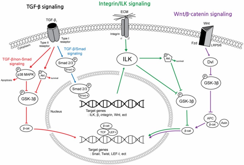
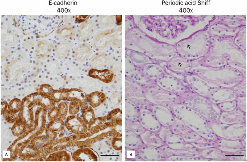
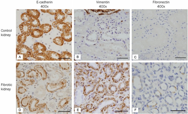
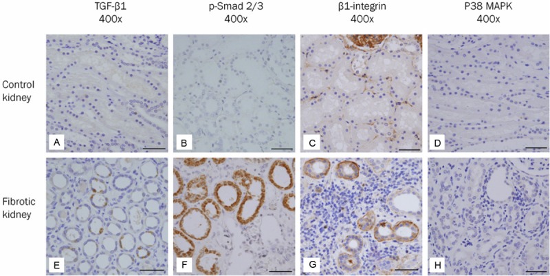
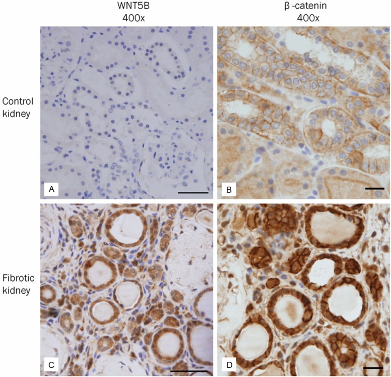
Epithelial-to-mesenchymal transition (EMT) is a process for fully differentiated epithelial cells to undergo a phenotypic change to fibroblasts via diverse intracellular signaling pathways. While the pivotal role of fibroblasts in renal fibrosis is widely accepted, their origin remains undefined. In addition, although a large number of studies have provided evidence of EMT in human kidney diseases, specific signaling pathways leading to EMT have not yet been discovered in humans. To evaluate the origin of interstitial fibroblasts and signaling pathways involved in the EMT process, we analyzed the differential expression of EMT-related molecules in paraffin-fixed sections from 19 human fibrotic kidneys and 4 control kidneys. In human fibrotic kidneys, tubular epithelial cells (TECs) with intact tubular basement membrane (TBM) showed loss or down-regulation of an epithelial marker (E-cadherin), de novo expression of mesenchymal markers (vimentin and fibronectin), and significant up-regulation of inducers and mediators controlling the EMT process (transforming growth factor-β1 (TGF-β1), p-Smad2/3, β1-integrin, p38 mitogen-activated protein kinase (MAPK), WNT5B and β-catenin) in the areas of interstitial inflammation and fibrosis, compared with their expression in control kidneys. In conclusion, the type II EMT process in humans is thought to be an adaptive response of TECs to chronic injury and is regulated by interconnections of TGF-β/Smad, integrin/integrin-linked kinase (ILK) and wnt/β-catenin signaling pathways.

Keywords: EMT; TGF-beta/Smad signaling; immunohistochemistry; integrin; renal fibrosis; wnt signaling.


Figures

Figure 1
Figure 1
A simplified schematic shows the major intracellular signal transduction pathways that regulate the epithelial-to-mesenchymal transition (EMT) process in fibrotic kidneys. EMT is a complex biological process through the TGF-β1, integrin/ILK or wnt/β-catenin signaling pathway. These pathways are intricately interconnected and converged by activation of β-catenin. TGF-β1 is a chief inducer of the EMT process, and p-Smad2/3, p38 MAPK and ILK function as mediators of the TGF-β1 signaling pathway. (TGF-β1, transforming growth factor-β1; p-Smad2/3, phosphorylated-Smad2/3; MAPK, mitogen-activated protein kinase; GSK-3β, glycogen synthase kinase-3β; ILK, integrin-linked kinase; Fzd, frizzled receptors; LRP 5/6, low density lipoprotein receptor-related protein 5/6; Dvl, Disheveled; APC, adenomatosis polyposis coli; TCF/LEF-1 complex, T cell factor/lymphoid enhancer-binding factor-1 complex).
Figure 2
Figure 2
Differential expression of E-cadherin in the proximal and distal tubules of human kidney. A: E-cadherin is widely expressed in the distal tubules and collecting ducts, but not in the proximal tubules. B: The proximal tubule demonstrates a characteristic brush border (periodic acid Shiff). Scale bars: 50 μm.
Figure 3
Figure 3
Expression of biomarkers suggesting phenotypic transition in human fibrotic kidneys. A-C: In the control kidney, E-cadherin (A), an epithelial biomarker, is strongly expressed in the cytoplasmic membrane of distal tubular and collecting duct epithelial cells. Vimentin (B) and fibronectin (C), mesenchymal biomarkers, are not expressed in tubular epithelial cells. A few scattered interstitial cells and smooth muscle cells of blood vessels express vimentin. D-F: In the fibrotic kidney, the expression of E-cadherin (D) is significantly decreased in cuboidal epithelial cells with intact tubular basement membrane surrounding interstitial inflammation and fibrosis. On the other hand, de novo expression of vimentin (E) and fibronectin (F) is observed in tubular epithelial cells in the areas of interstitial inflammation and fibrosis. Scale bars: 50 μm.
Figure 4
Figure 4
Expression of molecules involved in the TGF-β signaling and integrin/integrin-linked kinase (ILK) signaling pathways in human fibrotic kidneys. A-D: In the control kidney, tubular epithelial cells do not express TGF-β1 (A), p-Smad2/3 (B) and p38 MAPK (D). On the other hand, tubular epithelial cells weakly express β1-integrin (C). E-H: In the fibrotic kidney, expression of TGF-β1 (E), p-Smad2/3 (F) and β1-integrin (G) is observed in some cuboidal epithelial cells with intact tubular basement membrane surrounding interstitial inflammation and fibrosis. However, the expression of p38 MAPK (H) is not detected in tubular epithelial cells. Scale bars: 50 μm.
Figure 5
Figure 5
Expression of molecules involved in the wnt/β-catenin signaling pathway in human fibrotic kidneys. A, B: In the control kidney, tubular epithelial cells do not express WNT5B (A). Membranous expression of β-catenin (B) in tubular epithelial cells is characteristic. C, D: In the fibrotic kidney, a large number of tubular epithelial cells with intact basement membrane show up-regulation of WNT5B (C) and β-catenin (D) accompanied by positive nuclear staining. Several interstitial cells in the areas of interstitial inflammation and fibrosis are also positive for WNT5B and β-catenin. Scale bars: A, C = 50 μm, B, D = 20 μm.

References

    1. Eddy AA. Molecular basis of renal fibrosis. Pediatr Nephrol. 2000;15:290–301. - PubMed
    1. Strutz F, Okada H, Lo CW, Danoff T, Carone RL, Tomaszewski JE, Neilson EG. Identification and characterization of a fibroblast marker: FSP1. J Cell Biol. 1995;130:393–405. - PMC - PubMed
    1. Iwano M, Plieth D, Danoff TM, Xue C, Okada H, Neilson EG. Evidence that fibroblasts derive from epithelium during tissue fibrosis. J Clin Invest. 2002;110:341–350. - PMC - PubMed
    1. Jinde K, Nikolic-Paterson DJ, Huang XR, Sakai H, Kurokawa K, Atkins RC, Lan HY. Tubular phenotypic change in progressive tubulointerstitial fibrosis in human glomerulonephritis. Am J Kidney Dis. 2001;38:761–769. - PubMed
    1. Rastaldi MP, Ferrario F, Giardino L, Dell'Antonio G, Grillo C, Grillo P, Strutz F, Muller GA, Colasanti G, D'Amico G. Epithelial-mesenchymal transition of tubular epithelial cells in human renal biopsies. Kidney Int. 2002;62:137–146. - PubMed

Publication types

MeSH terms