Case Reports
. 2013 Aug;5 Suppl 3(Suppl 3):S234-45.
doi: 10.3978/j.issn.2072-1439.2013.07.30.

Uniportal video-assisted thoracoscopic lobectomy

Affiliations
Case Reports

Uniportal video-assisted thoracoscopic lobectomy

Diego Gonzalez-Rivas et al. J Thorac Dis. 2013 Aug.

Abstract

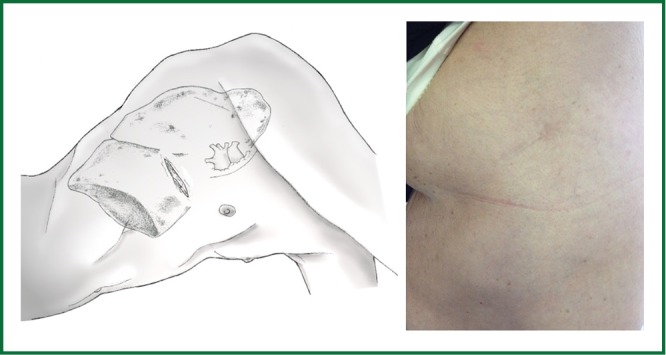
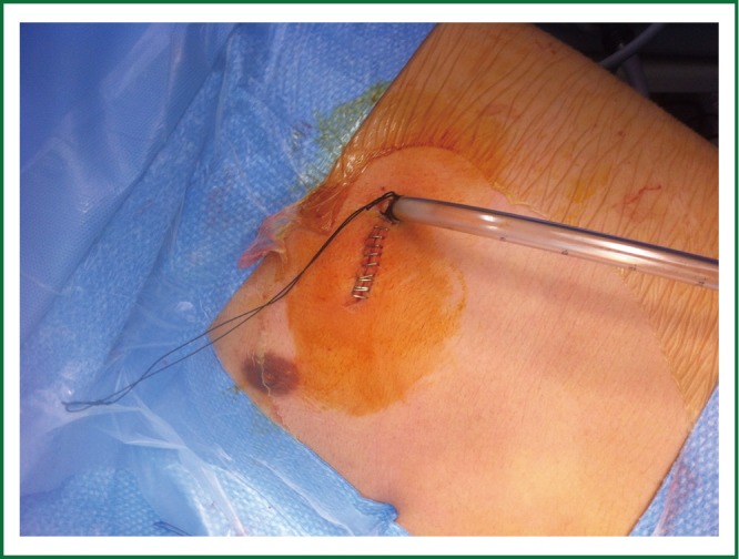
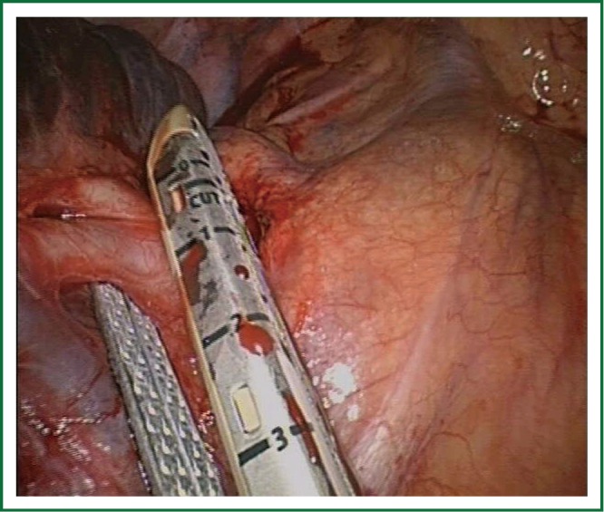
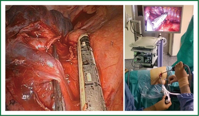
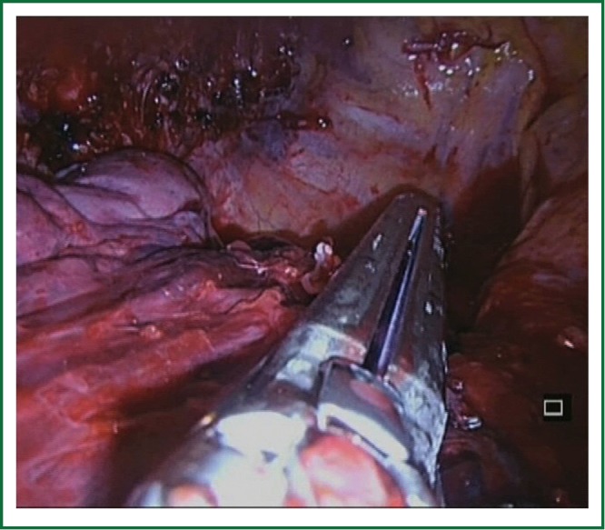
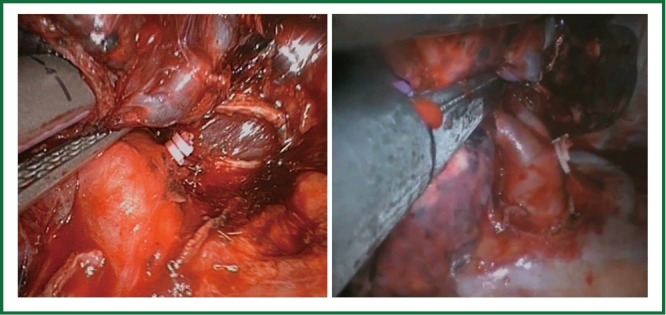
Over the past two decades, video-assisted thoracic surgery (VATS) has revolutionized the way thoracic surgeons diagnose and treat lung diseases. The major advance in VATS procedures is related to the major pulmonary resections. The best VATS technique for lobectomy has not been well defined yet. Most of the authors describe the VATS approach to lobectomy via 3 to 4 incisions but the surgery can be performed by only one incision with similar outcomes. This single incision is the same as we normally use for VATS lobectomies performed by double or triple port technique with no rib spreading. As our experience with VATS lobectomy has grown, we have gradually improved the technique for a less invasive approach. Consequently the greater the experience we gained, the more complex the cases we performed were, hence expanding the indications for single-incision thoracoscopic lobectomy.

Keywords: Video-assisted thoracic surgery (VATS); left lower lobectomy; left upper lobectomy; less invasive approach; lobectomies; lymphadenectomy; middle lobectomy; right lower lobectomy; right upper lobectomy.


Figures

Figure 1
Figure 1
Adapted instruments with proximal and distal articulation.
Figure 2
Figure 2
High definition 30° thoracoscope.
Figure 3
Figure 3
Surgeons positioned in front of the patient watching the same monitor. The scrub nurse is located on the opposite side.
Figure 4
Figure 4
Uniportal set of specific adapted instruments.
Figure 5
Figure 5
Drawing showing the placement of incision.
Video 1
Video 1
Insertion of staplers during upper lobectomies.
Video 2
Video 2
Incision for left upper lobectomy.
Video 3
Video 3
Use of vascular clips during lobectomy.
Figure 6
Figure 6
Single-chest tube placed in the posterior part of the incision.
Video 4
Video 4
Left lower lobectomy (artery exposed in the fissure).
Video 5
Video 5
Left lower lobectomy from bottom to top.
Video 6
Video 6
Right lower lobectomy with artery exposed in the fissure.
Video 7
Video 7
Right lower lobectomy from down to up.
Figure 7
Figure 7
Division of upper arterial trunk as first step.
Figure 8
Figure 8
Use of vascular clips when the angle for staplers is not optimal.
Video 8
Video 8
Left upper lobectomy (fissureless technique).
Figure 9
Figure 9
Division of the left superior pulmonary vein after division of truncus anterior.
Video 9
Video 9
Use of curved tip stapler to facilitate division of upper vein.
Figure 10
Figure 10
Division of anterior portion of fissure from a hilar view.
Video 10
Video 10
Different options for bronchus transection.
Figure 11
Figure 11
Use of TA linear stapler to transect the bronchus is case of complex fissure.
Figure 12
Figure 12
Division of boyden trunk as first step.
Figure 13
Figure 13
Division of upper lobe vein after arterial trunk is divided.
Figure 14
Figure 14
Division right upper lobe bronchus.
Figure 15
Figure 15
Division of the fissure in a right upper lobectomy from anterior to posterior as the last step of the lobectomy (fissureless technique).
Figure 16
Figure 16
Exposure of posterior division of main right bronchus.
Video 11
Video 11
Middle lobectomy.
Video 12
Video 12
Aortopulmonary window lymph node dissection.
Figure 17
Figure 17
Left subcarinal lymph node dissection after left lower lobectomy.
Video 13
Video 13
Left subcarinal lymph node dissection.
Video 14
Video 14
Right subcarinal lymph node dissection.
Figure 18
Figure 18
Surgical image of instrumentation during right paratracheal lymph node dissection.
Video 15
Video 15
Paratracheal lymph node dissection.
Video 16
Video 16
N1 station lymph node dissection after left upper lobectomy.
Figure 19
Figure 19
View of the single incison wound in a patient with a previous thoracothomy and double-port VATS. The patient was initially operated years ago by thoracotomy (wedge resection of a lower lobe metastasis). He was reoperated 3 years later by VATS 2 ports (atypical segmentectomy) and now reoperated by single incision to complete a lower lobectomy.
Figure 20
Figure 20
Lower lobe tumor with chest wall involvement operated by uniportal VATS for lobectomy and posterior incision for chest wall resection and reconstruction.
Figure 21
Figure 21
Sleeve anastomosis after left lower lobectomy.
Figure 22
Figure 22
Surgical instrumentation during vascular reconstruction.
Figure 23
Figure 23
Huge tumor operated by uniportal VATS: bilobectomy and anatomic segmentectomy S6 (right upper lobe, middle lobe and superior segment of lower lobe were involved).

References

    1. Roviaro G, Rebuffat C, Varoli F, et al. Videoendoscopic pulmonary lobectomy for cancer. Surg Laparosc Endosc 1992;2:244-7 - PubMed
    1. Rocco G, Internullo E, Cassivi SD, et al. The variability of practice in minimally invasive thoracic surgery for pulmonary resections. Thorac Surg Clin 2008;18:235-47 - PubMed
    1. Rocco G, Martin-Ucar A, Passera E.Uniportal VATS wedge pulmonary resections. Ann Thorac Surg 2004;77:726-8 - PubMed
    1. Gonzalez D, Paradela M, Garcia J, et al. Single-port video-assisted thoracoscopic lobectomy. Interact Cardiovasc Thorac Surg 2011;12:514-5 - PubMed
    1. McKenna RJ Jr, Mahtabifard A, Swanson SJ. eds. Atlas of minimally invasive thoracic surgery. Saunders 2011;63-5.

Publication types