. 2013 Sep 17;46(9):2182-90.
doi: 10.1021/ar400041a. Epub 2013 Jun 24.

Membrane protein structure determination in membrana

Affiliations

Membrane protein structure determination in membrana

Yi Ding et al. Acc Chem Res. .

Abstract

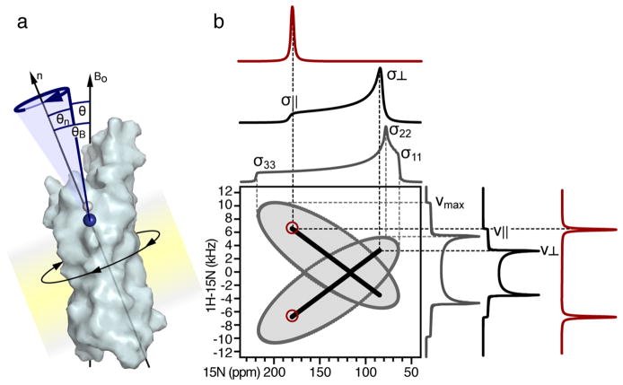
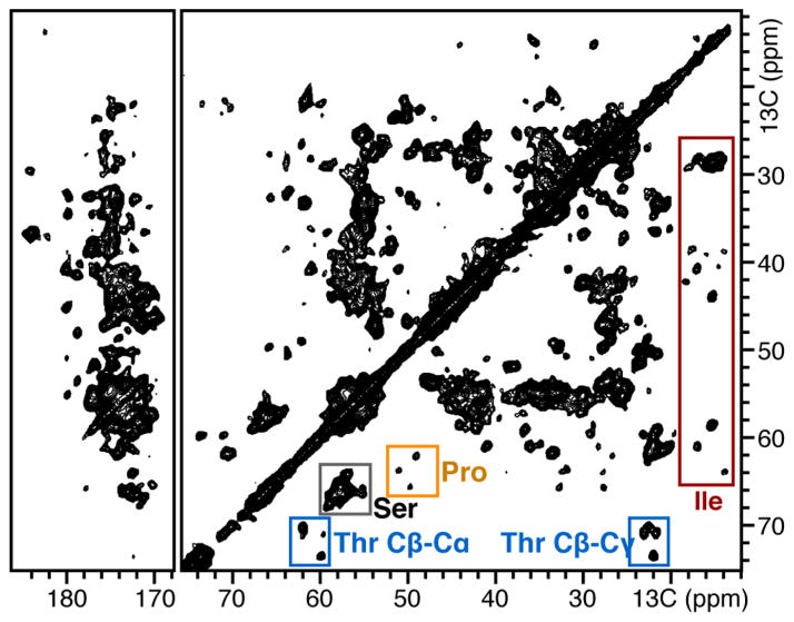
The two principal components of biological membranes, the lipid bilayer and the proteins integrated within it, have coevolved for specific functions that mediate the interactions of cells with their environment. Molecular structures can provide very significant insights about protein function. In the case of membrane proteins, the physical and chemical properties of lipids and proteins are highly interdependent; therefore structure determination should include the membrane environment. Considering the membrane alongside the protein eliminates the possibility that crystal contacts or detergent molecules could distort protein structure, dynamics, and function and enables ligand binding studies to be performed in a natural setting. Solid-state NMR spectroscopy is compatible with three-dimensional structure determination of membrane proteins in phospholipid bilayer membranes under physiological conditions and has played an important role in elucidating the physical and chemical properties of biological membranes, providing key information about the structure and dynamics of the phospholipid components. Recently, developments in the recombinant expression of membrane proteins, sample preparation, pulse sequences for high-resolution spectroscopy, radio frequency probes, high-field magnets, and computational methods have enabled a number of membrane protein structures to be determined in lipid bilayer membranes. In this Account, we illustrate solid-state NMR methods with examples from two bacterial outer membrane proteins (OmpX and Ail) that form integral membrane β-barrels. The ability to measure orientation-dependent frequencies in the solid-state NMR spectra of membrane-embedded proteins provides the foundation for a powerful approach to structure determination based primarily on orientation restraints. Orientation restraints are particularly useful for NMR structural studies of membrane proteins because they provide information about both three-dimensional structure and the orientation of the protein within the membrane. When combined with dihedral angle restraints derived from analysis of isotropic chemical shifts, molecular fragment replacement, and de novo structure prediction, orientation restraints can yield high-quality three-dimensional structures with few or no distance restraints. Using complementary solid-state NMR methods based on oriented sample (OS) and magic angle spinning (MAS) approaches, one can resolve and assign multiple peaks through the use of (15)N/(13)C labeled samples and measure precise restraints to determine structures.


Conflict of interest statement

The authors declare no competing financial interest.

Figures

FIGURE 1
FIGURE 1
Recent structures of membrane proteins determined in phospholipid bilayers by solid-state NMR. PDB codes: 1MZT, membrane-bound bacteriophage fd coat protein; 2L0J, pore-forming domain of influenza M2; 2LJ2, mercury transporter MerF; 2LNL, human chemokine receptor CXCR1. The phospholipid bilayer membrane is depicted in yellow.
FIGURE 2
FIGURE 2
Expression, purification, and refolding of Y. pestis Ail. (a) SDS PAGE analysis. Whole cells isolated before (−) or after (+) induction with IPTG show that induction of Ail yields a band near the predicted molecular weight (mw). Cell lysis and separation into supernatant (S), pellet (P), and inclusion bodies (IB) fractions show that inclusion bodies are enriched in Ail. (b) Isolated Ail inclusion bodies are white and fluffy. (c) SDS-PAGE analysis of Ail folded from urea into DPC micelles or DMPC bilayers. Unfolded Ail (U) migrates at an apparent molecular weight (mw) of 18 kDa; folded Ail (F) migrates near 12 kD. (d, e) Solution NMR 1H/15N HSQC spectra of Ail unfolded in urea (d) or folded in DPC (e). Tris-tricine gels were stained with Coomassie blue.
FIGURE 3
FIGURE 3
OS solid-state NMR spectra of 15N labeled OmpX in DMPC phospolipid bilayers. (a, b) One- and two-dimensional 1H/15N SLF spectra of OmpX in magnetically aligned DMPC/DHPC lipid bilayers with the bilayer normal parallel to the magnetic field. Peaks from phenylalanine sites were assigned as described. Peaks labeled in red resist 2H exchange. Spectra were obtained on a Bruker Avance 700 MHz spectrometer using a home-built radio frequency probe. (c) Crystal structure of OmpX aligned in the membrane on the basis of orientation restraints derived from 15N phenylalanine signals. Amide N atoms from phenylalanine are shown as spheres. Phenylalanine residues labeled in red define a 16 Å band of membrane-integrated, nonexchangeable H bonds.
FIGURE 4
FIGURE 4
SASR refinement of the transmembrane helix of fd coat protein in lipid bilayers aligned with the bilayer normal (n) parallel to the magnetic field., (a) Peaks in the 1H/15N SLF spectrum are first assigned to residue types leucine, alanine, and valine (red dots) by comparison with spectra from selectively 15N-Leu-, 15N-Ala-, and 15N-Val-labeled protein. Then AssignFit is used to specifically assign each peak to a residue number based on best fit of the observed signals (black) to the signals back-calculated from an ideal helix starting model (yellow circles). (b) Refinement of the ideal helix with the assigned DC and CSA restraints extracted from the experimental spectrum yields back-calculated signals (pink circles) that agree closely with the observed data (black). (c) The refined helix (pink) differs from the starting ideal model (yellow) and changes direction after Lys40.
FIGURE 5
FIGURE 5
Uniaxially ordered samples yield orientation-dependent solid-state NMR spectra. (a) A membrane protein undergoing rotational diffusion around an axis (n) normal to the membrane plane. The angle θB defines the membrane orientation relative to the magnetic field (Bo); the angle θn defines the orientation of an amide NH bond (blue) relative to the axis n. Rotational diffusion of the protein around n averages the angle θ between the NH bond and Bo. (b) Predicted solid-state NMR 1H/15N spectra. A static powder sample yields a butterfly-shaped two-dimensional SLF spectrum (gray) whose edges correspond to the maximum values of the 15N CS tensor (σ11, σ22, σ33) and 1H–15N DC (νmax). Rotational diffusion around n produces motionally averaged, axially symmetric powder spectra (black), whose edges correspond to parallel (σ||, ν||, θB = 0°) and perpendicular (σ, ν, θB = 90°) orientations of n relative to Bo. The two-dimensional SLF spectrum has a characteristic cross-like pattern, as observed for gramicidin. Uniaxial alignment of the sample (e.g., with n parallel to Bo) yields single line spectra (red) with 15N CS and 1H–15N DC frequencies that correspond to the parallel edges of rotationally averaged powder patterns.
FIGURE 6
FIGURE 6
Solid-state NMR spectroscopy of 15N/13C-labeled Ail in DMPC proteoliposomes. Two-dimensional 13C/13C correlation MAS solid-state NMR spectrum of 15N/13C-labeled Ail in DMPC liposomes. Peaks from 4 threonine (blue), 4 proline (gold), 16 serine (gray), and 7 isoleucine (red), are highlighted. Spectra were obtained on a Bruker Avance 700 MHz spectrometer using a Bruker MAS probe and controller.

References

    1. Mulkidjanian AY, Galperin MY, Koonin EV. Co-evolution of primordial membranes and membrane proteins. Trends Biochem Sci. 2009;34:206–215. - PMC - PubMed
    1. Cross TA, Sharma M, Yi M, Zhou HX. Influence of solubilizing environments on membrane protein structures. Trends Biochem Sci. 2011;36:117–125. - PMC - PubMed
    1. Hagn F, Etzkorn M, Raschle T, Wagner G. Optimized phospholipid bilayer nanodiscs facilitate high-resolution structure determination of membrane proteins. J Am Chem Soc. 2013;135:1919–1925. - PMC - PubMed
    1. McLaughlin AC, Cullis PR, Hemminga MA, Hoult DI, Radda GK, Ritchie GA, Seeley PJ, Richards RE. Application of 31P NMR to model and biological membrane systems. FEBS Lett. 1975;57:213–218. - PubMed
    1. Griffin RG. Letter: Observation of the effect of water on the 31P nuclear magnetic resonance spectra of dipalmitoyllecithin. J Am Chem Soc. 1976;98:851–853. - PubMed

Publication types

MeSH terms

Substances

LinkOut - more resources