. 2013 Nov 15;591(22):5599-609.
doi: 10.1113/jphysiol.2013.261289. Epub 2013 Sep 16.

Modulation of the autonomic nervous system and behaviour by acute glial cell Gq protein-coupled receptor activation in vivo

Affiliations

Modulation of the autonomic nervous system and behaviour by acute glial cell Gq protein-coupled receptor activation in vivo

Cendra Agulhon et al. J Physiol. .

Abstract

Glial fibrillary acidic protein (GFAP)-expressing cells (GFAP(+) glial cells) are the predominant cell type in the central and peripheral nervous systems. Our understanding of the role of GFAP(+) glial cells and their signalling systems in vivo is limited due to our inability to manipulate these cells and their receptors in a cell type-specific and non-invasive manner. To circumvent this limitation, we developed a transgenic mouse line (GFAP-hM3Dq mice) that expresses an engineered Gq protein-coupled receptor (Gq-GPCR) known as hM3Dq DREADD (designer receptor exclusively activated by designer drug) selectively in GFAP(+) glial cells. The hM3Dq receptor is activated solely by a pharmacologically inert, but bioavailable, ligand (clozapine-N-oxide; CNO), while being non-responsive to endogenous GPCR ligands. In GFAP-hM3Dq mice, CNO administration increased heart rate, blood pressure and saliva formation, as well as decreased body temperature, parameters that are controlled by the autonomic nervous system (ANS). Additionally, changes in activity-related behaviour and motor coordination were observed following CNO administration. Genetically blocking inositol 1,4,5-trisphosphate (IP3)-dependent Ca(2+) increases in astrocytes failed to interfere with CNO-mediated changes in ANS function, locomotor activity or motor coordination. Our findings reveal an unexpectedly broad role of GFAP(+) glial cells in modulating complex physiology and behaviour in vivo and suggest that these effects are not dependent on IP3-dependent increases in astrocytic Ca(2+).


Figures

Figure 1
Figure 1. GFAP+ glial cell-selective hM3Dq expression and function in GFAP-hM3Dq mice
A, schematic map of transgenic construct containing HA epitope tagged (HA)-hM3Dq driven by 2.2 kb human GFAP promoter (hGFAP). B, Western blots showing the pattern of expression of HA-tagged hM3Dq protein in neural and non-neural tissues of GFAP-hM3Dq vs. WT mice. Note that the salivary gland homogenate probably contained autonomic ganglia that are closely associated with salivary glands. The size of the hM3Dq in salivary glands is ∼65 kDa, which is the expected size of this receptor (Armbruster et al. 2007). In brain and spinal cord, the size is larger, suggesting glycosylation of the receptor. C, immunohistochemistry for HA-tagged hM3Dq protein showing HA immunoreactivity (red) overlapping with green fluorescent protein (GFP) staining (green) in highly ramified astrocytic processes in CA1 region of hippocampus of GFAP-hM3Dq/GFAP-GFP double transgenic mice. Arrowheads denote dense astrocytic processes expressing higher levels of hM3Dq (red) in a subpopulation of astrocytes. Arrows point to astrocytes expressing moderate levels of hM3Dq. C, lower panel, higher magnification images of the region delimited by the white box (upper panel). D, HA-tagged hM3Dq immunoreactivity (green) was not detected in NeuN-expressing cell bodies of CA1 pyramidal neurons (red) in GFAP-hM3Dq mice. Small arrows denote hM3Dq staining (green) in ramified astrocytic processes that envelope an unstained CA1 neuron proximal dendrite. E and F, cells loaded with the astrocytic marker SR101 (red), Ca2+ indicator OGB-1 (green) and merged SR101 and OGB-1 (upper). Lower panels show time courses of CNO- and cocktail-mediated Ca2+ responses (ΔF/F0) in the astrocytes (I–IV) and neurons (1–4) outlined in the upper panels. Confocal Ca2+ imaging showed that hM3Dq is functional and gives Ca2+ increases following CNO application in astrocytes but not in neurons of GFAP-hM3Dq mice (E, lower panel). No responses were evoked in astrocytes or neurons of WT littermate control mice (F, lower panel). A cocktail (DHPG, histamine and carbachol) of ligands to endogenous Gq-GPCRs was used as a positive control to ensure cell viability. Abbreviations in C–F: CA1, CA1 pyramidal neurons; c.c., corpus callosum; s.o., stratum oriens; s.r., stratum radiatum; v, vessel.
Figure 2
Figure 2. Effects of hM3Dq activation on cardiovascular function, saliva production and thermal homeostasis
A–C, significant increases in heart rate and blood pressure in CNO-treated GFAP-hM3Dq and GFAP-hM3Dq::IP3R2 KO mice relative to littermate controls. *P < 0.05 GFAP-hM3Dq vs. WT; +P < 0.05 GFAP-hM3Dq::R2 KO vs. R2 KO; #P < 0.05 GFAP-hM3Dq vs. GFAP-hM3Dq::R2 KO. D, saliva produced over 15 min measured in GFAP-hM3Dq, GFAP-hM3Dq::IP3R2 KO and littermate controls treated with saline (S) or CNO (C). E, body temperature changes measured 30 min after treatment with CNO (C) or saline (S) in GFAP-hM3Dq, GFAP-hM3Dq::IP3R2 KO and littermate controls. hM3Dq refers to GFAP-hM3Dq mice and R2KO refers to IP3R2 KO mice. Error bars, SEM. *P < 0.05 compared to control groups.
Figure 3
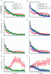
Figure 3. Activation of hM3Dq signalling affects activity-related behaviour
Locomotor activity measured in the open field chamber. Mice were injected with saline or CNO immediately before placing in the chamber. hM3Dq refers to GFAP-hM3Dq mice and R2KO refers to IP3R2 KO mice. *P < 0.05 for GFAP-hM3Dq or GFAP-hM3Dq::R2 KO injected with CNO vs. littermate controls.
Figure 4
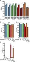
Figure 4. Activation of hM3Dq signalling in GFAP+ cells affects motor coordination, but not motor learning, and induces sedation
A, in contrast to controls, GFAP-hM3Dq and GFAP-hM3Dq::IP3R2 KO mice injected with CNO failed to improve their rotarod scores between trials 1 and 3 on day 1 of testing. B, on day 2 of testing, in the absence of CNO, GFAP-hM3Dq and GFAP-hM3Dq::IP3R2 KO mice performed similarly to control mice. C, on day 3 of testing, CNO depressed the rotarod performance of GFAP-hM3Dq and GFAP-hM3Dq::IP3R2 KO mice that were given saline on day 1 (S = saline; C = CNO). D, LORR was measured in GFAP-hM3Dq, GFAP-hM3Dq::IP3R2 KO and littermate control mice after CNO or saline injection in the presence of the GABAA agonist THIP (25 mg kg−1; a dose that alone failed to induce LORR) administration. *P < 0.05.

References

    1. Agulhon C, Fiacco TA, McCarthy KD. Hippocampal short- and long-term plasticity are not modulated by astrocyte Ca2+ signaling. Science. 2010;327:1250–1254. - PubMed
    1. Agulhon C, Petravicz J, McMullen AB, Sweger EJ, Minton SK, Taves SR, Casper KB, Fiacco TA, McCarthy KD. What is the role of astrocyte calcium in neurophysiology. Neuron. 2008;59:932–946. - PMC - PubMed
    1. Alexander GM, Rogan SC, Abbas AI, Armbruster BN, Pei Y, Allen JA, Nonneman RJ, Hartmann J, Moy SS, Nicolelis MA, McNamara JO, Roth BL. Remote control of neuronal activity in transgenic mice expressing evolved G protein-coupled receptors. Neuron. 2009;63:27–39. - PMC - PubMed
    1. Armbruster BN, Li X, Pausch MH, Herlitze S, Roth BL. Evolving the lock to fit the key to create a family of G protein-coupled receptors potently activated by an inert ligand. Proc Natl Acad Sci U S A. 2007;104:5163–5168. - PMC - PubMed
    1. Atasoy D, Betley JN, Su HH, Sternson SM. Deconstruction of a neural circuit for hunger. Nature. 2012;488:172–177. - PMC - PubMed

Publication types

MeSH terms