

Trends in biological joint resurfacing
- PMID: 24043640
- PMCID: PMC3776950
- DOI: 10.1302/2046-3758.29.2000189
Trends in biological joint resurfacing
Abstract
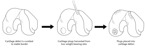
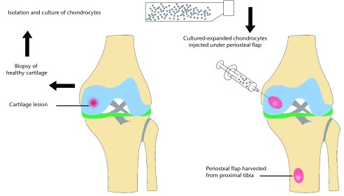
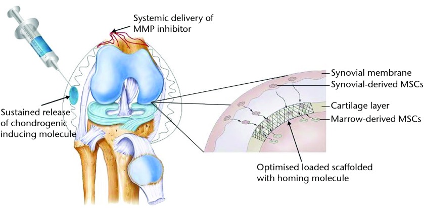
The treatment of osteochondral lesions and osteoarthritis remains an ongoing clinical challenge in orthopaedics. This review examines the current research in the fields of cartilage regeneration, osteochondral defect treatment, and biological joint resurfacing, and reports on the results of clinical and pre-clinical studies. We also report on novel treatment strategies and discuss their potential promise or pitfalls. Current focus involves the use of a scaffold providing mechanical support with the addition of chondrocytes or mesenchymal stem cells (MSCs), or the use of cell homing to differentiate the organism's own endogenous cell sources into cartilage. This method is usually performed with scaffolds that have been coated with a chemotactic agent or with structures that support the sustained release of growth factors or other chondroinductive agents. We also discuss unique methods and designs for cell homing and scaffold production, and improvements in biological joint resurfacing. There have been a number of exciting new studies and techniques developed that aim to repair or restore osteochondral lesions and to treat larger defects or the entire articular surface. The concept of a biological total joint replacement appears to have much potential. Cite this article: Bone Joint Res 2013;2:193-9.
Keywords: Biological joint resurfacing; Cartilage regeneration; Orthopaedic surgery; Regenerative medicine; Stem cells; Tissue engineering.
Conflict of interest statement
Figures
References
-
- Mano JF, Reis RL. Osteochondral defects: present situation and tissue engineering approaches. J Tissue Eng Regen Med 2007;1:261–273 - PubMed
-
- Swieszkowski W, Tuan BH, Kurzydlowski KJ, Hutmacher DW. Repair and regeneration of osteochondral defects in the articular joints. Biomol Eng 2007;24:489–495 - PubMed
LinkOut - more resources
Full Text Sources
Other Literature Sources
