. 2013 Sep 23;210(10):2071-86.
doi: 10.1084/jem.20130103. Epub 2013 Sep 16.

A Toxoplasma dense granule protein, GRA24, modulates the early immune response to infection by promoting a direct and sustained host p38 MAPK activation

Affiliations

A Toxoplasma dense granule protein, GRA24, modulates the early immune response to infection by promoting a direct and sustained host p38 MAPK activation

Laurence Braun et al. J Exp Med. .

Abstract

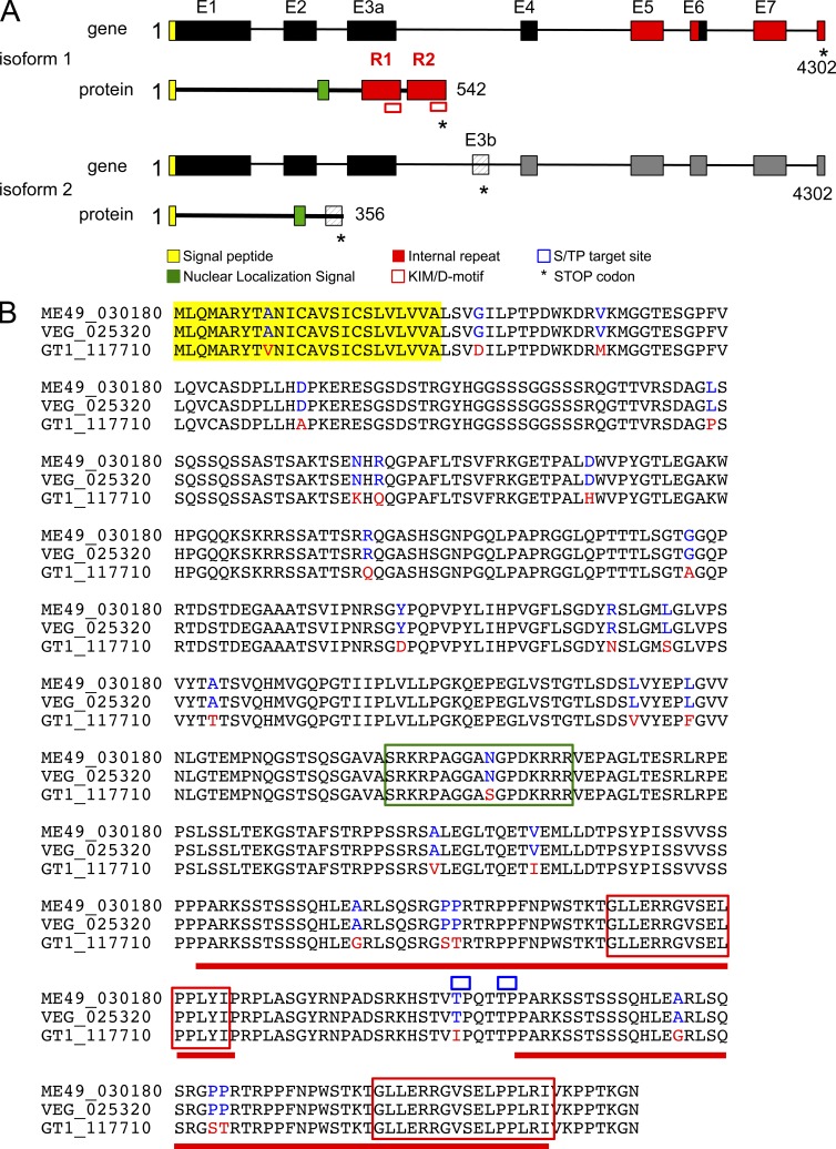
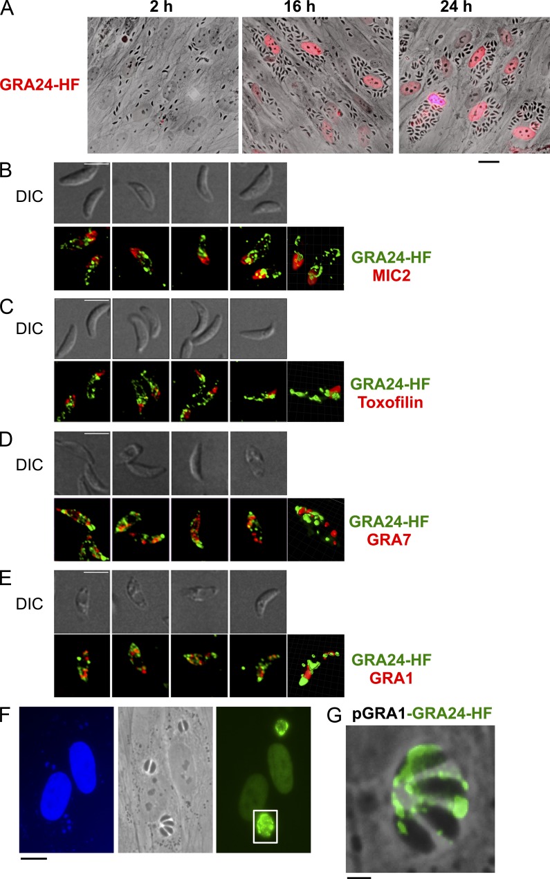
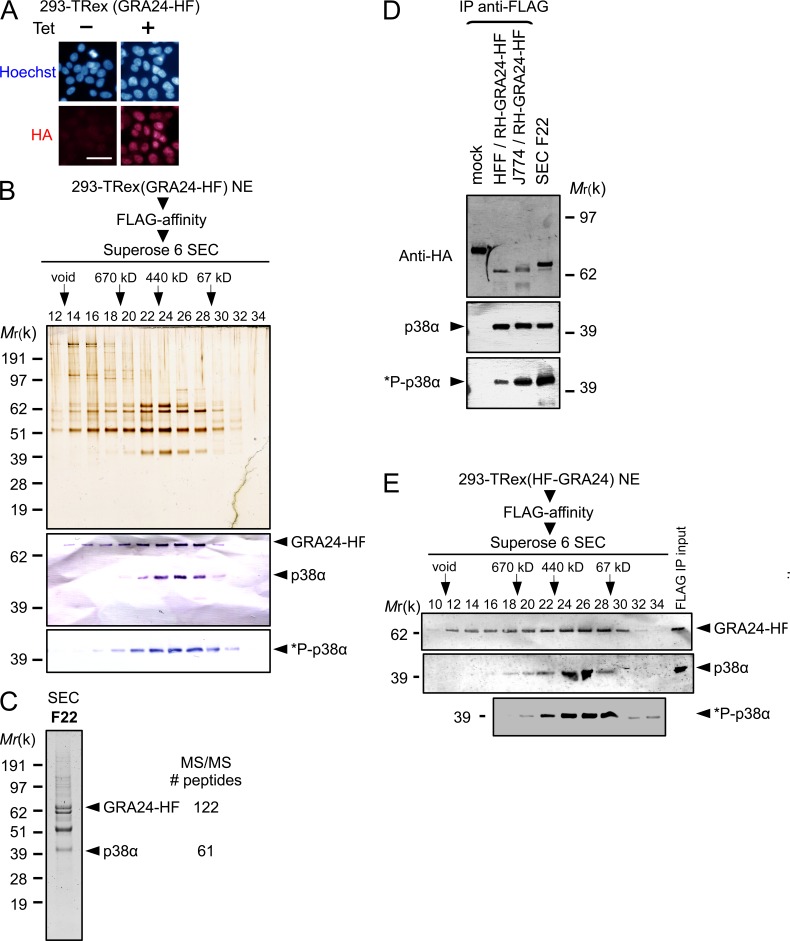
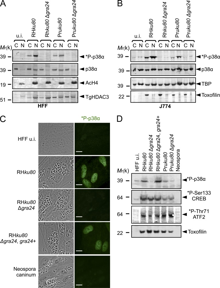
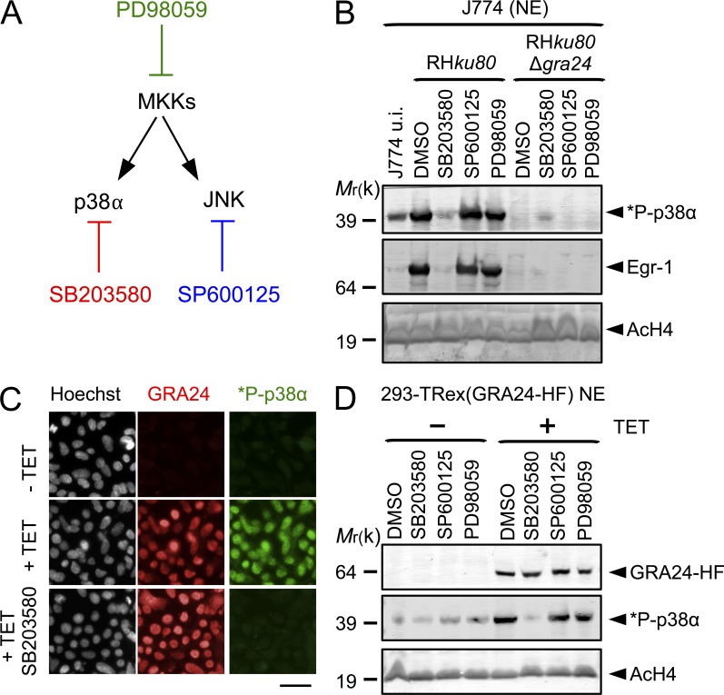
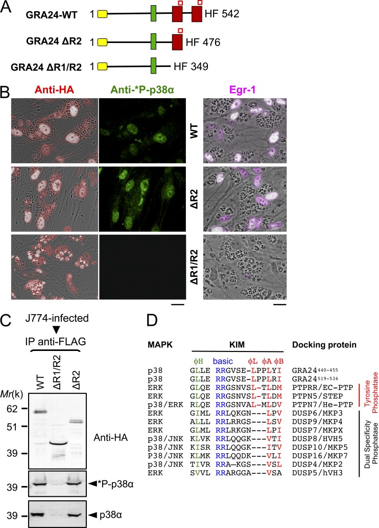
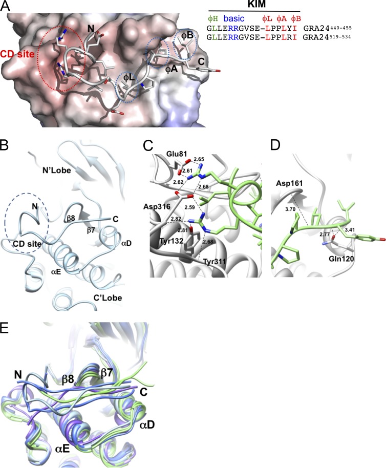
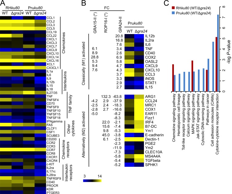
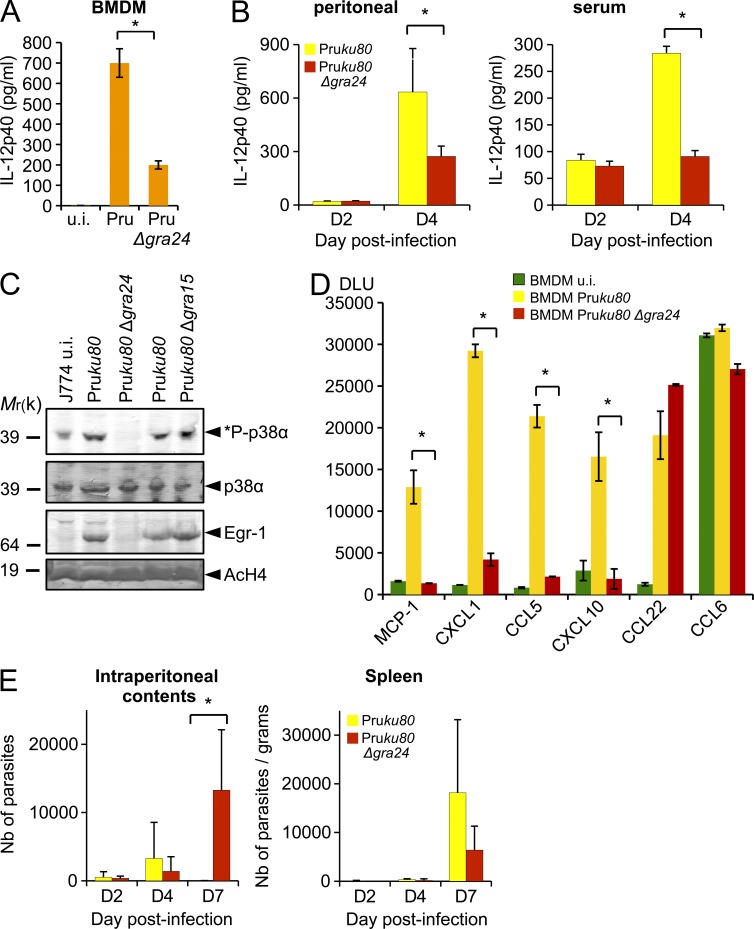
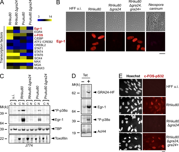
Toxoplasma gondii, the causative agent of toxoplasmosis, is an obligate intracellular protozoan parasite that resides inside a parasitophorous vacuole. During infection, Toxoplasma actively remodels the transcriptome of its hosting cells with profound and coupled impact on the host immune response. We report that Toxoplasma secretes GRA24, a novel dense granule protein which traffics from the vacuole to the host cell nucleus. Once released into the host cell, GRA24 has the unique ability to trigger prolonged autophosphorylation and nuclear translocation of the host cell p38α MAP kinase. This noncanonical kinetics of p38α activation correlates with the up-regulation of the transcription factors Egr-1 and c-Fos and the correlated synthesis of key proinflammatory cytokines, including interleukin-12 and the chemokine MCP-1, both known to control early parasite replication in vivo. Remarkably, the GRA24-p38α complex is defined by peculiar structural features and uncovers a new regulatory signaling path distinct from the MAPK signaling cascade and otherwise commonly activated by stress-related stimuli or various intracellular microbes.


Figures

Figure 1.
Figure 1.
The gene GRA24 generates splice variants encoding two distinct proteins. (A) Schematic representation of the GRA24 alternative splice variants and their corresponding GRA24 protein isoforms. The putative signal peptide is shown in yellow (the cleavage site is predicted by SignalP). A putative nuclear localization signal is in green (as predicted by PSORT). The KIM/D-motif (in red square) and the internal repeats (colored in red) are shown. (B) GRA24 amino acid alignment. The predicted amino acid sequence for the primary translation product of the GRA24 gene is shown for type I (TGGT1_117710), II (TGME49_030180), and III (TGVEG_025320) parasites. Single amino acid polymorphisms are indicated for type II and III (in blue) versus type I (in red). The alignment was done by ClustalW2 (EMBL-EBI).
Figure 2.
Figure 2.
GRA24 is a novel DG protein exported beyond the PV into the host cell nucleus. (A) Time course of GRA24 secretion and export to the host cell nucleus. Human fibroblasts (HFF) were infected with parasites expressing an HAFlag (HF)-tagged copy of GRA24 (RHku80 GRA24-HF), fixed, and stained with anti-HA antibodies (red). Images are representative of at least four experiments. Bar, 20 µm. (B) GRA24-HAFlag is contained in cytoplasmic organelles distinct from the apical micronemes and rhoptries and partially colocalizing with DG proteins. (B–E) Free parasites were fixed, permeabilized, and successively stained with antibodies raised against HA to label GRA24 (green) and then with antibodies (red) against the micronemal protein MIC2 (B), the rhoptry protein toxofilin (C), and the DG proteins GRA7 (D) and GRA1 (E). The top panels show the DIC images corresponding to the IFA (bottom panels), and the right frames show 3D images processed using MetaMorph and Imaris software. Images are representative of at least two experiments. Bars, 5 µm. (F) IFA of GRA24-HF expressed by the strong promoter of GRA1. HFF monolayers were infected with RHku80 parasites expressing ectopically pGRA1-GRA24-HF. 18 h after infection, cells were fixed and secretion of GRA24 was monitored using anti-HA antibodies (green). Images are representative of at least two experiments. Bar, 10 µm. (G) Higher magnification of a vacuole showing GRA24-HF signal in the vacuolar space. Bar, 2.5 µm.
Figure 3.
Figure 3.
GRA24 forms a dimeric complex with the p38α MAPK. (A) Immunofluorescence analysis (IFA) of GRA24-HAFlag ectopically and stably expressed in 293-TRex cell line. Cells were either left untreated (−) or treated with 1 µg/ml tetracycline for 20 h before fixation and staining with anti-HA antibodies (red) and Hoechst DNA-specific dye (blue). Bar, 2.5 µm. (B) GRA24-associated polypeptides were purified from nuclear extracts of 293-TRex cells tetracycline-induced to express GRA24-HF. Size exclusion chromatography (SEC) of GRA24-containing complexes after Flag affinity selection. Fractions were analyzed by silver staining and immunoblotting to detect GRA24-HF (anti-HA), total p38α, and Thr180/Tyr182 phosphorylated *P-p38α. Data are representative of two experiments. (C) Mass spectrometry analysis of SEC fraction 22. Identity of the proteins with their respective number of peptides is indicated on the right. (D) GRA24-p38α association detected in HFF and J774 MØ infected by RHku80 GRA24-HF (18 h). Tagged proteins were Flag-immunoprecipitated from extracts and eluates were analyzed by immunoblotting. Data are representative of two experiments. (E) GRA24-associated polypeptides were purified from nuclear extracts of 293-TRex cells induced to express an N-terminal HAFlag-tagged GRA24. SEC Fractions were analyzed by immunoblotting to detect HAFlag-GRA24 (anti-HA), total p38α, and Thr180/Tyr182 phosphorylated *P-p38α. Data are representative of two experiments.
Figure 4.
Figure 4.
GRA24 elicits phosphorylation and relocation of host p38α in the nucleus. (A and B) Immunoblotting detection of *P-p38α and p38α in HFF (A) and J774 MØ (B) uninfected (u.i.) or infected (24 h) with RHku80, RHku80 Δgra24, Pruku80, and Pruku80 Δgra24 strains. Cytosolic (C) and nuclear (N) cell lysates were probed with the indicated antibodies. Histone H4 acetylated (K5-K8-K12-K16) (nuclear), TBP (host-specific), and TgHDAC3 (parasite-specific) levels are shown as loading controls. (C) Subcellular in situ detection of *P-p38α (green) in HFF uninfected (u.i.) or infected (24 h) with RHku80, RHku80 Δgra24, RHku80 Δgra24, GRA24+, and Neospora caninum strains. Data are representative of at least three experiments. Bars, 10 µm. (D) Immunoblotting detection of *P-p38α, *P-Ser133-CREB, and *P-Thr71-ATF2 in nuclear fraction and Toxofilin (parasite-specific) in cytosolic fraction from HFF uninfected (u.i.) or infected (24 h) with the aforementioned strains and Neospora caninum. Data are representative of two experiments.
Figure 5.
Figure 5.
GRA24 promotes p38α autophosphorylation. (A) Diagram showing the enzymes and their respective inhibitor. (B) Specific pharmacological inhibition of GRA24-dependent p38α phosphorylation in J774 MØ. Cells were incubated for 1 h with p38 (SB203580, 15 µM), SAPK/JNK (SP600125, 25 µM), and MKK (PD98059, 15 µM) inhibitors or DMSO vehicle before infection in presence of the drug with RHku80 and RHku80 Δgra24 strains (18 h). Nuclear fractions were immunoblotted with the indicated antibodies. Data are representative of two experiments. (C) In situ *P-p38α (green) detection in 293-TRex cells before (−Tet) or after (+Tet) conditional GRA24-HF expression (18 h; red). SB203580 impedes p38α phosphorylation. Images are representative of at least three experiments. Bar, 2.5 µm. (D) p38α phosphorylation status in 293-TRex cells pretreated by inhibitors (see B) and induced (+Tet) or not (−Tet) to express GRA24-HF. Nuclear fractions were immunoblotted with the indicated antibodies. Data are representative of two experiments.
Figure 6.
Figure 6.
A singular KIM motif in GRA24 repeats is required for p38α autophosphorylation. (A) Diagram of GRA24 C-terminal truncations. Signal peptide (yellow), nuclear localization signal (green), internal repeats (red), and KIM (red square) sequences are shown. (B) IFA of GRA24 chimeric proteins (red), subcellular localization, and p38α phosphorylation status (green) in HFF infected with parasites expressing HF-tagged GRA24 full-length or truncates (see A). Data are representative of three experiments. Bars, 20 µm. (C) J774 MØ were infected (24 h) with parasites expressing HF-tagged GRA24 full-length or truncates. Tagged proteins were Flag-immunoprecipitated and eluates were analyzed by immunoblotting. Data are representative of two experiments. (D) Sequence alignment of KIMs from GRA24 and phosphatases. The N-terminal hydrophobic residue (φH), positively charged residues (basic), and φL-X-φA-X-φB motif are indicated.
Figure 7.
Figure 7.
Structural features of the GRA24-KIM1 peptide main chain docked into p38α. (A and B) GRA24-KIM1 peptide docked into p38α (1LEW). The N-terminal side binds to the charged CD site while C-terminal hydrophobic residues insert into hydrophobic pockets ϕL, ϕA, and ϕB. The hydrophobic pocket is formed by the loop between β7 and β8 and the loop joining αD and αE helices. (C and D) GRA24-KIM1 peptide docked and energy minimized (see Materials and methods) into p38 KIM-binding domain. (C) Hydrogen bond network of the two arginines at the CD domain. (D) Peptide hydrophobic C terminus flanked by loops 159–163 and 118–125. (E) Cα-backbone KIM binding site area superposition of p38α, ERK2, and JNK models demonstrating local displacement upon binding. p38α peptides (MEF2A-1lew and MKK3b-1lez green), ERK2 peptides (MKP3-2fys and HePTP-2gph, blue), and JNK (pepJIP1-1ukh, purple) and p38α-GRA24/KIM1 docked peptide (light blue) are shown.
Figure 8.
Figure 8.
GRA24 alters the host-cell transcriptome. C57BL/6 BMDMs were infected with either type I RHku80 Δgra24 or type II Pruku80 Δgra24 mutant parasites and compared with infection with their respective parental Toxoplasma strains. Cells were infected at an MOI of 1:3 and total RNAs were extracted 18–20 h after infection. cDNA synthesis, labeling, and differential gene expression analysis are detailed in Materials and methods. The differentially regulated genes were identified after normalization of the raw data and filtering genes with a fold change cutoff of twofold or more and p-value ≤0.05. (A) Heat map of the GRA24-regulated cytokine-related genes identified by KEGG analysis. Mean log2 gene expression values were median-centered and genes were clustered according to the biological pathways identified. The complete set of genes is listed in GEO dataset accession no. GSE38782. (B) Heat map representation of the GRA24-regulated genes associated with classical (M1) or alternative (M2) MØ phenotypes after infection of BMDM with type II strains. Gene symbols and the fold change (FC, at least twofold mean difference) in gene expression are listed for GRA24II-dependent genes for comparison with those reported by Jensen et al. (2011) as ROP16I- and GRA15II-regulated. (C) KEGG analysis of the differentially expressed genes when comparing WT versus Δgra24-infected cells. Genes listed in GEO dataset accession no. GSE38782 (fold change cutoff twofold or more) were analyzed to determine pathways that were statistically overrepresented (p-value <0.05).
Figure 9.
Figure 9.
GRA24 promotes chemokine secretion and contributes to the control of early parasite replication in vivo at the site of infection. (A) IL-12p40 cytokine production by uninfected (u.i.) or 24-h-infected (Pruku80 Δgra24 or parental strains) BMDM measured using ELISA. Means of three independent experiments ± SD are shown (*, P < 0.005, two sample Student’s t tests). (B) Peritoneal lavage fluid and serum were collected on days 2 and 4 after infection of C57BL/6 mice that had received an i.p. dose of 103 Pruku80 WT or Pruku80 Δgra24 tachyzoites. Concentrations of IL-12p40 were determined by ELISA. Data shown are means ± SD with n = 3 individual mice per parasite genotype at each time point. Error bars represent SD from one experiment. *: significantly different from WT (P < 0.05, Student’s t test). (C) Subcellular detection in J774 infected with Δgra24 and Δgra15 mutants demonstrated that *P-p38α and Egr-1 induction is dependent of GRA24 and independent of GRA15. Data are representative of two experiments. (D) Comparison of GRA24-dependent and -independent chemokine expression profiles after BMDM infection by Toxoplasma. Quantification is given in digital light units (DLU) drawn from the whole chemokine array. Error bars represent SD (two sample Student’s t tests; *, P < 0.006). (E) WT C57BL/6 mice were i.p. infected with a dose of 103 Pruku80 WT or Pruku80 Δgra24 tachyzoites. Parasites were enumerated using recovered i.p. contents or from homogenates of spleen at the indicated times after infection. Data shown are means ± SD (from three mice per parasite genotype) and representative of two independent experiments. *: significantly different from WT (P < 0.05, Student’s t test).
Figure 10.
Figure 10.
GRA24 triggers up-regulated expression of the host transcription factors Egr-1 and c-FOS. (A) Heatmap representation of host transcription factors up-regulated in a GRA24-dependent fashion in BMDM infected by the aforementioned strains. Egr-1 and c-FOS are indicated in red. (B) IFA of GRA24-dependent induction of Egr-1 (red) in uninfected (u.i.) or infected (24 h) HFF by the indicated strains. Images are representative of at least four experiments. Bar, 20 µm. (C) Cellular fractionation of J774 cells left uninfected (u.i.) or infected (24 h) by the indicated strains. Cytoplasmic (C) and nuclear (N) fractions were analyzed by Western blotting using indicated antibodies. TBP (host-specific) and toxofilin (parasite-specific) are shown as loading controls. Data are representative of three experiments. (D) Western blot analysis of nuclear fractions of 293-TRex cell line expressing GRA24-HF using indicated antibodies. Cells were either left untreated (− Tet) or treated (+ Tet) with 1 µg/ml tetracycline for 20 h. AcH4 was used to assess host nuclear protein enrichment. Data are representative of three experiments. (E) IFA showing the serine 32 phosphorylation status of c-FOS (in red) in serum-starved confluent HFF left uninfected (u.i.) or after 24 h of infection by RHku80, RHku80 Δgra24, and RHku80 Δgra24, GRA24+ parasites. Images are representative of two experiments. Bar, 20 µm.

References

    1. Adachi O., Kawai T., Takeda K., Matsumoto M., Tsutsui H., Sakagami M., Nakanishi K., Akira S. 1998. Targeted disruption of the MyD88 gene results in loss of IL-1- and IL-18-mediated function. Immunity. 9:143–150 10.1016/S1074-7613(00)80596-8 - DOI - PubMed
    1. Ashwell J.D. 2006. The many paths to p38 mitogen-activated protein kinase activation in the immune system. Nat. Rev. Immunol. 6:532–540 10.1038/nri1865 - DOI - PubMed
    1. Blader I.J., Manger I.D., Boothroyd J.C. 2001. Microarray analysis reveals previously unknown changes in Toxoplasma gondii-infected human cells. J. Biol. Chem. 276:24223–24231 10.1074/jbc.M100951200 - DOI - PubMed
    1. Bougdour A., Maubon D., Baldacci P., Ortet P., Bastien O., Bouillon A., Barale J.C., Pelloux H., Ménard R., Hakimi M.A. 2009. Drug inhibition of HDAC3 and epigenetic control of differentiation in Apicomplexa parasites. J. Exp. Med. 206:953–966 10.1084/jem.20082826 - DOI - PMC - PubMed
    1. Bougdour A., Durandau E., Brenier-Pinchart M.P., Ortet P., Barakat M., Kieffer S., Curt-Varesano A., Curt-Bertini R.L., Bastien O., Coute Y., et al. 2013. Host cell subversion by Toxoplasma GRA16, an exported dense granule protein that targets the host cell nucleus and alters gene expression. Cell Host Microbe. 13:489–500 10.1016/j.chom.2013.03.002 - DOI - PubMed

Publication types

MeSH terms

Substances