. 2013 Oct 1;110(40):16073-8.
doi: 10.1073/pnas.1315828110. Epub 2013 Sep 16.

Programmed cell death 1 inhibits inflammatory helper T-cell development through controlling the innate immune response

Affiliations

Programmed cell death 1 inhibits inflammatory helper T-cell development through controlling the innate immune response

Yuxiang Rui et al. Proc Natl Acad Sci U S A. .

Abstract

Programmed cell death 1 (PD-1) is an inhibitory coreceptor on immune cells and is essential for self-tolerance because mice genetically lacking PD-1 (PD-1(-/-)) develop spontaneous autoimmune diseases. PD-1(-/-) mice are also susceptible to severe experimental autoimmune encephalomyelitis (EAE), characterized by a massive production of effector/memory T cells against myelin autoantigen, the mechanism of which is not fully understood. We found that an increased primary response of PD-1(-/-) mice to heat-killed mycobacteria (HKMTB), an adjuvant for EAE, contributed to the enhanced production of T-helper 17 (Th17) cells. Splenocytes from HKMTB-immunized, lymphocyte-deficient PD-1(-/-) recombination activating gene (RAG)2(-/-) mice were found to drive antigen-specific Th17 cell differentiation more efficiently than splenocytes from HKMTB-immunized PD-1(+/+) RAG2(-/-) mice. This result suggested PD-1's involvement in the regulation of innate immune responses. Mice reconstituted with PD-1(-/-) RAG2(-/-) bone marrow and PD-1(+/+) CD4(+) T cells developed more severe EAE compared with the ones reconstituted with PD-1(+/+) RAG2(-/-) bone marrow and PD-1(+/+) CD4(+) T cells. We found that upon recognition of HKMTB, CD11b(+) macrophages from PD-1(-/-) mice produced very high levels of IL-6, which helped promote naive CD4(+) T-cell differentiation into IL-17-producing cells. We propose a model in which PD-1 negatively regulates antimycobacterial responses by suppressing innate immune cells, which in turn prevents autoreactive T-cell priming and differentiation to inflammatory effector T cells.

Keywords: autoimmunity; inflammation.


Conflict of interest statement

The authors declare no conflict of interest.

Figures

Fig. 1.
Fig. 1.
Augmented EAE with suboptimal immunization of PD-1−/− mice. (A) EAE scores of PD-1+/+ (n = 12) and PD-1−/− (n = 12) mice (C57BL/6 background) after immunization with MOG/CFA/HKMTB in the presence of PTX on day 0 and day 2. (B) EAE scores of PD-1+/+ (n = 13) and PD-1−/− (n = 12) mice after immunization with MOG/CFA/HKMTB. (C) EAE scores of PD-1+/+ (n = 13) and PD-1−/− (n = 11) mice after immunization with MOG/CFA. Data presented are combined from two independent experiments (means ± SEM). Statistical analysis was performed using a two-tailed Student t test. *P < 0.05 and **P < 0.01.
Fig. 2.
Fig. 2.
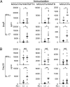
EAE in PD-1−/− mice associates with enhanced inflammatory cytokine production. (A) PD-1+/+ and PD-1−/− mice (C57BL/6 background) were immunized with MOG/CFA/HKMTB/PTX (n = 12), MOG/CFA/HKMTB (n = 11), or MOG/CFA (n = 6) and analyzed 8 d later. (B) PD-1+/+ and PD-1−/− mice were immunized with MOG/CFA/HKMTB/PTX (n = 7), MOG/CFA/HKMTB (n = 7), or MOG/CFA (n = 13) and analyzed 30 d later. (A and B) Sorted CD4+ T cells were restimulated with MOG35–55, and the cytokine concentrations were determined by ELISA. Statistical significance was determined by Student t test. Not significant (NS) P > 0.05, *P < 0.05, and **P < 0.01.
Fig. 3.
Fig. 3.
Splenocytes from HKMTB-immunized PD-1−/− RAG2−/− mice facilitate Th17 induction in vitro. Splenocytes from nonimmunized or HKMTB/CFA-immunized PD-1+/+ RAG2−/− and PD-1−/− RAG2−/− mice (C57BL/6 background) were cultured with naive CD4+ T cells from OTII TCR Tg mice in the presence of OVA323–339 (5 μg/mL) under conditions indicated. Development of the (A) IL-17–producing or (B) IFN-γ–producing CD4+ T cells was examined by intracellular cytokine staining. Essentially the same data were obtained from experiments immunizing PD-1−/− RAG2−/− mice (BALB/c background) and naive CD4+ cells from DO11.10 TCR Tg mice (BALB/c background) (Fig. S2).
Fig. 4.
Fig. 4.
Nonlymphocyte populations of PD-1−/− mice cause enhanced Th17 development and exacerbation of EAE. (A) Lethally irradiated C57BL/6 mice were reconstituted by bone marrow transplantation and adoptive transfer of CD4+ cells as indicated. Mice were immunized with MOG/CFA/HKMTB, and the EAE score was evaluated. Data presented are combined from two independent experiments (total of 18 mice included in each group) (mean ± SEM). Statistical significance was determined by Student t test (*P < 0.05 and **P < 0.01). (B) CD4+ T cells were harvested at day 30 and restimulated by MOG35–55 as indicated in Materials and Methods. IL-17 and IFN-γ were measured. Each point represents an individual mouse, with the black bar indicating the mean value. Mice reconstituted with PD-1−/− RAG2−/− BM showed significantly higher IL-17 than their PD-1+/+ RAG2−/− counterparts by one-tailed Student t test. *P = 0.04.
Fig. 5.
Fig. 5.
CD11b+ cells from PD-1−/− mice efficiently induce Th17 cell development upon HKMTB stimulation. CD11b+ cells were purified from the splenocytes of (A) PD-1+/+ and PD-1−/− mice or (C) PD-1+/+ RAG2−/− and PD-1−/− RAG2−/− mice (all in C57BL/6 background) and were either untreated (ctrl) or stimulated by HKMTB (10 and 100 μg/mL). Three days later, the stimulated cells were cocultured with CD4+ T cells from OTII TCR Tg mice in the presence of OVA323–339 (5 μg/mL) for 4 d. IL-17–producing OTII+ CD4+ T cells (PD-1+/+) were quantified by intracellular cytokine staining. (B) Summary of the data shown in A. Note that CD11b cells from PD-1+/+ and PD-1−/− mice did not induce Th17 by two independent experiments. Data shown for CD11b+ cells are mean ± SD from six independent experiments (for ctrl and HKMTB 100-μg/mL condition) and four experiments (for HKMTB 10-μg/mL condition). Statistical significance was determined by Student t test. *P < 0.05 and **P < 0.01.
Fig. 6.
Fig. 6.
Enhanced production of IL-6 by CD11b+ cells from PD-1−/− mice causes strong Th17 development. CD11b+ cells purified from the splenocytes from PD-1+/+ and PD-1−/− mice (C57BL/6 background) were either untreated (ctrl) or stimulated with HKMTB (10 and 100 μg/mL) or HKLM (10 and 100 μg/mL). Three days later, the production of (A) IL-6, (B) TNF-α, and (C) IL-10 was evaluated. Data presented are combined from three independent experiments (mean ± SD). Significance was determined by Student t test. *P < 0.05. (D) HKMTB-stimulated CD11b+ cells were cocultured in the presence of OTII+CD4+ T cells and OVA323–339 together with rat IgG (200 ng/mL) or anti–IL-6R (as shown) under neutral condition for 4 d. IL-17–producing OTII+ CD4+ T cells were quantified by intracellular cytokine staining. Data shown are representative of three independent experiments.

References

    1. Okazaki T, Honjo T. PD-1 and PD-1 ligands: From discovery to clinical application. Int Immunol. 2007;19(7):813–824. - PubMed
    1. Nishimura H, et al. Autoimmune dilated cardiomyopathy in PD-1 receptor-deficient mice. Science. 2001;291(5502):319–322. - PubMed
    1. Nishimura H, Nose M, Hiai H, Minato N, Honjo T. Development of lupus-like autoimmune diseases by disruption of the PD-1 gene encoding an ITIM motif-carrying immunoreceptor. Immunity. 1999;11(2):141–151. - PubMed
    1. Wang J, et al. PD-1 deficiency results in the development of fatal myocarditis in MRL mice. Int Immunol. 2010;22(6):443–452. - PubMed
    1. Prokunina L, et al. A regulatory polymorphism in PDCD1 is associated with susceptibility to systemic lupus erythematosus in humans. Nat Genet. 2002;32(4):666–669. - PubMed

Publication types

MeSH terms